• 文献检索
  • 文档翻译
  • 深度研究
  • 学术资讯
  • Suppr Zotero 插件Zotero 插件
  • 邀请有礼
  • 套餐&价格
  • 历史记录
应用&插件
Suppr Zotero 插件Zotero 插件浏览器插件Mac 客户端Windows 客户端微信小程序
定价
高级版会员购买积分包购买API积分包
服务
文献检索文档翻译深度研究API 文档MCP 服务
关于我们
关于 Suppr公司介绍联系我们用户协议隐私条款
关注我们

Suppr 超能文献

核心技术专利:CN118964589B侵权必究
粤ICP备2023148730 号-1Suppr @ 2026

文献检索

告别复杂PubMed语法,用中文像聊天一样搜索,搜遍4000万医学文献。AI智能推荐,让科研检索更轻松。

立即免费搜索

文件翻译

保留排版,准确专业,支持PDF/Word/PPT等文件格式,支持 12+语言互译。

免费翻译文档

深度研究

AI帮你快速写综述,25分钟生成高质量综述,智能提取关键信息,辅助科研写作。

立即免费体验

程序性细胞死亡配体1(PD-L1)在弥漫性大B细胞淋巴瘤中的表达及临床价值:一项回顾性研究

Expression and clinical value of programmed cell death-ligand 1 (PD-L1) in diffuse large B cell lymphoma: a retrospective study.

作者信息

Hu Li-Yang, Xu Xiao-Lu, Rao Hui-Lan, Chen Jie, Lai Ren-Chun, Huang Hui-Qiang, Jiang Wen-Qi, Lin Tong-Yu, Xia Zhong-Jun, Cai Qing-Qing

机构信息

State Key Laboratory of Oncology in South China, Collaborative Innovation Center of Cancer Medicine, Sun Yat-sen University Cancer Center, Guangzhou, 510060, Guangdong, P. R. China.

Department of Medical Oncology, Sun Yat-sen University Cancer Center, Guangzhou, 510060, Guangdong, P. R. China.

出版信息

Chin J Cancer. 2017 Dec 16;36(1):94. doi: 10.1186/s40880-017-0262-z.

DOI:10.1186/s40880-017-0262-z
PMID:29246182
原文链接:https://pmc.ncbi.nlm.nih.gov/articles/PMC5732416/
Abstract

BACKGROUND

The programmed cell death-1 (PD-1)/programmed cell death-ligand 1 (PD-L1) pathway inhibits the activation of T cells and plays a crucial role in the negative regulation of cellular and humoral immune responses. Diffuse large B-cell lymphoma (DLBCL) is the most common lymphoid malignancy in adults. In the present study, we aimed to detect the expression of PD-L1 in DLBCL and to analyze its relationship with prognosis.

METHODS

We reviewed medical records of 204 newly diagnosed DLBCL patients in Sun Yat-sen University Cancer Center between October 2005 and August 2012. The expression of PD-L1 in tumor tissues from these 204 patients was detected using immunohistochemical (IHC) assay. The expression of anaplastic lymphoma kinase (ALK), CD5, CD30, and C-Myc in tumor specimens from 109 patients was detected using IHC, and Epstein-Barr virus (EBV)-encoded RNAs (EBERs) were detected using fluorescence in situ hybridization. The Spearman method was used for correlation analysis. The Kaplan-Meier method with log-rank test was used for univariate analysis. Cox proportional hazards model was used for multivariate analysis.

RESULTS

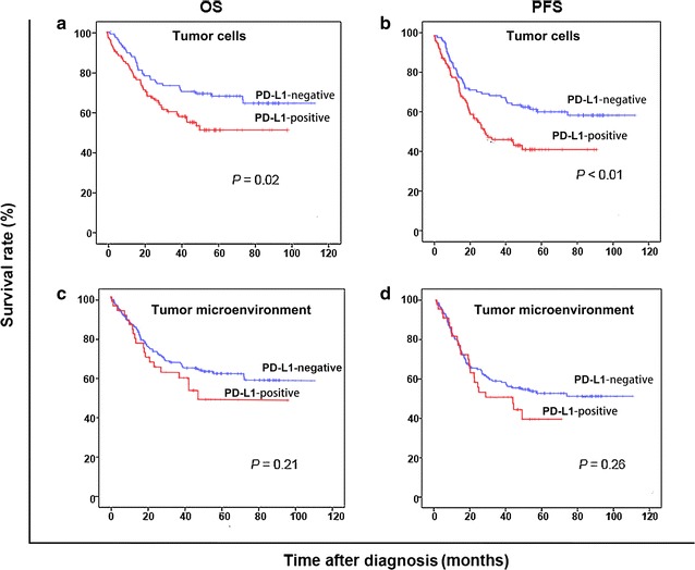
Of the 204 patients, 100 (49.0%) were PD-L1-positive in tumor cells and 44 (21.6%) were PD-L1-positive in tumor microenvironment. PD-L1 expression in tumor cells and tumor microenvironment were more common in the non-germinal center B-cell-like (GCB) subtype than in the GCB subtype (P = 0.02 and P = 0.04). Patients with PD-L1 expression in tumor microenvironment were more likely to be resistant to first-line chemotherapy when compared with the patients without PD-L1 expression in tumor microenvironment (P = 0.03). PD-L1 expression in tumor microenvironment was negatively correlated with C-Myc expression (r = - 0.20, P = 0.04). No correlations were detected between PD-L1 expression and the expression of ALK, CD5, and CD30 as well as EBERs. The 5-year overall survival (OS) rates were 50.0% and 67.3% in patients with and without PD-L1 expression in tumor cells (P = 0.02). PD-L1 expression in tumor cells was an independent risk predictor for OS (P < 0.01).

CONCLUSIONS

PD-L1 expression is more common in the non-GCB subtype than in the GCB subtype. PD-L1 expression in tumor microenvironment has a negative correlation with C-Myc. PD-L1 positivity predicts short survival in DLBCL patients. For patients with PD-L1 expression, more strategy such as anti-PD-L1 antibody treatment should be recommended.

摘要

背景

程序性细胞死亡蛋白1(PD-1)/程序性细胞死亡配体1(PD-L1)通路可抑制T细胞的激活,并在细胞免疫和体液免疫反应的负调控中起关键作用。弥漫性大B细胞淋巴瘤(DLBCL)是成人中最常见的淋巴系统恶性肿瘤。在本研究中,我们旨在检测DLBCL中PD-L1的表达,并分析其与预后的关系。

方法

我们回顾了2005年10月至2012年8月在中山大学肿瘤防治中心新诊断的204例DLBCL患者的病历。采用免疫组织化学(IHC)检测这204例患者肿瘤组织中PD-L1的表达。采用IHC检测109例患者肿瘤标本中间变性淋巴瘤激酶(ALK)、CD5、CD30和C-Myc的表达,采用荧光原位杂交检测爱泼斯坦-巴尔病毒(EBV)编码RNA(EBERs)。采用Spearman法进行相关性分析。采用Kaplan-Meier法和对数秩检验进行单因素分析。采用Cox比例风险模型进行多因素分析。

结果

204例患者中,100例(49.0%)肿瘤细胞中PD-L1呈阳性,44例(21.6%)肿瘤微环境中PD-L1呈阳性。肿瘤细胞和肿瘤微环境中PD-L1的表达在非生发中心B细胞样(GCB)亚型中比在GCB亚型中更常见(P = 0.02和P = 0.04)。与肿瘤微环境中无PD-L1表达的患者相比,肿瘤微环境中有PD-L1表达的患者对一线化疗更可能耐药(P = 0.03)。肿瘤微环境中PD-L1的表达与C-Myc的表达呈负相关(r = -0.20,P = 0.04)。未检测到PD-L1表达与ALK、CD5、CD30的表达以及EBERs之间存在相关性。肿瘤细胞中有和无PD-L1表达的患者5年总生存率(OS)分别为50.0%和67.3%(P = 0.02)。肿瘤细胞中PD-L1的表达是OS的独立风险预测因子(P < 0.01)。

结论

PD-L1的表达在非GCB亚型中比在GCB亚型中更常见。肿瘤微环境中PD-L1的表达与C-Myc呈负相关。PD-L1阳性预示DLBCL患者生存期短。对于有PD-L1表达的患者,应推荐更多策略,如抗PD-L1抗体治疗。

https://cdn.ncbi.nlm.nih.gov/pmc/blobs/b0a7/5732416/71d0a08dde45/40880_2017_262_Fig1_HTML.jpg

相似文献

1
Expression and clinical value of programmed cell death-ligand 1 (PD-L1) in diffuse large B cell lymphoma: a retrospective study.程序性细胞死亡配体1(PD-L1)在弥漫性大B细胞淋巴瘤中的表达及临床价值:一项回顾性研究
Chin J Cancer. 2017 Dec 16;36(1):94. doi: 10.1186/s40880-017-0262-z.
2
Clinicopathologic implication of PD-L1 and phosphorylated STAT3 expression in diffuse large B cell lymphoma.PD-L1 和磷酸化 STAT3 表达在弥漫性大 B 细胞淋巴瘤中的临床病理意义。
J Transl Med. 2018 Nov 20;16(1):320. doi: 10.1186/s12967-018-1689-y.
3
Clinicopathological analysis of primary intestinal diffuse large B-cell lymphoma: Prognostic evaluation of CD5, PD-L1, and Epstein-Barr virus on tumor cells.原发性肠道弥漫性大 B 细胞淋巴瘤的临床病理分析:肿瘤细胞 CD5、PD-L1 和 Epstein-Barr 病毒的预后评估。
Cancer Med. 2018 Dec;7(12):6051-6063. doi: 10.1002/cam4.1875. Epub 2018 Nov 18.
4
PD-L1 expression in EBV-negative diffuse large B-cell lymphoma: clinicopathologic features and prognostic implications.EBV 阴性弥漫性大 B 细胞淋巴瘤中 PD-L1 的表达:临床病理特征及预后意义
Oncotarget. 2016 Sep 13;7(37):59976-59986. doi: 10.18632/oncotarget.11045.
5
Co-expression of PD-L1 and p-AKT is associated with poor prognosis in diffuse large B-cell lymphoma via PD-1/PD-L1 axis activating intracellular AKT/mTOR pathway in tumor cells.PD-L1与p-AKT的共表达通过激活肿瘤细胞内的AKT/mTOR通路的PD-1/PD-L1轴,与弥漫性大B细胞淋巴瘤的不良预后相关。
Oncotarget. 2016 May 31;7(22):33350-62. doi: 10.18632/oncotarget.9061.
6
Expression of programmed cell death 1/programmed cell death ligand 1 in the tumor microenvironments of primary gastrointestinal diffuse large B cell lymphomas.程序性细胞死亡蛋白1/程序性细胞死亡配体1在原发性胃肠道弥漫性大B细胞淋巴瘤肿瘤微环境中的表达
Pathol Res Pract. 2018 Apr;214(4):507-512. doi: 10.1016/j.prp.2018.03.001. Epub 2018 Mar 12.
7
Comparison of PD-L1 detection assays and corresponding significance in evaluation of diffuse large B-cell lymphoma.PD-L1 检测分析方法的比较及其在弥漫性大 B 细胞淋巴瘤评估中的意义。
Cancer Med. 2019 Jul;8(8):3831-3845. doi: 10.1002/cam4.2316. Epub 2019 May 31.
8
Expression of programmed cell death ligand 1 is associated with poor overall survival in patients with diffuse large B-cell lymphoma.程序性细胞死亡配体1的表达与弥漫性大B细胞淋巴瘤患者的总生存期较差相关。
Blood. 2015 Nov 5;126(19):2193-201. doi: 10.1182/blood-2015-02-629600. Epub 2015 Aug 3.
9
PD-L1 expression is low in large B-cell lymphoma with MYC or double-hit translocation.PD-L1 表达在 MYC 或双打击易位的大 B 细胞淋巴瘤中较低。
Hematol Oncol. 2019 Oct;37(4):375-382. doi: 10.1002/hon.2664. Epub 2019 Aug 25.
10
EBV-positive diffuse large B-cell lymphoma features PD-L1 protein but not mRNA overexpression.EBV 阳性弥漫性大 B 细胞淋巴瘤表现为 PD-L1 蛋白过表达,但 mRNA 不表达。
Pathology. 2018 Dec;50(7):725-729. doi: 10.1016/j.pathol.2018.08.011. Epub 2018 Oct 30.

引用本文的文献

1
PD-L1 Expression and Tumor Microenvironment Dynamics in Diffuse Large B-Cell Lymphoma: Immunophenotypic Insights.弥漫性大B细胞淋巴瘤中PD-L1表达与肿瘤微环境动态变化:免疫表型见解
J Med Life. 2025 May;18(5):463-471. doi: 10.25122/jml-2025-0078.
2
Exploring ferroptosis and miRNAs: implications for cancer modulation and therapy.探索铁死亡与微小RNA:对癌症调控和治疗的意义
Mol Cell Biochem. 2025 Jan 27. doi: 10.1007/s11010-024-05169-9.
3
Programmed Death Ligand 1 (PD-L1) Expression in Lymphomas: State of the Art.程序性死亡配体 1(PD-L1)在淋巴瘤中的表达:最新进展。

本文引用的文献

1
Prognostic value of programmed death-1, programmed death-ligand 1, programmed death-ligand 2 expression, and CD8(+) T cell density in primary tumors and metastatic lymph nodes from patients with stage T1-4N+M0 gastric adenocarcinoma.程序性死亡蛋白1、程序性死亡配体1、程序性死亡配体2表达以及CD8(+) T细胞密度在T1-4N+M0期胃腺癌患者原发性肿瘤和转移性淋巴结中的预后价值
Chin J Cancer. 2017 Jul 29;36(1):61. doi: 10.1186/s40880-017-0226-3.
2
PD-1/PD-L1 inhibitors in haematological malignancies: update 2017.血液系统恶性肿瘤中的PD-1/PD-L1抑制剂:2017年更新
Immunology. 2017 Nov;152(3):357-371. doi: 10.1111/imm.12788. Epub 2017 Aug 4.
3
Int J Mol Sci. 2024 Jun 11;25(12):6447. doi: 10.3390/ijms25126447.
4
Is There an Immunohistochemical PD-L1 Cut-Off Point That Serves as a Prognostic Indicator for Large B-Cell Lymphomas?是否存在一个免疫组化PD-L1切点可作为大B细胞淋巴瘤的预后指标?
Diagnostics (Basel). 2024 May 31;14(11):1167. doi: 10.3390/diagnostics14111167.
5
The Prognostic Impact of Tumor Microenvironment and Checkpoint Blockade-Associated Molecules (PD-1, PD-L1, CD163 and CD14) in Nodal Diffuse Large B-cell Lymphoma, NOS.肿瘤微环境和检查点阻断相关分子(PD-1、PD-L1、CD163和CD14)在结内弥漫性大B细胞淋巴瘤(NOS)中的预后影响
Indian J Hematol Blood Transfus. 2024 Apr;40(2):340-345. doi: 10.1007/s12288-023-01667-w. Epub 2023 May 11.
6
The prognostic significance of programmed cell death protein 1 and its ligand on lymphoma cells and tumor-immune cells in diffuse large B-cell lymphoma, not otherwise specified.程序性细胞死亡蛋白 1 及其配体对弥漫性大 B 细胞淋巴瘤、非特指型中淋巴瘤细胞和肿瘤免疫细胞的预后意义。
Radiol Oncol. 2024 Feb 21;58(1):99-109. doi: 10.2478/raon-2024-0010. eCollection 2024 Mar 1.
7
Presence of Epstein-Barr virus (EBV) antigens detected by sensitive methods has no influence on local immune environment in diffuse large B cell lymphoma.采用灵敏方法检测到的爱泼斯坦-巴尔病毒(EBV)抗原的存在,对弥漫性大B细胞淋巴瘤的局部免疫环境没有影响。
Cancer Immunol Immunother. 2024 Jan 27;73(2):29. doi: 10.1007/s00262-023-03617-x.
8
Activation of STING by SAMHD1 Deficiency Promotes PANoptosis and Enhances Efficacy of PD-L1 Blockade in Diffuse Large B-cell Lymphoma.SAMHD1 缺陷激活 STING 促进弥漫性大 B 细胞淋巴瘤的 PANoptosis 并增强 PD-L1 阻断的疗效。
Int J Biol Sci. 2023 Aug 28;19(14):4627-4643. doi: 10.7150/ijbs.85236. eCollection 2023.
9
Immunohistochemical Expression of Programmed Death Ligand 1 in Oral Extranodal Diffuse Large B Cell Lymphoma.程序性死亡配体1在口腔结外弥漫性大B细胞淋巴瘤中的免疫组化表达
Eur J Dent. 2023 May;17(2):424-430. doi: 10.1055/s-0042-1747951. Epub 2022 Aug 9.
10
Diagnostic Performance of PD-L1 versus PD-1 Expression in Circulating CD20 Cells in Diffuse Large B-Cell Lymphoma.弥漫性大B细胞淋巴瘤中循环CD20细胞的PD-L1与PD-1表达的诊断性能
Antibodies (Basel). 2022 Feb 16;11(1):15. doi: 10.3390/antib11010015.
Tumor Microenvironment and Checkpoint Molecules in Primary Cutaneous Diffuse Large B-Cell Lymphoma-New Therapeutic Targets.
原发性皮肤弥漫性大B细胞淋巴瘤中的肿瘤微环境与检查点分子——新的治疗靶点
Am J Surg Pathol. 2017 Jul;41(7):998-1004. doi: 10.1097/PAS.0000000000000851.
4
Checkpoint inhibitors in hematological malignancies.血液系统恶性肿瘤中的检查点抑制剂
J Hematol Oncol. 2017 May 8;10(1):103. doi: 10.1186/s13045-017-0474-3.
5
The expression and clinical relevance of PD-1, PD-L1, and TP63 in patients with diffuse large B-cell lymphoma.弥漫性大B细胞淋巴瘤患者中PD-1、PD-L1和TP63的表达及临床相关性
Medicine (Baltimore). 2017 Apr;96(15):e6398. doi: 10.1097/MD.0000000000006398.
6
PD-1-PD-L1 immune-checkpoint blockade in B-cell lymphomas.PD-1-PD-L1 免疫检查点阻断在 B 细胞淋巴瘤中的应用。
Nat Rev Clin Oncol. 2017 Apr;14(4):203-220. doi: 10.1038/nrclinonc.2016.168. Epub 2016 Nov 2.
7
The T-cell Receptor Repertoire Influences the Tumor Microenvironment and Is Associated with Survival in Aggressive B-cell Lymphoma.T 细胞受体谱影响肿瘤微环境,并与侵袭性 B 细胞淋巴瘤的生存相关。
Clin Cancer Res. 2017 Apr 1;23(7):1820-1828. doi: 10.1158/1078-0432.CCR-16-1576. Epub 2016 Sep 20.
8
New risk factors and new tendency for central nervous system relapse in patients with diffuse large B-cell lymphoma: a retrospective study.弥漫性大B细胞淋巴瘤患者中枢神经系统复发的新危险因素及新趋势:一项回顾性研究
Chin J Cancer. 2016 Sep 13;35(1):87. doi: 10.1186/s40880-016-0150-y.
9
Atezolizumab in combination with bevacizumab enhances antigen-specific T-cell migration in metastatic renal cell carcinoma.阿替利珠单抗联合贝伐珠单抗增强转移性肾细胞癌的抗原特异性 T 细胞迁移。
Nat Commun. 2016 Aug 30;7:12624. doi: 10.1038/ncomms12624.
10
PD-L1 expression in EBV-negative diffuse large B-cell lymphoma: clinicopathologic features and prognostic implications.EBV 阴性弥漫性大 B 细胞淋巴瘤中 PD-L1 的表达:临床病理特征及预后意义
Oncotarget. 2016 Sep 13;7(37):59976-59986. doi: 10.18632/oncotarget.11045.