Zhang Honghao, Louie Ke'ale W, Kulkarni Anshul K, Zapien-Guerra Karen, Yang Jingwen, Mishina Yuji
Department of Biologic and Materials Sciences & Prosthodontics, School of Dentistry University of Michigan Ann Arbor MI USA.
JBMR Plus. 2021 Dec 24;6(2):e10589. doi: 10.1002/jbm4.10589. eCollection 2022 Feb.
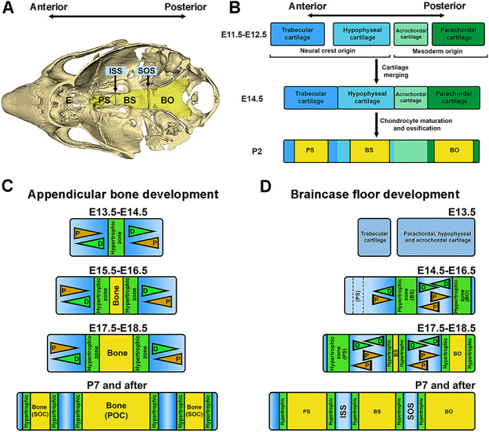
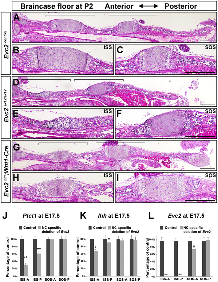
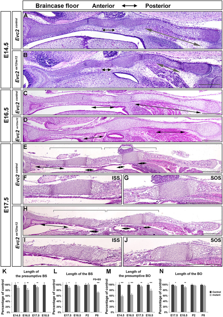
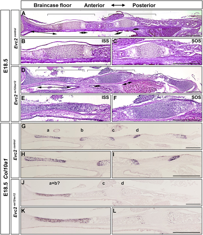
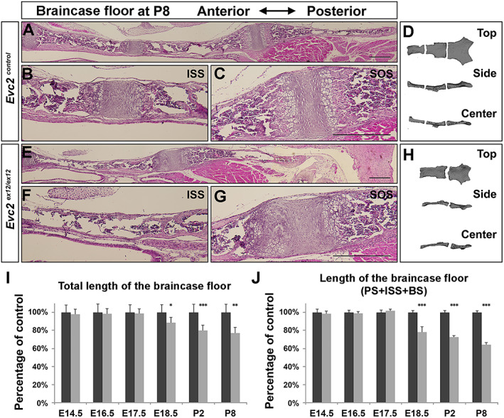
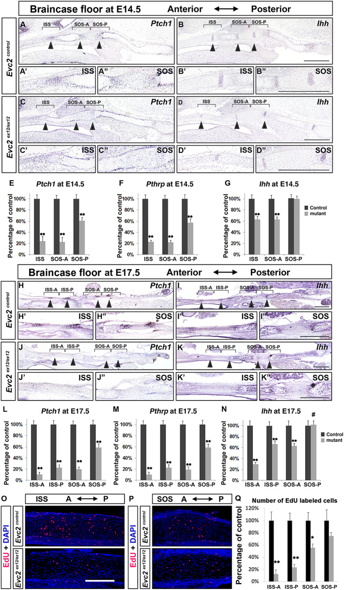
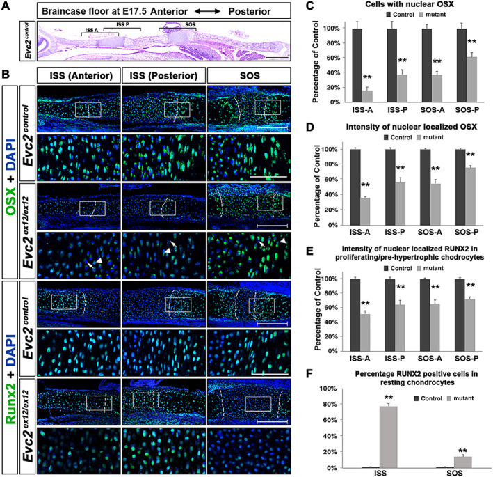
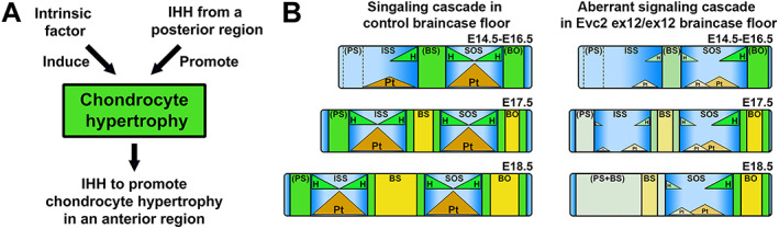
The cranial base is a critical structure in the head, which is composed of endoskeletal and dermal skeletal. The braincase floor, part of the cranial base, is a midline structure of the head. Because it is a midline structure connecting the posterior skull with the facial region, braincase floor is critical for the orientation of the facial structure. Shortened braincase floor leads to mid-facial hypoplasia and malocclusions. During embryonic development, elongation of the braincase floor occurs through endochondral ossification in the parachordal cartilage, hypophyseal cartilage, and trabecular cartilage, which leads to formation of basioccipital (BO), basisphenoid (BS), and presphenoid (PS) bones, respectively. Currently, little is known about whether maturation of parachordal cartilage, hypophyseal cartilage, and trabecular cartilage occurs in a simultaneous or sequential manner and if the formation of one impacts the others. Our previous studies demonstrated that loss of function of ciliary protein leads to premature fusion in the intersphenoid synchondrosis (ISS). In this study, we take advantage of mutant mice to delineate the mechanism governing synchondrosis formation. Our analysis supports a cascade mechanism on the spatiotemporal regulation of the braincase floor development that the hypertrophy of parachordal cartilage (posterior side) impacts the hypertrophy of hypophyseal cartilage (middle) and trabecular cartilage (anterior side) in a sequential manner. The cascade mechanism well explains the premature fusion of the ISS in mutant mice and is instructive to understand the specifically shortened anterior end of the braincase floor in various types of genetic syndromes. © 2021 The Authors. published by Wiley Periodicals LLC on behalf of American Society for Bone and Mineral Research.
颅底是头部的一个关键结构,由内骨骼和皮骨骼组成。颅底的一部分——脑颅底部,是头部的中线结构。由于它是连接后颅骨和面部区域的中线结构,脑颅底部对于面部结构的定向至关重要。脑颅底部缩短会导致面中部发育不全和咬合不正。在胚胎发育过程中,脑颅底部的伸长通过副索软骨、垂体软骨和小梁软骨的软骨内成骨发生,分别导致枕骨基部(BO)、蝶骨基部(BS)和蝶骨前部(PS)的形成。目前,对于副索软骨、垂体软骨和小梁软骨的成熟是同时还是顺序发生,以及其中一个的形成是否会影响其他软骨,人们知之甚少。我们之前的研究表明,睫状蛋白功能丧失会导致蝶骨间软骨结合(ISS)过早融合。在本研究中,我们利用突变小鼠来阐明软骨结合形成的机制。我们的分析支持了一种关于脑颅底部发育时空调节的级联机制,即副索软骨(后侧)的肥大依次影响垂体软骨(中间)和小梁软骨(前侧)的肥大。这种级联机制很好地解释了突变小鼠中ISS的过早融合,并且对于理解各种类型遗传综合征中脑颅底部前端特异性缩短具有指导意义。© 2021作者。由Wiley Periodicals LLC代表美国骨与矿物质研究学会出版。