Department of Biosciences, Rice University, Houston, United States.
Elife. 2022 Mar 3;11:e74307. doi: 10.7554/eLife.74307.
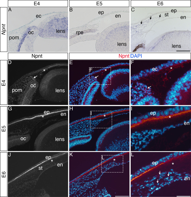
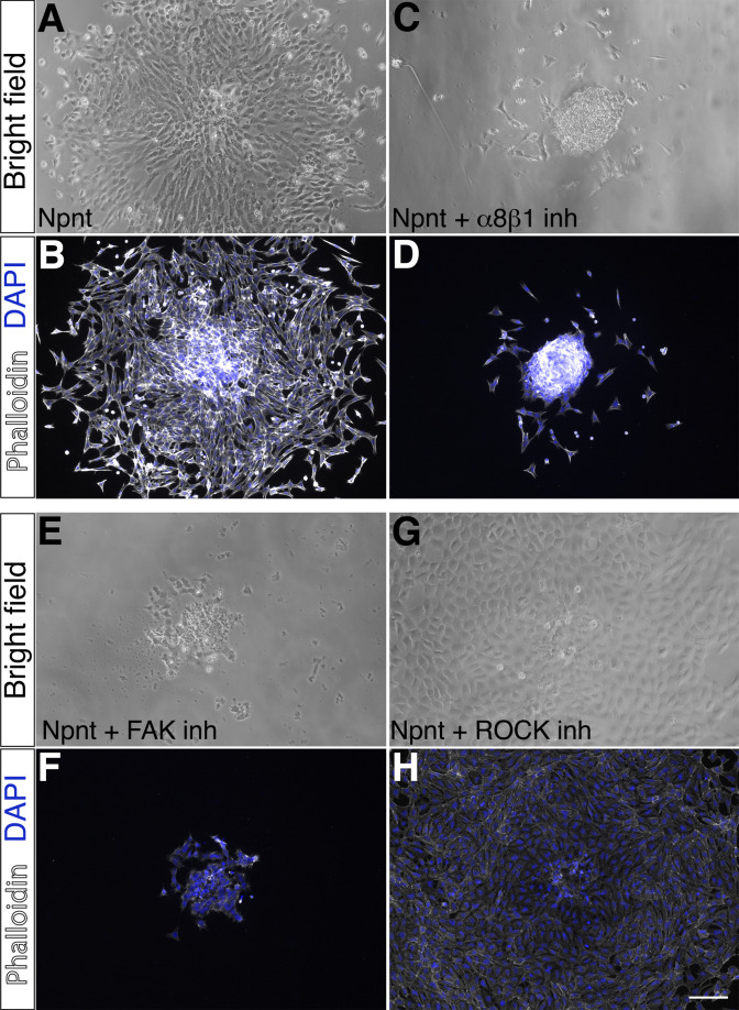
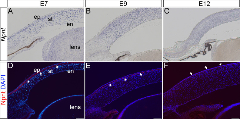
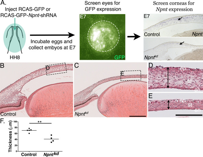
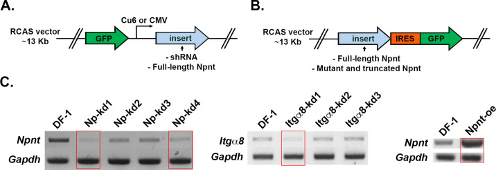
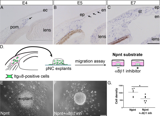
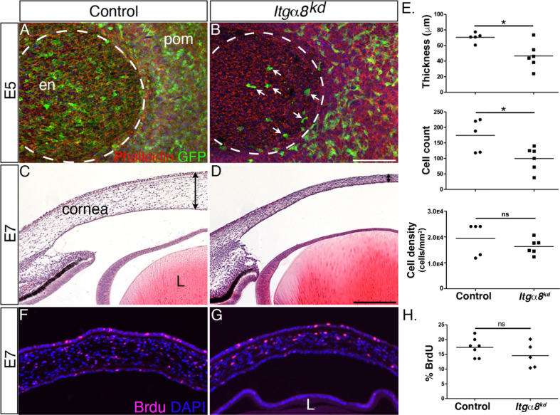
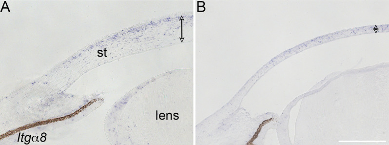
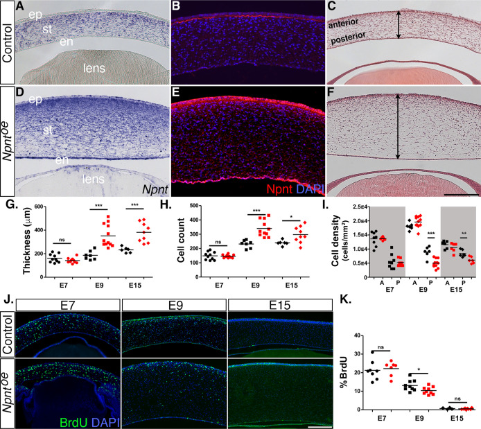
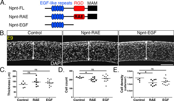
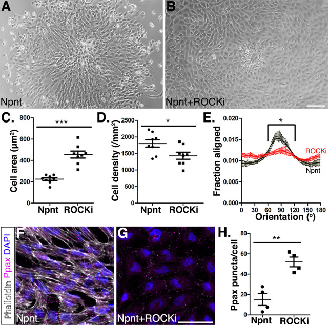
During development, cells aggregate at tissue boundaries to form normal tissue architecture of organs. However, how cells are segregated into tissue precursors remains largely unknown. Cornea development is a perfect example of this process whereby neural crest cells aggregate in the periocular region prior to their migration and differentiation into corneal cells. Our recent RNA-seq analysis identified upregulation of nephronectin (Npnt) transcripts during early stages of corneal development where its function has not been investigated. We found that Npnt mRNA and protein are expressed by various ocular tissues, including the migratory periocular neural crest (pNC), which also express the integrin alpha 8 (Itgα8) receptor. Knockdown of either or attenuated cornea development, whereas overexpression of resulted in cornea thickening. Moreover, overexpression of Npnt variants lacking RGD-binding sites did not affect corneal thickness. Neither the knockdown nor augmentation of Npnt caused significant changes in cell proliferation, suggesting that Npnt directs pNC migration into the cornea. In vitro analyses showed that Npnt promotes pNC migration from explanted periocular mesenchyme, which requires Itgα8, focal adhesion kinase, and Rho kinase. Combined, these data suggest that Npnt augments cell migration into the presumptive cornea extracellular matrix by functioning as a substrate for Itgα8-positive pNC cells.
在发育过程中,细胞聚集在组织边界处形成器官的正常组织结构。然而,细胞如何被分隔成组织前体在很大程度上仍然未知。角膜发育就是一个很好的例子,神经嵴细胞聚集在眼眶区域,然后迁移并分化为角膜细胞。我们最近的 RNA 测序分析表明,在角膜发育的早期阶段,纤连蛋白(Npnt)转录本上调,但其功能尚未得到研究。我们发现 Npnt mRNA 和蛋白在各种眼部组织中表达,包括迁移的眼眶神经嵴(pNC),其也表达整合素 alpha 8(Itgα8)受体。或 的敲低减弱了角膜发育,而过表达 则导致角膜增厚。此外,缺乏 RGD 结合位点的 Npnt 变体的过表达不会影响角膜厚度。Npnt 的敲低或过表达均未导致细胞增殖发生显著变化,这表明 Npnt 指导 pNC 迁移到角膜中。体外分析表明,Npnt 促进源自眼眶间充质的 pNC 迁移,这需要 Itgα8、粘着斑激酶和 Rho 激酶。综合这些数据表明,Npnt 通过作为 Itgα8 阳性 pNC 细胞的基质,增强细胞向假定的角膜细胞外基质的迁移。