• 文献检索
  • 文档翻译
  • 深度研究
  • 学术资讯
  • Suppr Zotero 插件Zotero 插件
  • 邀请有礼
  • 套餐&价格
  • 历史记录
应用&插件
Suppr Zotero 插件Zotero 插件浏览器插件Mac 客户端Windows 客户端微信小程序
定价
高级版会员购买积分包购买API积分包
服务
文献检索文档翻译深度研究API 文档MCP 服务
关于我们
关于 Suppr公司介绍联系我们用户协议隐私条款
关注我们

Suppr 超能文献

核心技术专利:CN118964589B侵权必究
粤ICP备2023148730 号-1Suppr @ 2026

文献检索

告别复杂PubMed语法,用中文像聊天一样搜索,搜遍4000万医学文献。AI智能推荐,让科研检索更轻松。

立即免费搜索

文件翻译

保留排版,准确专业,支持PDF/Word/PPT等文件格式,支持 12+语言互译。

免费翻译文档

深度研究

AI帮你快速写综述,25分钟生成高质量综述,智能提取关键信息,辅助科研写作。

立即免费体验

子宫上皮样平滑肌瘤:一例伴有磁共振成像表现的病例报告

Epithelioid leiomyoma of the uterus: A case report with magnetic resonance imaging findings.

作者信息

Kita Ayaka, Maeda Tetsuo, Kitajima Kazuhiro, Murakoshi Homare, Watanabe Takahiro, Inagaki Mieko, Yoshida Shigeki

机构信息

Department of Gynecology and Obstetrics, Chibune General Hospital, 3-2-39 Fukumachi, Nishiyodogawa-ku, Osaka City, Osaka 555-0034, Japan.

Department of Radiology, Chibune General Hospital, 3-2-39 Fukumachi, Nishiyodogawa-ku, Osaka City, Osaka 555-0034, Japan.

出版信息

Case Rep Womens Health. 2022 Jan 20;34:e00386. doi: 10.1016/j.crwh.2022.e00386. eCollection 2022 Apr.

DOI:10.1016/j.crwh.2022.e00386
PMID:35242596
原文链接:https://pmc.ncbi.nlm.nih.gov/articles/PMC8873549/
Abstract

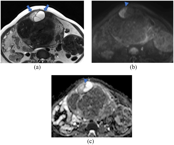
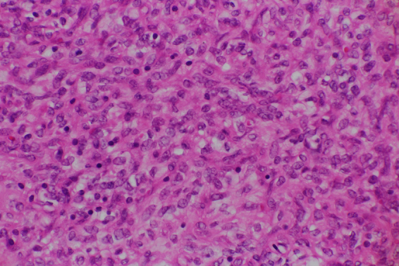
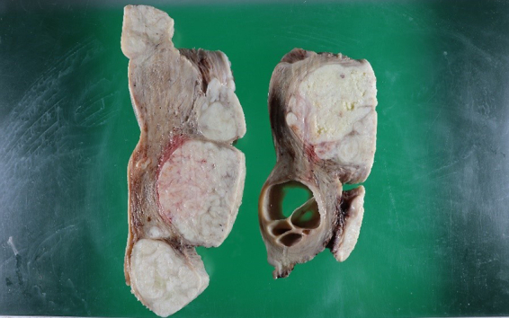
Epithelioid leiomyoma of the uterus is rare, and its prognostic factors have not been well established. Moreover, radiologic findings of this disease have not been previously documented. This is a case of a 49-year-old woman with epithelioid leiomyoma of the uterus. Magnetic resonance imaging (MRI) revealed a heterogeneous high-intensity mass with multiple ordinary uterine leiomyomas. The mass showed a slightly diffusion-restricted site. Since benign tumors could not be confidently diagnosed using these MRI findings, total abdominal hysterectomy with bilateral salpingectomy was performed, and a pathological diagnosis of epithelioid leiomyoma of the uterus was established. Microscopically, this lesion showed edematous changes and cyst formation, causing a heterogeneous appearance on T2-weighted images. In addition, the diffusion-restricted site is considered to be consistent with areas of solid and dense proliferation of tumor cells. The patient survived and was well 10 months after the surgery. It is important to recognize this benign variant of leiomyoma with an unusual appearance, to provide appropriate therapeutic management.

摘要

子宫上皮样平滑肌瘤较为罕见,其预后因素尚未完全明确。此外,此前尚无该疾病的放射学表现记录。本文报告一例49岁患有子宫上皮样平滑肌瘤的女性病例。磁共振成像(MRI)显示一个异质性高强度肿块,伴有多个普通子宫平滑肌瘤。该肿块显示有轻微扩散受限部位。由于仅凭这些MRI表现无法确诊为良性肿瘤,遂行全腹子宫切除术及双侧输卵管切除术,术后病理诊断为子宫上皮样平滑肌瘤。显微镜下,该病变呈现水肿改变及囊肿形成,导致在T2加权图像上表现为异质性外观。此外,扩散受限部位被认为与肿瘤细胞实性密集增殖区域相符。患者术后存活,术后10个月情况良好。认识这种具有不寻常表现的平滑肌瘤良性变体,对于提供恰当的治疗管理很重要。

https://cdn.ncbi.nlm.nih.gov/pmc/blobs/6252/8873549/70f33bea02f2/gr3.jpg
https://cdn.ncbi.nlm.nih.gov/pmc/blobs/6252/8873549/fcade55c1efd/gr1.jpg
https://cdn.ncbi.nlm.nih.gov/pmc/blobs/6252/8873549/9c293a74eca0/gr2.jpg
https://cdn.ncbi.nlm.nih.gov/pmc/blobs/6252/8873549/70f33bea02f2/gr3.jpg
https://cdn.ncbi.nlm.nih.gov/pmc/blobs/6252/8873549/fcade55c1efd/gr1.jpg
https://cdn.ncbi.nlm.nih.gov/pmc/blobs/6252/8873549/9c293a74eca0/gr2.jpg
https://cdn.ncbi.nlm.nih.gov/pmc/blobs/6252/8873549/70f33bea02f2/gr3.jpg

相似文献

1
Epithelioid leiomyoma of the uterus: A case report with magnetic resonance imaging findings.子宫上皮样平滑肌瘤:一例伴有磁共振成像表现的病例报告
Case Rep Womens Health. 2022 Jan 20;34:e00386. doi: 10.1016/j.crwh.2022.e00386. eCollection 2022 Apr.
2
Epithelioid cotyledonoid leiomyoma of uterus.子宫上皮样子叶样平滑肌瘤
BMJ Case Rep. 2014 Jan 10;2014:bcr2013202434. doi: 10.1136/bcr-2013-202434.
3
An unusual case of uterine cotyledonoid dissecting leiomyoma with adenomyosis.一例罕见的伴有子宫腺肌病的子宫叶状剥脱性平滑肌瘤。
Diagn Pathol. 2016 Aug 4;11(1):69. doi: 10.1186/s13000-016-0523-1.
4
A subserosal, pedunculated, multilocular uterine leiomyoma with ovarian tumor-like morphology and histological architecture of adenomatoid tumors: a case report and review of the literature.一例具有腺瘤样瘤的卵巢肿瘤样形态和组织学结构的浆膜下带蒂多房性子宫平滑肌瘤:病例报告及文献复习
J Med Case Rep. 2016 Dec 20;10(1):352. doi: 10.1186/s13256-016-1167-1.
5
MRI appearance of primary giant ovarian leiomyoma in a hysterectomised woman.子宫切除术后女性原发性巨大卵巢平滑肌瘤的磁共振成像表现
Br J Radiol. 2006 Oct;79(946):e126-8. doi: 10.1259/bjr/54090919.
6
Hydropic leiomyoma: A radiologic pathologic correlation of a rare uterine tumor.水肿性平滑肌瘤:一种罕见子宫肿瘤的放射学与病理学相关性研究
Radiol Case Rep. 2022 Jun 27;17(9):3151-3156. doi: 10.1016/j.radcr.2022.06.008. eCollection 2022 Sep.
7
Giant cystic leiomyoma of the uterus occupying the retroperitoneal space.占据腹膜后间隙的子宫巨大囊性平滑肌瘤。
J Radiol Case Rep. 2013 Dec 1;7(12):35-40. doi: 10.3941/jrcr.v7i12.1447. eCollection 2013 Dec.
8
Hydropic leiomyoma of the uterus presenting as a giant abdominal mass.表现为巨大腹部肿块的子宫水肿性平滑肌瘤。
BMJ Case Rep. 2015 Sep 8;2015:bcr2015211929. doi: 10.1136/bcr-2015-211929.
9
The Management of the Cotyledonoid Leiomyoma of the Uterus: A Narrative Review of the Literature.子宫胎叶状平滑肌瘤的处理:文献回顾性叙述。
Int J Environ Res Public Health. 2021 Aug 12;18(16):8521. doi: 10.3390/ijerph18168521.
10
Epithelioid Angiosarcoma Arising from a Huge Leiomyoma: A Case Report and a Literature Review.源于巨大平滑肌瘤的上皮样血管肉瘤:一例报告及文献复习
Case Rep Obstet Gynecol. 2018 Jun 4;2018:7591769. doi: 10.1155/2018/7591769. eCollection 2018.

引用本文的文献

1
Epithelioid Leiomyoma of the Uterus: A Rare Tumor Mimicking Malignancy.子宫上皮样平滑肌瘤:一种酷似恶性肿瘤的罕见肿瘤。
Cureus. 2025 Jul 24;17(7):e88675. doi: 10.7759/cureus.88675. eCollection 2025 Jul.

本文引用的文献

1
Uterine leiomyoblastoma.子宫平滑肌瘤样细胞瘤
BMJ Case Rep. 2021 Feb 18;14(2):e241533. doi: 10.1136/bcr-2020-241533.
2
Diagnostic Value of FDG PET/MRI in Females With Pelvic Malignancy-A Systematic Review of the Literature.18F-氟脱氧葡萄糖正电子发射断层显像/磁共振成像对女性盆腔恶性肿瘤的诊断价值——文献系统综述
Front Oncol. 2020 Sep 29;10:519440. doi: 10.3389/fonc.2020.519440. eCollection 2020.
3
Magnetic resonance imaging features of uterine sarcoma and mimickers.磁共振成像在子宫肉瘤及类似病变中的特征表现。
Abdom Radiol (NY). 2017 Jun;42(6):1762-1772. doi: 10.1007/s00261-017-1076-9.
4
Comparison of the diagnostic accuracy of contrast-enhanced MRI and diffusion-weighted MRI in the differentiation between uterine leiomyosarcoma / smooth muscle tumor with uncertain malignant potential and benign leiomyoma.对比增强磁共振成像(MRI)与扩散加权磁共振成像在子宫平滑肌肉瘤/恶性潜能不确定的平滑肌肿瘤与良性平滑肌瘤鉴别诊断中的诊断准确性比较。
J Magn Reson Imaging. 2016 Feb;43(2):333-42. doi: 10.1002/jmri.24998. Epub 2015 Sep 18.
5
Present and future role of FDG-PET/CT imaging in the management of gynecologic malignancies.氟代脱氧葡萄糖正电子发射断层扫描/计算机断层扫描(FDG-PET/CT)成像在妇科恶性肿瘤管理中的当前及未来作用
Jpn J Radiol. 2014 Jun;32(6):313-23. doi: 10.1007/s11604-014-0317-x. Epub 2014 Apr 18.
6
Clinical application of diffusion-weighted imaging for preoperative differentiation between uterine leiomyoma and leiomyosarcoma.弥散加权成像在子宫肌瘤与平滑肌肉瘤术前鉴别诊断中的临床应用。
Am J Obstet Gynecol. 2014 Apr;210(4):368.e1-368.e8. doi: 10.1016/j.ajog.2013.12.028. Epub 2013 Dec 22.
7
How to differentiate benign from malignant myometrial tumours using MR imaging.如何通过磁共振成像区分良性和恶性子宫肌肿瘤。
Eur Radiol. 2013 Aug;23(8):2306-14. doi: 10.1007/s00330-013-2819-9. Epub 2013 Apr 8.
8
Atypical leiomyomas of the uterus: a clinicopathologic study of 51 cases.子宫非典型性平滑肌瘤:51 例临床病理研究。
Am J Surg Pathol. 2013 May;37(5):643-9. doi: 10.1097/PAS.0b013e3182893f36.
9
Cell cycle regulatory markers in uterine atypical leiomyoma and leiomyosarcoma: immunohistochemical study of 68 cases with clinical follow-up.68 例具有临床随访的子宫非典型平滑肌瘤和平滑肌肉瘤中细胞周期调控标志物的免疫组织化学研究。
Am J Surg Pathol. 2013 May;37(5):634-42. doi: 10.1097/PAS.0b013e318287779c.
10
An unusual case of an osteosarcoma arising in a leiomyoma of the uterus.子宫平滑肌瘤中发生骨肉瘤的罕见病例。
Ann Saudi Med. 2012 Sep-Oct;32(5):544-6. doi: 10.5144/0256-4947.2012.23.5.1111.