Department of Neuroimaging and Interventional Neuroradiology, Apollo Hospital, Bangalore, India.
Department of Neuroimaging and Interventional Neuroradiology, St. Johns Medical College & Hospital, Bangalore, India.
World Neurosurg. 2022 Jun;162:e131-e140. doi: 10.1016/j.wneu.2022.02.107. Epub 2022 Mar 4.
Mucormycosis infection of the maxillofacial region and brain has been associated with coronavirus disease 2019 (COVID-19) infection. Mucormycosis was relatively a rare infection before COVID-19, and imaging findings are not very well described.
A retrospective imaging study of 101 patients diagnosed with COVID-19-associated mucormycosis by histopathology and/or culture was performed. All patients underwent computed tomography and/or magnetic resonance imaging based on the clinical condition of the patient and on consensus decision by the team of treating physicians. A simple 3-stage classification system based on imaging findings was adopted.
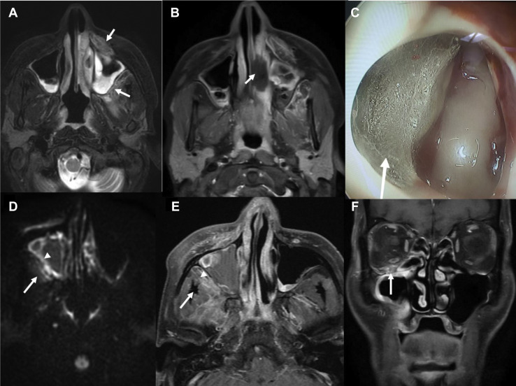
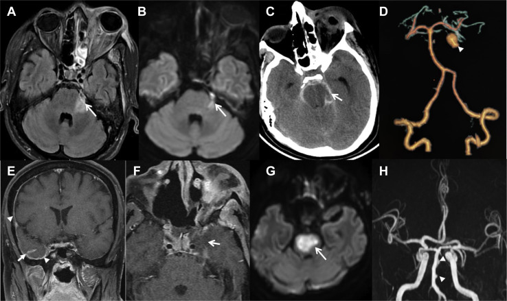
One hundred one cases were included in the final analysis (mean age = 55.1 years; male/female ratio = 67:34). The affected patients had diabetes in 94% of the instances (n = 95), 80.1% (n = 81) received steroids), whereas 59.4% (n = 60) patients received supplemental oxygen. The majority underwent surgical intervention, whereas in 6 cases, patients were treated with antibiotic regimens. Sixty subjects improved following therapy, whereas 18 eventually succumbed to the illness. We noted a significant positive correlation between the imaging stage and outcomes. No association was seen between other clinical parameters and final clinical outcomes. Salient imaging findings include lack of normal sinonasal mucosal enhancement, perisinus inflammation, ischemic optic neuropathy, perineural spread, pachymeningeal enhancement, and presence of strokes.
We describe the imaging findings in the largest cohort of patients with rhino-orbito-cerebral mucormycosis in the context of the current COVID-19 pandemic. A simplified staging system described here is helpful for standardized reporting and carries prognostic information.
新冠肺炎(COVID-19)感染与颌面部和脑部的毛霉菌感染有关。在 COVID-19 之前,毛霉菌病相对较少见,影像学表现也不是很明确。
对经组织病理学和/或培养诊断为 COVID-19 相关毛霉菌病的 101 例患者进行回顾性影像学研究。所有患者均根据患者的临床情况以及治疗医生团队的共识决定进行计算机断层扫描和/或磁共振成像。采用了一种基于影像学表现的简单 3 期分类系统。
最终分析纳入 101 例病例(平均年龄 55.1 岁;男女比例 67:34)。受影响的患者中有 94%(95 例)患有糖尿病,80.1%(81 例)接受了皮质类固醇治疗,59.4%(60 例)接受了补充氧气治疗。大多数患者接受了手术干预,而有 6 例患者仅接受了抗生素治疗方案。60 例患者经治疗后病情改善,而 18 例最终死亡。我们发现影像学分期与预后之间存在显著的正相关。其他临床参数与最终临床结局之间未见关联。主要的影像学表现包括:正常鼻窦黏膜增强缺失、窦周炎症、缺血性视神经病变、神经周围播散、硬脑膜增强和中风的存在。
我们描述了当前 COVID-19 大流行背景下最大的一组鼻眶脑毛霉菌病患者的影像学表现。这里描述的简化分期系统有助于标准化报告,并具有预后信息。