Zhang Shuang, Liao Xue-Jing, Wang Jia, Shen Yi, Shi Han-Fen, Zou Yan, Ma Chong-Yang, Wang Xue-Qian, Wang Qing-Guo, Wang Xu, Xu Ming-Yang, Cheng Fa-Feng, Bai Wan-Zhu
Beijing Key Laboratory, School of Basic Medical Sciences, Beijing University of Chinese Medicine, Beijing, China.
Institute of Acupuncture and Moxibustion, China Academy of Chinese Medical Sciences, Beijing, China.
Neural Regen Res. 2022 Oct;17(10):2247-2252. doi: 10.4103/1673-5374.336876.
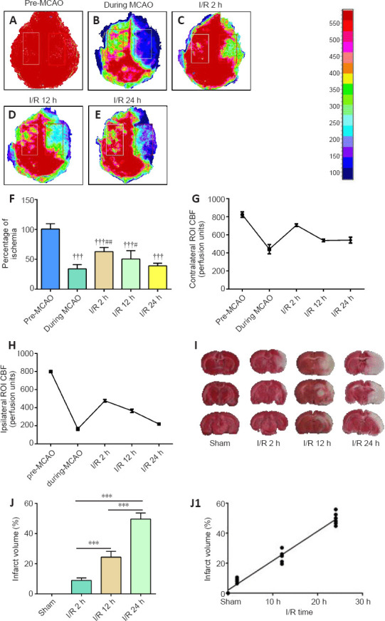
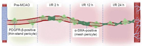
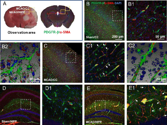
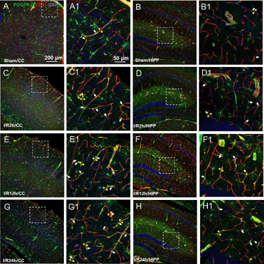
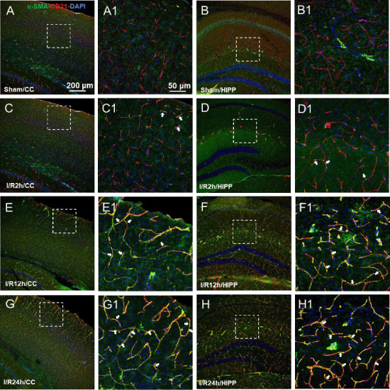
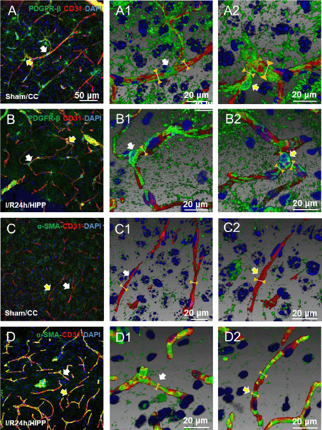
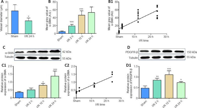
Pericytes, as the mural cells surrounding the microvasculature, play a critical role in the regulation of microcirculation; however, how these cells respond to ischemic stroke remains unclear. To determine the temporal alterations in pericytes after ischemia/reperfusion, we used the 1-hour middle cerebral artery occlusion model, which was examined at 2, 12, and 24 hours after reperfusion. Our results showed that in the reperfused regions, the cerebral blood flow decreased and the infarct volume increased with time. Furthermore, the pericytes in the infarct regions contracted and acted on the vascular endothelial cells within 24 hours after reperfusion. These effects may result in incomplete microcirculation reperfusion and a gradual worsening trend with time in the acute phase. These findings provide strong evidence for explaining the "no-reflow" phenomenon that occurs after recanalization in clinical practice.
周细胞作为围绕微脉管系统的壁细胞,在微循环调节中起关键作用;然而,这些细胞对缺血性中风如何反应仍不清楚。为了确定缺血/再灌注后周细胞的时间变化,我们使用了1小时大脑中动脉闭塞模型,并在再灌注后2小时、12小时和24小时进行检查。我们的结果表明,在再灌注区域,脑血流量随时间减少,梗死体积随时间增加。此外,梗死区域的周细胞在再灌注后24小时内收缩并作用于血管内皮细胞。这些作用可能导致微循环再灌注不完全,并在急性期随时间逐渐恶化。这些发现为解释临床实践中再通后出现的“无复流”现象提供了有力证据。