Department of Microbiology and Immunology at the Peter Doherty Institute for Infection and Immunity, The University of Melbourne, Parkville, VIC, 3010, Australia.
Australian Research Council Centre of Excellence for Advanced Molecular Imaging, University of Melbourne, Parkville, VIC, 3010, Australia.
Sci Rep. 2022 Mar 8;12(1):4034. doi: 10.1038/s41598-022-07704-4.
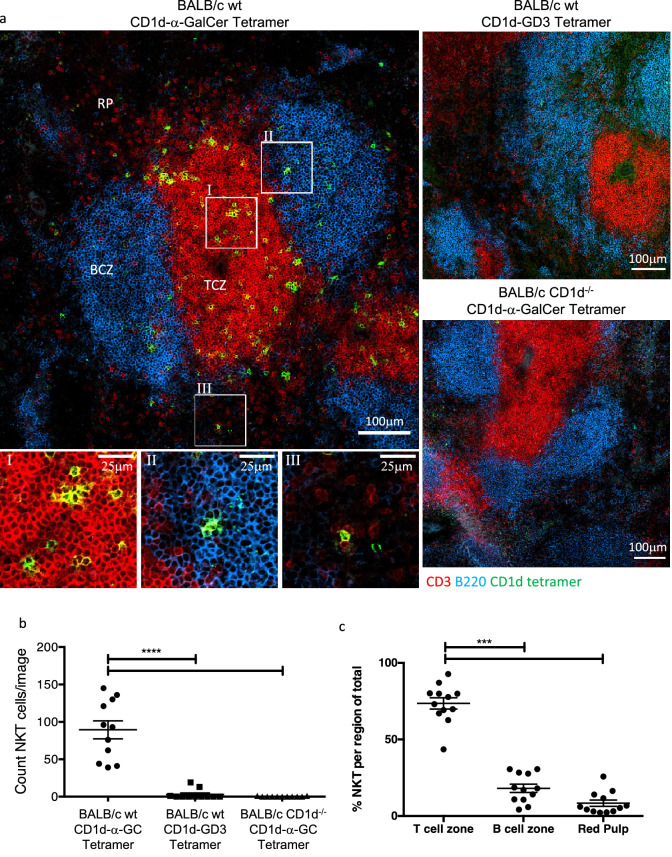
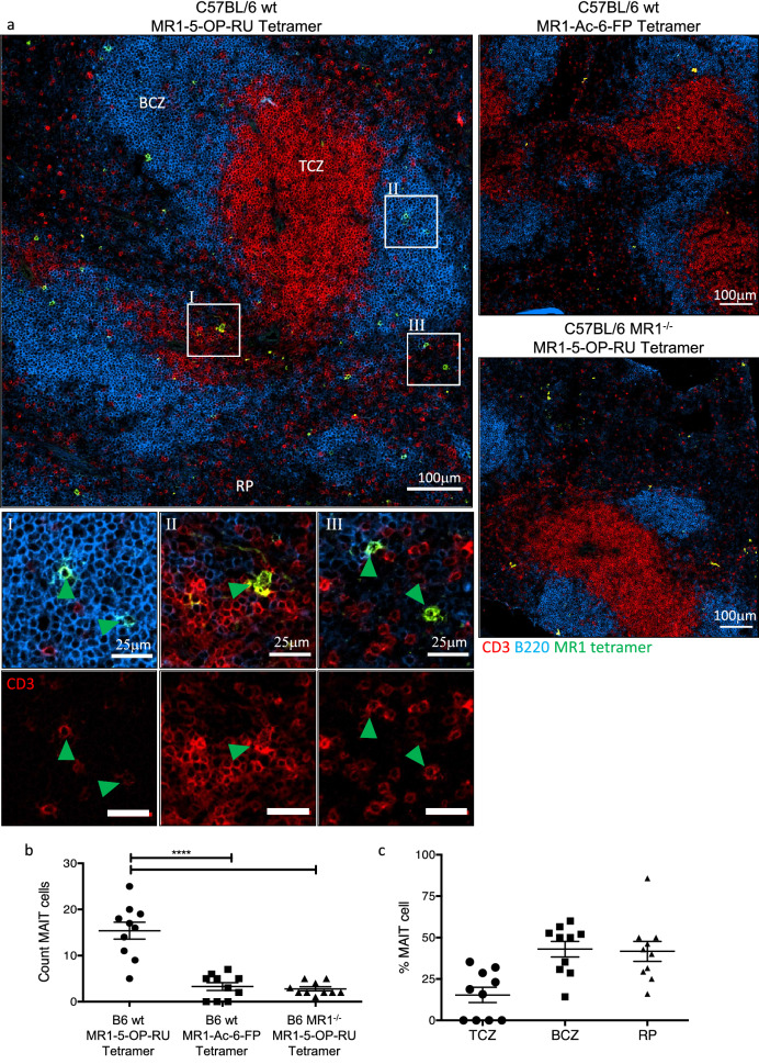
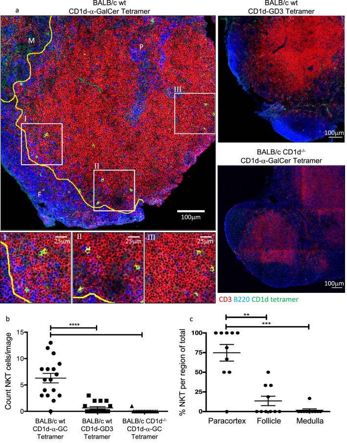
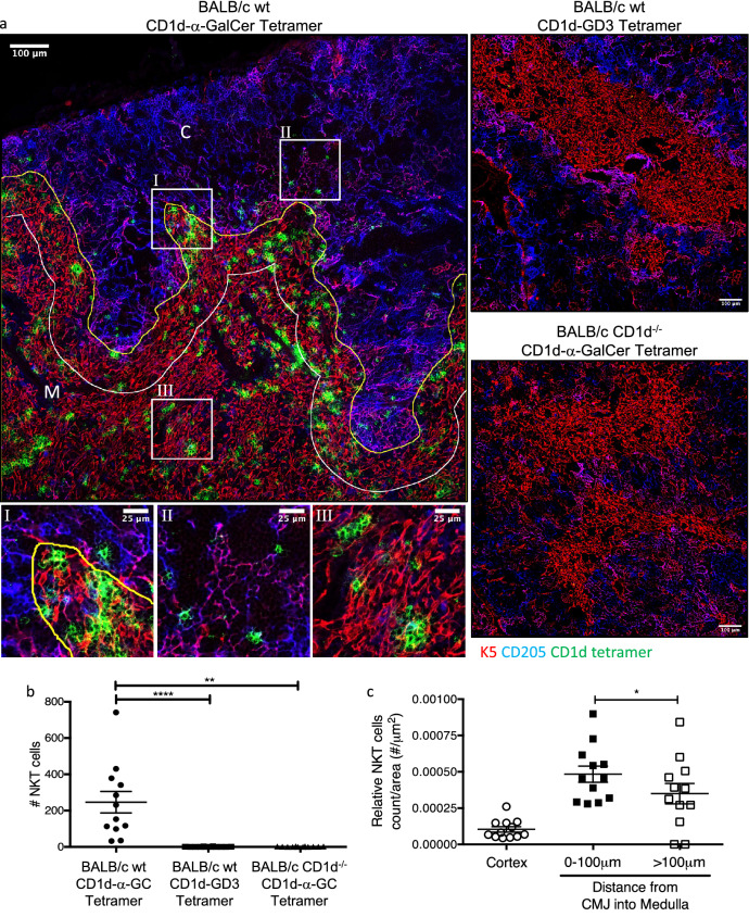
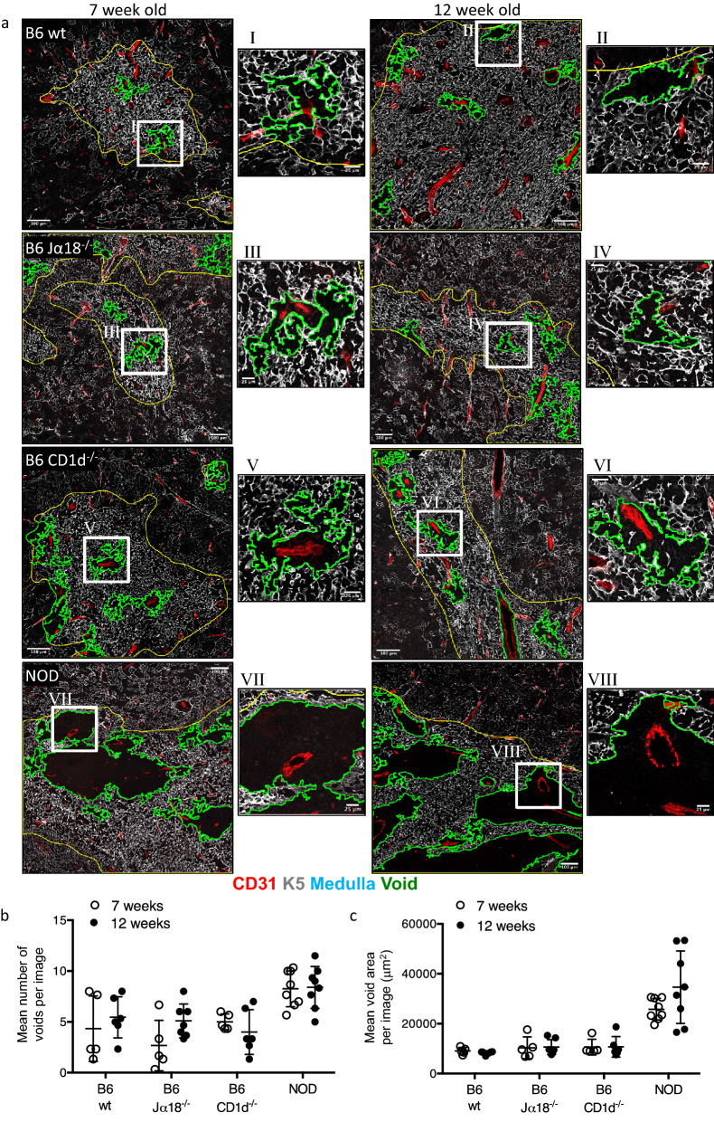
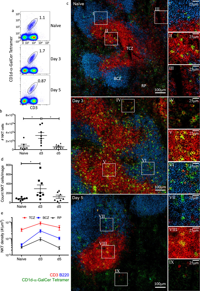
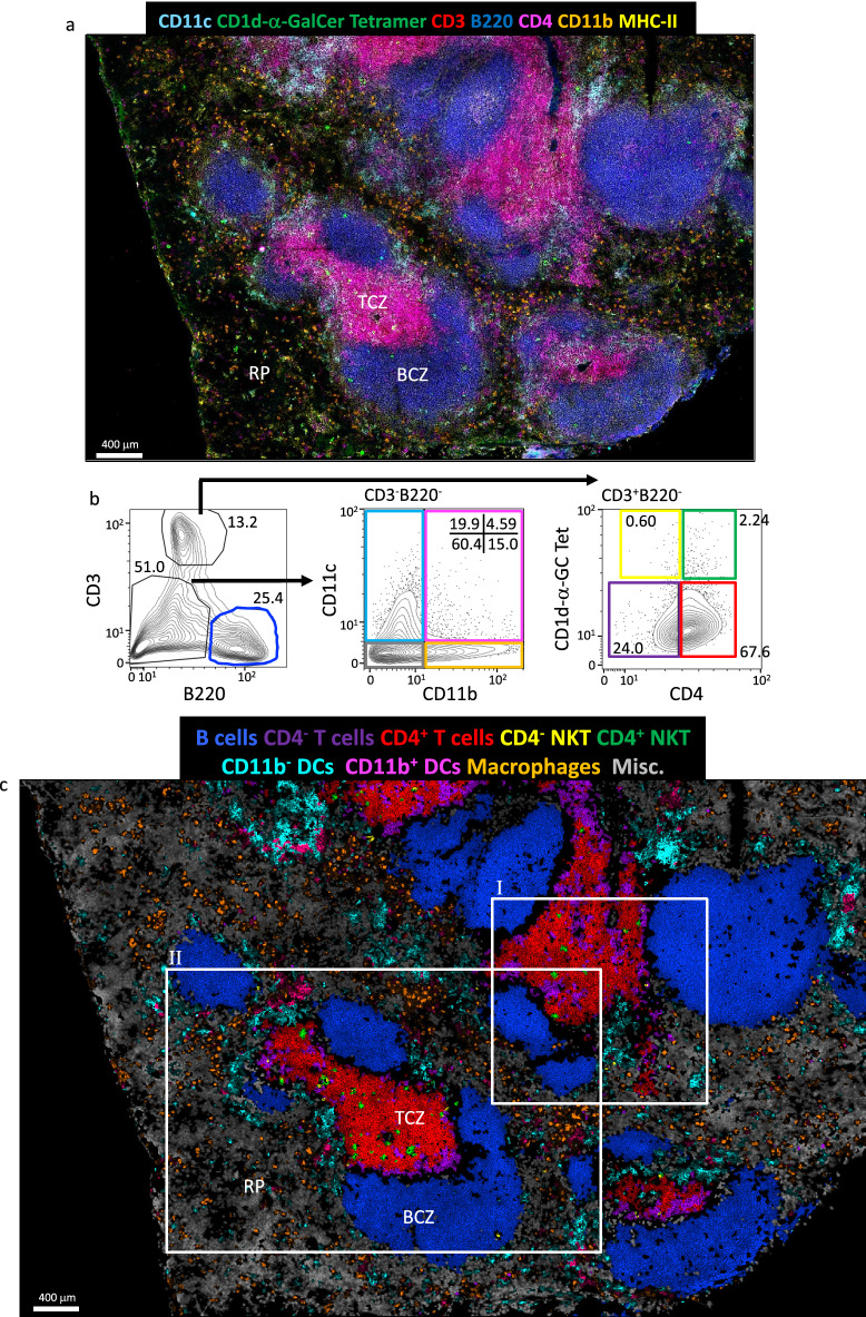
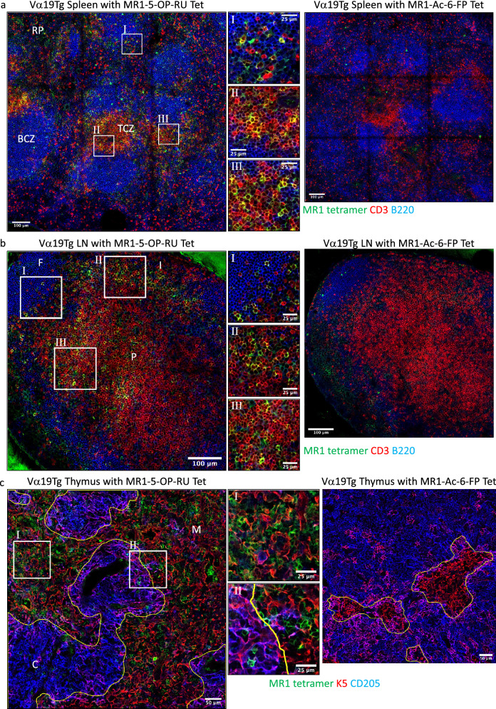
Natural Killer T (NKT) cells and Mucosal-Associated Invariant T (MAIT) cells are innate-like T cells that express semi-invariant αβ T cell receptors (TCRs) through which they recognise CD1d and MR1 molecules, respectively, in complex with specific ligands. These cells play important roles in health and disease in many organs, but their precise intra-organ location is not well established. Here, using CD1d and MR1 tetramer staining techniques, we describe the precise location of NKT and MAIT cells in lymphoid and peripheral organs. Within the thymus, NKT cells were concentrated in the medullary side of the corticomedullary junction. In spleen and lymph nodes, NKT cells were mainly localised within T cell zones, although following in vivo activation with the potent NKT-cell ligand α-GalCer, they expanded throughout the spleen. MAIT cells were clearly detectable in Vα19 TCR transgenic mice and were rare but detectable in lymphoid tissue of non-transgenic mice. In contrast to NKT cells, MAIT cells were more closely associated with the B cell zone and red pulp of the spleen. Accordingly, we have provided an extensive analysis of the in situ localisation of NKT and MAIT cells and suggest differences between the intra-organ location of these two cell types.
自然杀伤 T (NKT) 细胞和黏膜相关不变 T (MAIT) 细胞是先天样 T 细胞,它们通过表达半不变的 αβ T 细胞受体 (TCR) 分别识别 CD1d 和 MR1 分子,与特定配体形成复合物。这些细胞在许多器官的健康和疾病中发挥着重要作用,但它们在器官内的确切位置尚未得到很好的确定。在这里,我们使用 CD1d 和 MR1 四聚体染色技术,描述了 NKT 和 MAIT 细胞在淋巴器官和外周器官中的精确位置。在胸腺中,NKT 细胞集中在皮质髓质交界处的髓质侧。在脾脏和淋巴结中,NKT 细胞主要位于 T 细胞区,但在体内用有效的 NKT 细胞配体 α-GalCer 激活后,它们在整个脾脏中扩增。在 Vα19 TCR 转基因小鼠中可以清楚地检测到 MAIT 细胞,在非转基因小鼠的淋巴组织中也可以检测到,但数量较少。与 NKT 细胞不同,MAIT 细胞与脾脏的 B 细胞区和红髓更密切相关。因此,我们对 NKT 和 MAIT 细胞的原位定位进行了广泛分析,并提出了这两种细胞类型在器官内位置的差异。