Lin Shuai, Sheng Qianwen, Ma Xiaobin, Li Shanli, Xu Peng, Dai Cong, Wang Meng, Kang Huafeng, Dai Zhijun
Department of Oncology, The Second Affiliated Hospital of Xi'an Jiaotong University, Xi'an 710004, China.
Department of Oncology, Xi'an International Medical Center Hospital, Xi'an, China.
Evid Based Complement Alternat Med. 2022 Mar 4;2022:7354700. doi: 10.1155/2022/7354700. eCollection 2022.
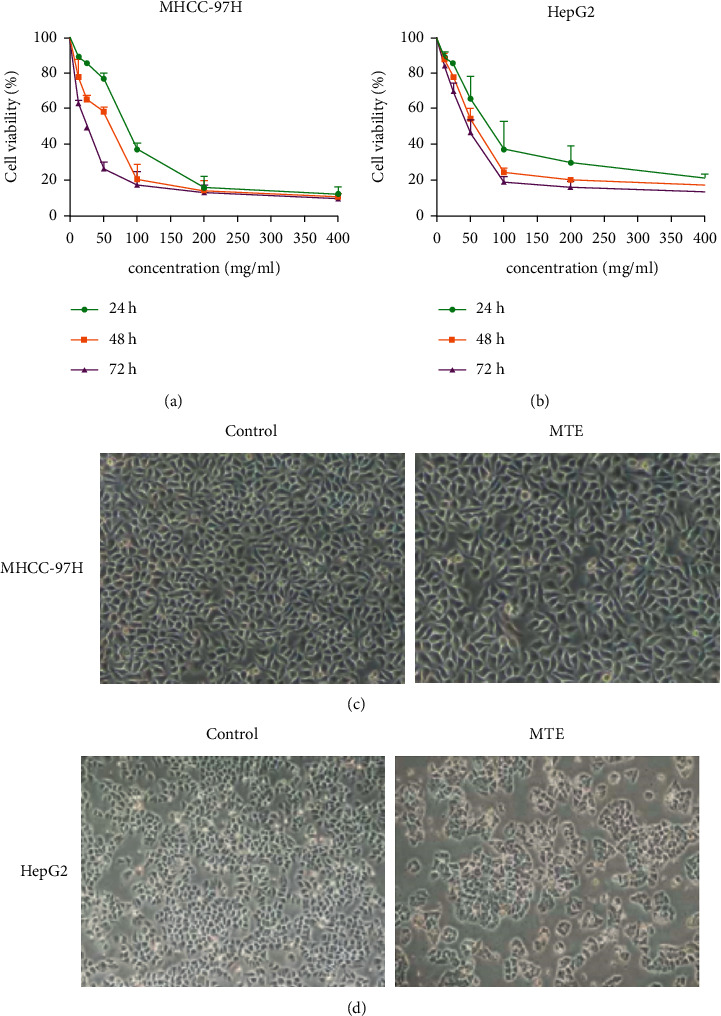
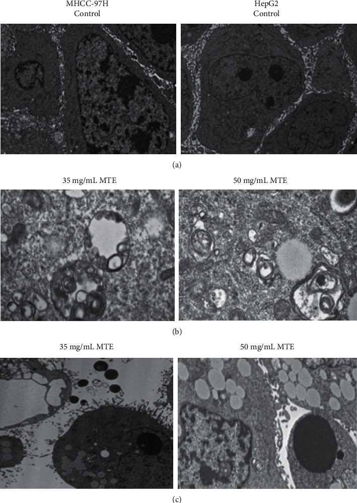
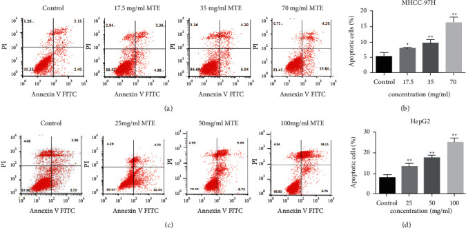
Hepatocellular carcinoma (HCC) seriously endangers humans. In traditional Chinese medicine, (MTE) has anti-inflammatory, antiasthmatic, antihypertensive, and anticancer effects. This study reveals the antiproliferative effect of MTE on the HCC cells and provides a theoretical basis for the development and clinical application of anti-HCC agents. . MHCC-97H and HepG2 cells were cultured and exposed to various concentrations and durations of MTE, and an MTT assay was used to detect the effects of MTE on cell proliferation. Transmission electron microscopy revealed the morphological changes in the two cell lines after MTE stimulation. The MTE effects on the apoptosis and cell cycle distribution of the cell lines were detected by flow cytometry. Western blotting and qRT-PCR were used to detect target gene expression at the protein and mRNA levels, respectively. . MTE reduced the viability of the MHCC-97H and HepG2 cells in a dose- and time-dependent manners ( < 0.05). Autophagic vesicles and apoptotic bodies were found in the MHCC-97H and HepG2 cells after MTE incubation, and the Annexin V-PI assay showed that the apoptotic rates of the cell lines increased with increasing MTE concentration ( < 0.05). Autophagy inducer rapamycin promoted the MTE-induced apoptotic rates of the cell lines, whereas autophagy inhibitor chloroquine inhibited the apoptotic rates. More cells in the S phase were found in the two cell lines after MTE treatment ( < 0.05). After MTE incubation, MIF, CD47, and beclin-1 protein levels significantly increased. Furthermore, in the MTE group, Akt, mTOR, and caspase3 expressions decreased; however, LC 3 expression increased, which was significantly different from the control group ( < 0.05). . MTE inhibited proliferation and induced autophagy, apoptosis, and S phase cell cycle arrest in the MHCC-97H and HepG2 cells. These effects might be related to the activation of MIF and mTOR signaling inhibition.
肝细胞癌(HCC)严重危害人类健康。在传统中医中,(MTE)具有抗炎、抗哮喘、抗高血压和抗癌作用。本研究揭示了MTE对肝癌细胞的抗增殖作用,并为抗肝癌药物的开发和临床应用提供了理论依据。. 培养MHCC - 97H和HepG2细胞,并使其暴露于不同浓度和作用时间的MTE中,采用MTT法检测MTE对细胞增殖的影响。透射电子显微镜观察MTE刺激后两种细胞系的形态变化。通过流式细胞术检测MTE对细胞系凋亡和细胞周期分布的影响。分别采用蛋白质印迹法和qRT - PCR检测靶基因在蛋白质和mRNA水平的表达。. MTE以剂量和时间依赖性方式降低MHCC - 97H和HepG2细胞的活力(<0.05)。MTE作用后,在MHCC - 97H和HepG2细胞中发现自噬泡和凋亡小体,Annexin V - PI检测显示细胞系的凋亡率随MTE浓度增加而升高(<0.05)。自噬诱导剂雷帕霉素促进MTE诱导的细胞系凋亡率,而自噬抑制剂氯喹抑制凋亡率。MTE处理后,两个细胞系中处于S期的细胞增多(<0.05)。MTE作用后,MIF、CD47和beclin - 1蛋白水平显著升高。此外,在MTE组中,Akt、mTOR和caspase3表达降低;然而,LC 3表达升高,与对照组相比有显著差异(<0.05)。. MTE抑制MHCC - 97H和HepG2细胞的增殖,并诱导自噬、凋亡和S期细胞周期阻滞。这些作用可能与MIF的激活和mTOR信号通路的抑制有关。