Ahmad Wasim, Ahmad Mushtaq, Umar Khayam Sahibzada Muhammad, Khusro Ameer, Emran Talha Bin, Muhammedali Alnasrawi Abeer, Alkahtani Jawaher, Elshikh Mohamed S
Department of Biotechnology, University of Science and Technology, Bannu, Khyber Pakhtunkhwa, Pakistan.
Department of Pharmacy, Sarhad University of Science and Information Technology, Peshawar, Khyber Pakhtunkhwa, Pakistan.
Saudi J Biol Sci. 2022 Mar;29(3):1887-1892. doi: 10.1016/j.sjbs.2021.10.033. Epub 2021 Oct 22.
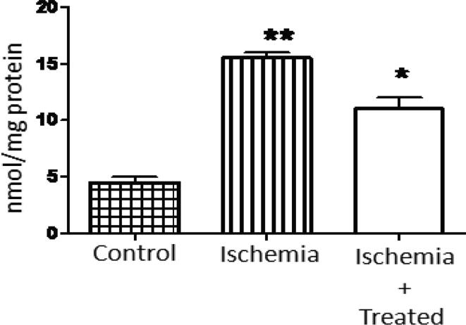
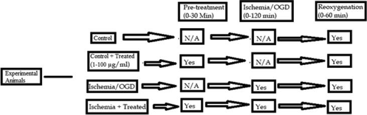
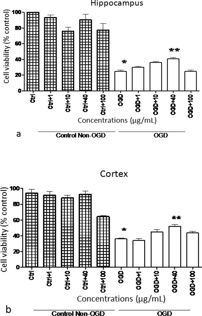
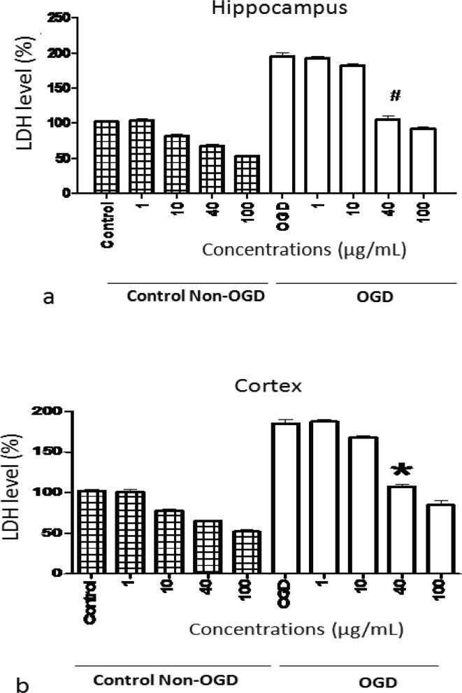
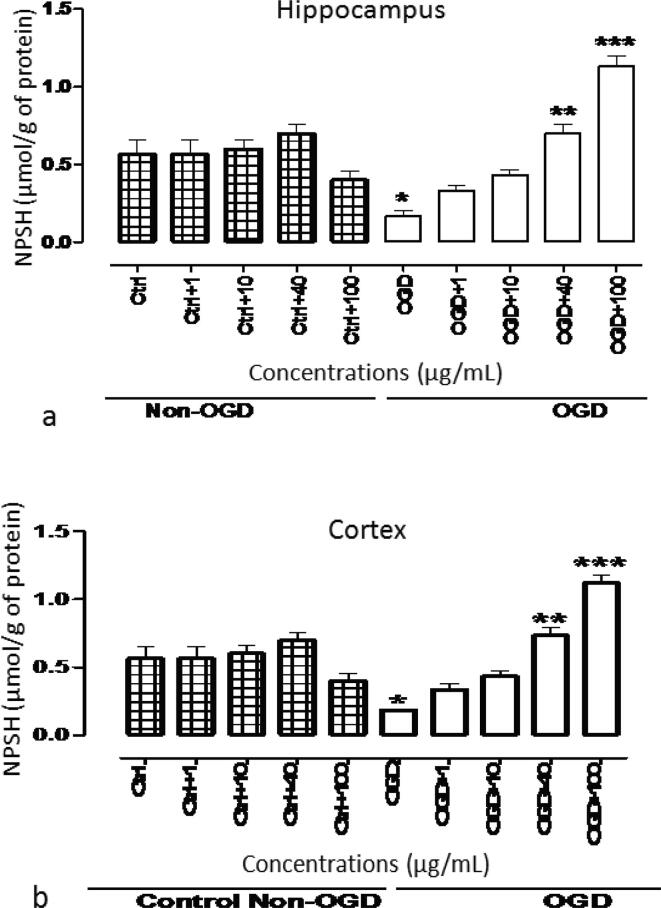
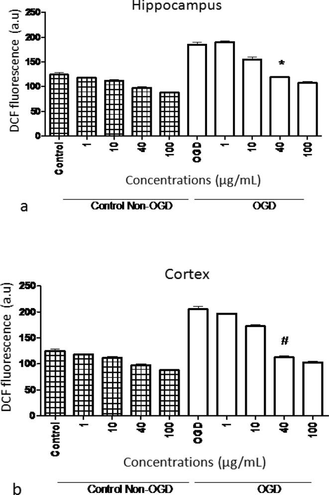
This study was aimed to determine the neuroprotective influence of in terms of restoring normal state of the rat's hippocampus and cortex after oxidative insult caused by ischemia and reperfusion. Cell viability and membrane integrity were assessed using MTT and lactate dehydrogenase (LDH) assay, respectively. Ischemic insult was introduced in the rat brain's hippocampal and cortical slivers by exposing oxygen and glucose deficiency (OGD) for 2 h, followed by 1 h of re-perfusion. Cellular oxidative stress levels were quantified by incorporating 2',7'-dichlorofluorescein diacetate fluorescent probes. Additionally, the lipid peroxidation was assessed using TBARS assay. Findings revealed significant neuroprotection against OGD-induced mitochondrial impairment at 40 µg/mL of in rat's hippocampal and cortical slices. The LDH levels were decreased significantly ( < 0.001) during pre-incubation and reoxygenation periods using varied concentrations of extract. Cellular oxidative stress levels results showed significant ( < 0.001) reduction in dichlorofluorescein fluorescence in slices homogenate of hippocampus and cortex using extract. The lipid peroxidation assay results showed decreased ( < 0.01) levels of malondialdehyde in liver tissues of treated rats treated (200 mg/kg body weight) when compared to the ischemic animal. In summary, findings clearly indicated the neuroprotective effects of extract against ischemia in brain hippocampal and cortex slivers. could undoubtedly be utilized as a healing agent in preventing neuronal cells' loss during is chemic-reperfusion process.
本研究旨在确定[具体物质]在恢复大鼠海马体和皮质因缺血再灌注引起的氧化损伤后的正常状态方面的神经保护作用。分别使用MTT和乳酸脱氢酶(LDH)测定法评估细胞活力和膜完整性。通过暴露于缺氧缺糖(OGD)2小时,然后再灌注1小时,在大鼠脑海马体和皮质切片中引入缺血性损伤。通过加入2',7'-二氯荧光素二乙酸荧光探针来量化细胞氧化应激水平。此外,使用硫代巴比妥酸反应物(TBARS)测定法评估脂质过氧化。研究结果显示,在大鼠海马体和皮质切片中,40μg/mL的[具体物质]对OGD诱导的线粒体损伤具有显著的神经保护作用。在预孵育和复氧期间,使用不同浓度的[具体物质]提取物,LDH水平显著降低(P<0.001)。细胞氧化应激水平结果显示,使用[具体物质]提取物时,海马体和皮质切片匀浆中的二氯荧光素荧光显著降低(P<0.001)。脂质过氧化测定结果显示,与缺血动物相比,经治疗的大鼠(200mg/kg体重)肝脏组织中的丙二醛水平降低(P<0.01)。总之,研究结果清楚地表明了[具体物质]提取物对脑海马体和皮质切片缺血的神经保护作用。[具体物质]无疑可作为一种治疗剂,用于预防缺血再灌注过程中神经元细胞的损失。