Koenigkam-Santos Marcel, Wada Danilo Tadao, Benatti Maira Nilson, Siyuan Li, Batah Sabrina Setembre, Cetlin Andrea Antunes, de Menezes Marcelo Bezerra, Fabro Alexandre Todorovic
Department of Medical Images, Hematology and Oncology, Ribeirao Preto Medical School, University of Sao Paulo, Campus Universitario Monte Alegre, Ribeirao Preto, SP, Brazil.
Department of Internal Medicine, Ribeirao Preto Medical School, University of Sao Paulo, Campus Universitario Monte Alegre, Ribeirao Preto, SP, Brazil.
Ann Transl Med. 2022 Feb;10(3):140. doi: 10.21037/atm-21-4354.
Correlation between pathology and imaging of the new SARS-Cov-2 disease (COVID-19) is scarce. This study aimed to characterize SARS-Cov-2 pneumonia on imaging of patients submitted to minimally invasive autopsy (MIA).
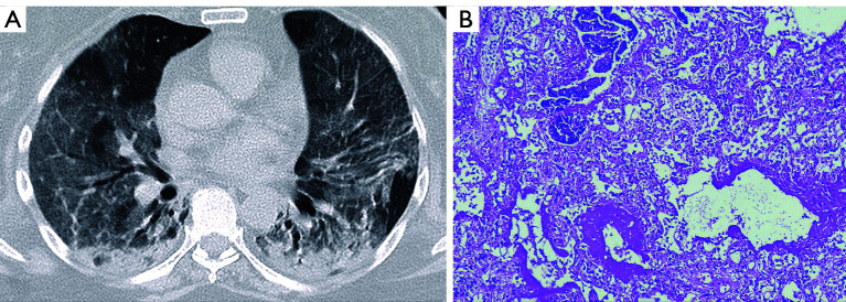
This unicentric retrospective observational study included 46 consecutive patients with confirmed COVID-19 who underwent MIA. All clinical chest images were reviewed and classified for the presence and grade of viral pneumonia, as well as disease evolution. On CT, phenotypes were described as consistent with mild, moderate, or severe viral pneumonia, with or without radiological signs of organizing pneumonia (OP). In severe pneumonia, CT could also be classified as diffuse progressive OP or radiological diffuse alveolar damage (DAD). Specific features on CT were noted, including fibroproliferative signs that could indicate potential or initial fibrosis.
MIA showed a heterogeneous panel of alterations, with a high prevalence of OP and acute fibrinous and organizing pneumonia (AFOP). Also, signs of interstitial fibrosis corresponded to the most prevalent pathological feature. Initial chest radiography (CXR) findings were mainly consistent with moderate or severe viral pneumonia. Most patients showed stability or improvement (reduction of opacities) on imaging. CTs were performed on 15 patients. Consolidations were found in most patients, frequently showing features consistent with an OP phenotype. Fibroproliferative changes were also prevalent on CT.
In this study, SARS-Cov-2 pneumonia showed heterogeneous radiological and pathological patterns. Signs of organization and potential or initial fibrosis were prevalent on both imaging and pathology. Imaging phenotyping may help to predict post-infection fibrosing interstitial pneumonitis in COVID-19.
新型严重急性呼吸综合征冠状病毒2型疾病(COVID-19)的病理学与影像学之间的相关性尚少。本研究旨在通过对接受微创尸检(MIA)患者的影像学检查来描述SARS-CoV-2肺炎的特征。
这项单中心回顾性观察性研究纳入了46例连续接受MIA的确诊COVID-19患者。对所有临床胸部影像进行回顾,并根据病毒性肺炎的存在情况、分级以及疾病进展进行分类。在CT上,将表型描述为与轻度、中度或重度病毒性肺炎相符,有无机化性肺炎(OP)的放射学征象。在重症肺炎中,CT还可分类为弥漫性进行性OP或放射学上的弥漫性肺泡损伤(DAD)。记录CT上的特定特征,包括可能提示潜在或初始纤维化的纤维增生性征象。
MIA显示出一系列异质性改变,OP以及急性纤维素性和机化性肺炎(AFOP)的发生率很高。此外,间质纤维化征象是最常见的病理特征。最初的胸部X线摄影(CXR)结果主要与中度或重度病毒性肺炎相符。大多数患者在影像学上显示病情稳定或改善(opacity减少)。15例患者进行了CT检查。大多数患者发现有实变,常表现出与OP表型相符的特征。纤维增生性改变在CT上也很常见。
在本研究中,SARS-CoV-2肺炎表现出异质性的放射学和病理学模式。机化以及潜在或初始纤维化的征象在影像学和病理学上均很常见。影像学表型分析可能有助于预测COVID-19感染后纤维化性间质性肺炎。