• 文献检索
  • 文档翻译
  • 深度研究
  • 学术资讯
  • Suppr Zotero 插件Zotero 插件
  • 邀请有礼
  • 套餐&价格
  • 历史记录
应用&插件
Suppr Zotero 插件Zotero 插件浏览器插件Mac 客户端Windows 客户端微信小程序
定价
高级版会员购买积分包购买API积分包
服务
文献检索文档翻译深度研究API 文档MCP 服务
关于我们
关于 Suppr公司介绍联系我们用户协议隐私条款
关注我们

Suppr 超能文献

核心技术专利:CN118964589B侵权必究
粤ICP备2023148730 号-1Suppr @ 2026

文献检索

告别复杂PubMed语法,用中文像聊天一样搜索,搜遍4000万医学文献。AI智能推荐,让科研检索更轻松。

立即免费搜索

文件翻译

保留排版,准确专业,支持PDF/Word/PPT等文件格式,支持 12+语言互译。

免费翻译文档

深度研究

AI帮你快速写综述,25分钟生成高质量综述,智能提取关键信息,辅助科研写作。

立即免费体验

单分子荧光成像和深度学习揭示了淀粉样蛋白-β 42 的高度异质聚集。

Single-molecule fluorescence imaging and deep learning reveal highly heterogeneous aggregation of amyloid-β 42.

机构信息

Laboratory of Chemical Physics, National Institute of Diabetes and Digestive and Kidney Diseases, NIH, Bethesda, MD 20892.

出版信息

Proc Natl Acad Sci U S A. 2022 Mar 22;119(12):e2116736119. doi: 10.1073/pnas.2116736119. Epub 2022 Mar 15.

DOI:10.1073/pnas.2116736119
PMID:35290118
原文链接:https://pmc.ncbi.nlm.nih.gov/articles/PMC8944908/
Abstract

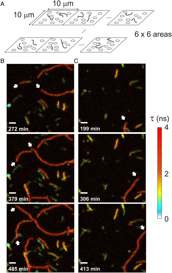
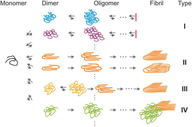
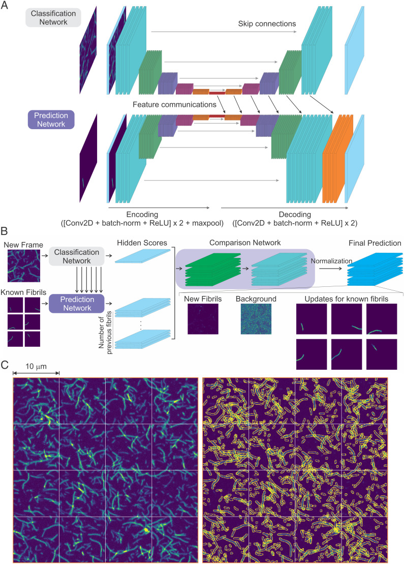
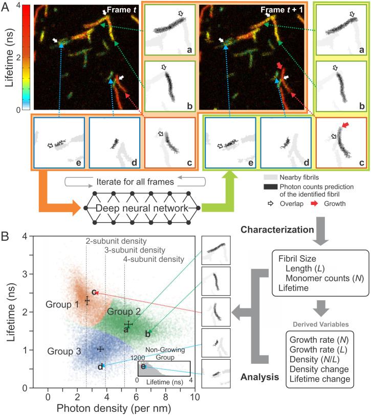
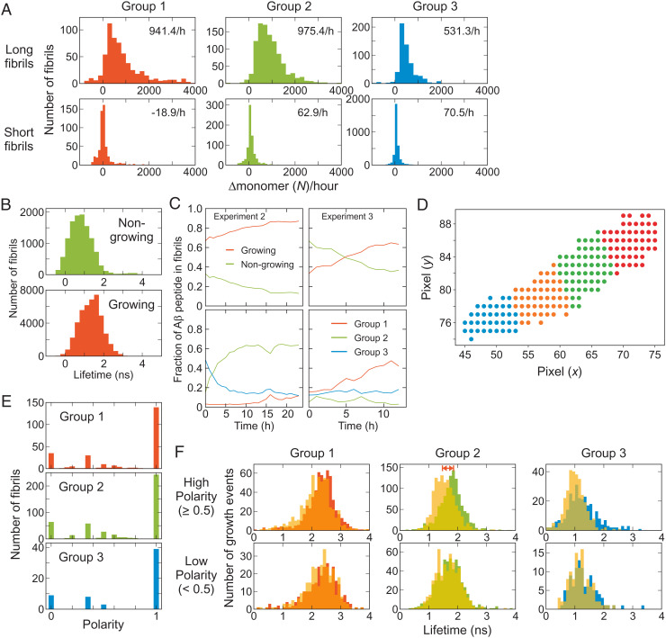
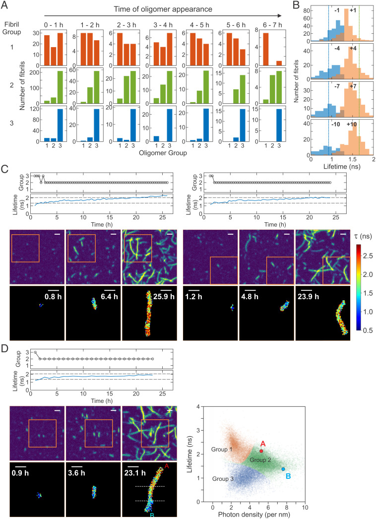
Polymorphism in the structure of amyloid fibrils suggests the existence of many different assembly pathways. Characterization of this heterogeneity is the key to understanding the aggregation mechanism and toxicity, but in practice it is extremely difficult to probe individual aggregation pathways in a mixture. Here, we present development of a method combining single-molecule fluorescence lifetime imaging and deep learning for monitoring individual fibril formation in real time and their high-throughput analysis. A deep neural network (FNet) separates an image of highly overlapping fibrils into single fibril images, which allows for tracking the growth and changes in characteristics of individual fibrils. Using this method, we investigated aggregation of the 42-residue amyloid-β peptide (Aβ42). We demonstrate that highly heterogeneous fibril formation can be quantitatively characterized in terms of the number of cross-β subunits, elongation speed, growth polarity, and conformation of fibrils. Tracking individual fibril formation and growth also leads to the discovery of a general nucleation mechanism (termed heterogeneous secondary nucleation), where a fibril is formed on the surface of an oligomer with a different structure. Our development will be broadly applicable to characterization of heterogeneous aggregation processes of other proteins.

摘要

淀粉样纤维结构的多态性表明存在许多不同的组装途径。这种异质性的特征描述是理解聚集机制和毒性的关键,但实际上,在混合物中探测单个聚集途径极其困难。在这里,我们提出了一种将单分子荧光寿命成像和深度学习相结合的方法,用于实时监测和高通量分析单个纤维的形成。一个深度神经网络(FNet)将高度重叠的纤维的图像分离成单个纤维的图像,这允许跟踪单个纤维的生长和特征的变化。使用这种方法,我们研究了 42 个残基的淀粉样β肽(Aβ42)的聚集。我们证明,高度异质的纤维形成可以根据交叉-β亚基的数量、伸长速度、生长极性和纤维构象进行定量描述。跟踪单个纤维的形成和生长也导致了一种普遍的成核机制(称为异质二次成核)的发现,其中纤维在具有不同结构的寡聚物的表面上形成。我们的开发将广泛适用于其他蛋白质的异质聚集过程的特征描述。

https://cdn.ncbi.nlm.nih.gov/pmc/blobs/f0fa/8944908/023def6412e6/pnas.2116736119fig06.jpg
https://cdn.ncbi.nlm.nih.gov/pmc/blobs/f0fa/8944908/14c75f941659/pnas.2116736119fig01.jpg
https://cdn.ncbi.nlm.nih.gov/pmc/blobs/f0fa/8944908/b83569a378af/pnas.2116736119fig02.jpg
https://cdn.ncbi.nlm.nih.gov/pmc/blobs/f0fa/8944908/0df6d009b4cf/pnas.2116736119fig03.jpg
https://cdn.ncbi.nlm.nih.gov/pmc/blobs/f0fa/8944908/4fe0d38ee977/pnas.2116736119fig04.jpg
https://cdn.ncbi.nlm.nih.gov/pmc/blobs/f0fa/8944908/408602b67ce5/pnas.2116736119fig05.jpg
https://cdn.ncbi.nlm.nih.gov/pmc/blobs/f0fa/8944908/023def6412e6/pnas.2116736119fig06.jpg

相似文献

1
Single-molecule fluorescence imaging and deep learning reveal highly heterogeneous aggregation of amyloid-β 42.单分子荧光成像和深度学习揭示了淀粉样蛋白-β 42 的高度异质聚集。
Proc Natl Acad Sci U S A. 2022 Mar 22;119(12):e2116736119. doi: 10.1073/pnas.2116736119. Epub 2022 Mar 15.
2
Direct evidence for self-propagation of different amyloid-β fibril conformations.直接证据表明不同淀粉样β纤维构象的自我传播。
Neurodegener Dis. 2014;14(3):151-9. doi: 10.1159/000363623. Epub 2014 Oct 4.
3
Computational studies of protein aggregation mediated by amyloid: Fibril elongation and secondary nucleation.淀粉样蛋白介导的蛋白质聚集的计算研究:原纤维的延伸和二次成核。
Prog Mol Biol Transl Sci. 2020;170:461-504. doi: 10.1016/bs.pmbts.2019.12.008. Epub 2020 Jan 10.
4
Conserved S/T Residues of the Human Chaperone DNAJB6 Are Required for Effective Inhibition of Aβ42 Amyloid Fibril Formation.人类伴侣蛋白DNAJB6的保守丝氨酸/苏氨酸残基是有效抑制Aβ42淀粉样纤维形成所必需的。
Biochemistry. 2018 Aug 14;57(32):4891-4902. doi: 10.1021/acs.biochem.8b00353. Epub 2018 Jul 30.
5
Interactions of Alzheimer amyloid-beta peptides with glycosaminoglycans effects on fibril nucleation and growth.阿尔茨海默病淀粉样β肽与糖胺聚糖的相互作用对纤维成核和生长的影响。
Eur J Biochem. 1999 Dec;266(3):1101-10. doi: 10.1046/j.1432-1327.1999.00957.x.
6
AlphaB-crystallin, a small heat-shock protein, prevents the amyloid fibril growth of an amyloid beta-peptide and beta2-microglobulin.αB晶状体蛋白,一种小分子热休克蛋白,可阻止淀粉样β肽和β2微球蛋白的淀粉样原纤维生长。
Biochem J. 2005 Dec 15;392(Pt 3):573-81. doi: 10.1042/BJ20050339.
7
Structural variation in amyloid-β fibrils from Alzheimer's disease clinical subtypes.阿尔茨海默病临床亚型中淀粉样β纤维的结构变异
Nature. 2017 Jan 12;541(7636):217-221. doi: 10.1038/nature20814. Epub 2017 Jan 4.
8
Understanding Amyloid-β Oligomerization at the Molecular Level: The Role of the Fibril Surface.在分子水平上理解β-淀粉样蛋白寡聚化:原纤维表面的作用。
Chemistry. 2016 Jun 20;22(26):8768-72. doi: 10.1002/chem.201601701. Epub 2016 May 24.
9
New Mechanism of Amyloid Fibril Formation.淀粉样纤维形成的新机制。
Curr Protein Pept Sci. 2019;20(6):630-640. doi: 10.2174/1389203720666190125160937.
10
Automatic identification of crossovers in cryo-EM images of murine amyloid protein A fibrils with machine learning.使用机器学习技术对鼠类淀粉样蛋白 A 纤维的冷冻电镜图像中的交叉进行自动识别。
J Microsc. 2020 Jan;277(1):12-22. doi: 10.1111/jmi.12858. Epub 2019 Dec 29.

引用本文的文献

1
Three-color single-molecule maximum likelihood analysis of folding and binding of diffusing molecules.扩散分子折叠与结合的三色单分子最大似然分析
Biophys J. 2025 Sep 2;124(17):2909-2926. doi: 10.1016/j.bpj.2025.07.033. Epub 2025 Jul 30.
2
design of a mechano-pharmaceutical screening platform against formation of individual beta-amyloid oligomers.针对单个β-淀粉样寡聚体形成的机械药理学筛选平台的设计
Cell Rep Phys Sci. 2024 Dec 18;5(12). doi: 10.1016/j.xcrp.2024.102336.
3
Characterizing heterogeneity in amyloid formation processes.

本文引用的文献

1
Mechanism of Secondary Nucleation at the Single Fibril Level from Direct Observations of Aβ42 Aggregation.从直接观察 Aβ42 聚集看单纤维水平的二次成核机制。
J Am Chem Soc. 2021 Oct 13;143(40):16621-16629. doi: 10.1021/jacs.1c07228. Epub 2021 Sep 28.
2
Aβ receptors specifically recognize molecular features displayed by fibril ends and neurotoxic oligomers.Aβ 受体特异性识别纤维末端和神经毒性寡聚物所呈现的分子特征。
Nat Commun. 2021 Jun 8;12(1):3451. doi: 10.1038/s41467-021-23507-z.
3
Hierarchical chemical determination of amyloid polymorphs in neurodegenerative disease.
表征淀粉样蛋白形成过程中的异质性。
Curr Opin Struct Biol. 2024 Dec;89:102951. doi: 10.1016/j.sbi.2024.102951. Epub 2024 Nov 19.
4
Fluorescence-Based Monitoring of Early-Stage Aggregation of Amyloid-β, Amylin Peptide, Tau, and α-Synuclein Proteins.基于荧光的淀粉样β、胰岛淀粉样多肽、tau 和 α-突触核蛋白早期聚集的监测。
ACS Chem Neurosci. 2024 Sep 4;15(17):3113-3123. doi: 10.1021/acschemneuro.4c00097. Epub 2024 Aug 16.
5
Structure of cytotoxic amyloid oligomers generated during disaggregation.解聚过程中生成的细胞毒性淀粉样寡聚物的结构。
J Biochem. 2024 May 31;175(6):575-585. doi: 10.1093/jb/mvae023.
6
A Perspective on How Fibrinaloid Microclots and Platelet Pathology May be Applied in Clinical Investigations.纤维蛋白原样微血栓和血小板病理如何应用于临床研究的观点。
Semin Thromb Hemost. 2024 Jun;50(4):537-551. doi: 10.1055/s-0043-1774796. Epub 2023 Sep 25.
7
Single-molecule FRET and molecular diffusion analysis characterize stable oligomers of amyloid-β 42 of extremely low population.单分子荧光共振能量转移和分子扩散分析表征了极低丰度的淀粉样β-蛋白42稳定寡聚体。
PNAS Nexus. 2023 Aug 1;2(8):pgad253. doi: 10.1093/pnasnexus/pgad253. eCollection 2023 Aug.
8
Co-translational formation of disulfides guides folding of the SARS-CoV-2 receptor binding domain.共翻译形成二硫键指导 SARS-CoV-2 受体结合域的折叠。
Biophys J. 2023 Aug 22;122(16):3238-3253. doi: 10.1016/j.bpj.2023.07.002. Epub 2023 Jul 7.
9
Energy landscapes of Aβ monomers are sculpted in accordance with Ostwald's rule of stages.Aβ 单体的能量景观是按照奥斯特瓦尔德阶段规则塑造的。
Sci Adv. 2023 Mar 22;9(12):eadd6921. doi: 10.1126/sciadv.add6921.
10
Deep learning in single-molecule imaging and analysis: recent advances and prospects.单分子成像与分析中的深度学习:最新进展与展望
Chem Sci. 2022 Sep 22;13(41):11964-11980. doi: 10.1039/d2sc02443h. eCollection 2022 Oct 26.
在神经退行性疾病中淀粉样蛋白多形体的分级化学测定。
Nat Chem Biol. 2021 Mar;17(3):237-245. doi: 10.1038/s41589-020-00708-z. Epub 2021 Jan 11.
4
Differences in the free energies between the excited states of A40 and A42 monomers encode their aggregation propensities.A40 和 A42 单体激发态之间的自由能差异编码了它们的聚集倾向。
Proc Natl Acad Sci U S A. 2020 Aug 18;117(33):19926-19937. doi: 10.1073/pnas.2002570117. Epub 2020 Jul 30.
5
Structures of α-synuclein filaments from multiple system atrophy.多系统萎缩中α-突触核蛋白丝的结构。
Nature. 2020 Sep;585(7825):464-469. doi: 10.1038/s41586-020-2317-6. Epub 2020 May 27.
6
Complete aggregation pathway of amyloid β (1-40) and (1-42) resolved on an atomically clean interface.在原子清洁界面上解析的淀粉样β(1-40)和(1-42)的完整聚集途径。
Sci Adv. 2020 Apr 8;6(15):eaaz6014. doi: 10.1126/sciadv.aaz6014. eCollection 2020 Apr.
7
Single-Molecule FRET of Intrinsically Disordered Proteins.单分子荧光共振能量转移技术在无序蛋白质研究中的应用
Annu Rev Phys Chem. 2020 Apr 20;71:391-414. doi: 10.1146/annurev-physchem-012420-104917. Epub 2020 Feb 25.
8
Physical determinants of the self-replication of protein fibrils.蛋白质原纤维自我复制的物理决定因素。
Nat Phys. 2016 Sep;12(9):874-880. doi: 10.1038/nphys3828. Epub 2016 Jul 18.
9
Novel tau filament fold in chronic traumatic encephalopathy encloses hydrophobic molecules.慢性创伤性脑病中的新型 tau 丝折叠包裹疏水分子。
Nature. 2019 Apr;568(7752):420-423. doi: 10.1038/s41586-019-1026-5. Epub 2019 Mar 20.
10
U-Net: deep learning for cell counting, detection, and morphometry.U-Net:用于细胞计数、检测和形态测量学的深度学习。
Nat Methods. 2019 Jan;16(1):67-70. doi: 10.1038/s41592-018-0261-2. Epub 2018 Dec 17.