• 文献检索
  • 文档翻译
  • 深度研究
  • 学术资讯
  • Suppr Zotero 插件Zotero 插件
  • 邀请有礼
  • 套餐&价格
  • 历史记录
应用&插件
Suppr Zotero 插件Zotero 插件浏览器插件Mac 客户端Windows 客户端微信小程序
定价
高级版会员购买积分包购买API积分包
服务
文献检索文档翻译深度研究API 文档MCP 服务
关于我们
关于 Suppr公司介绍联系我们用户协议隐私条款
关注我们

Suppr 超能文献

核心技术专利:CN118964589B侵权必究
粤ICP备2023148730 号-1Suppr @ 2026

文献检索

告别复杂PubMed语法,用中文像聊天一样搜索,搜遍4000万医学文献。AI智能推荐,让科研检索更轻松。

立即免费搜索

文件翻译

保留排版,准确专业,支持PDF/Word/PPT等文件格式,支持 12+语言互译。

免费翻译文档

深度研究

AI帮你快速写综述,25分钟生成高质量综述,智能提取关键信息,辅助科研写作。

立即免费体验

新型冠状病毒肺炎侵袭性毛霉病的放射学表现谱

Radiological spectrum of invasive mucormycosis in COVID-19.

作者信息

Passi Nandini, Wadhwa Anshu C, Naik Swati

机构信息

Department of Radio-diagnosis, Batra Hospital and Medical Research Centre (BHMRC), New Delhi, India.

出版信息

BJR Case Rep. 2022 Mar 9;7(6):20210111. doi: 10.1259/bjrcr.20210111. eCollection 2022 Mar.

DOI:10.1259/bjrcr.20210111
PMID:35300232
原文链接:https://pmc.ncbi.nlm.nih.gov/articles/PMC8906162/
Abstract

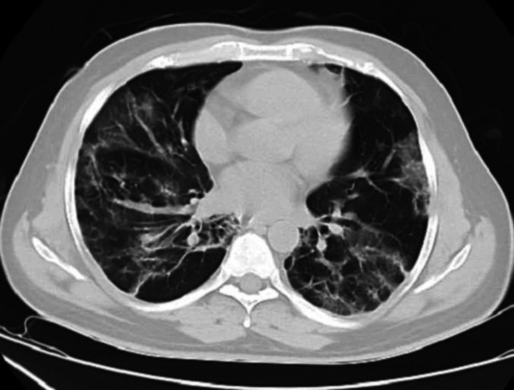
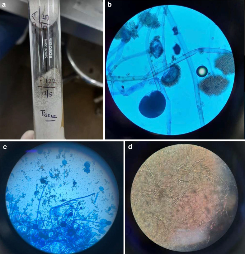
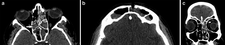
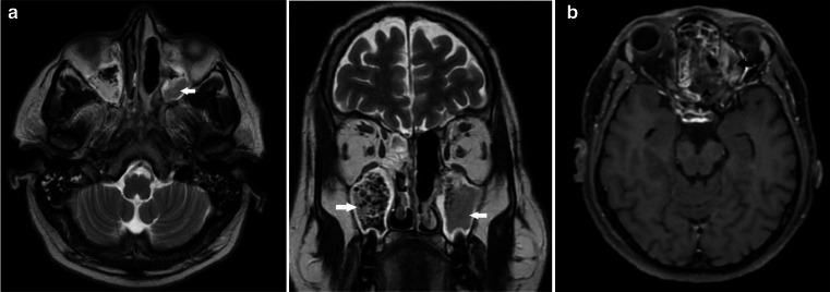
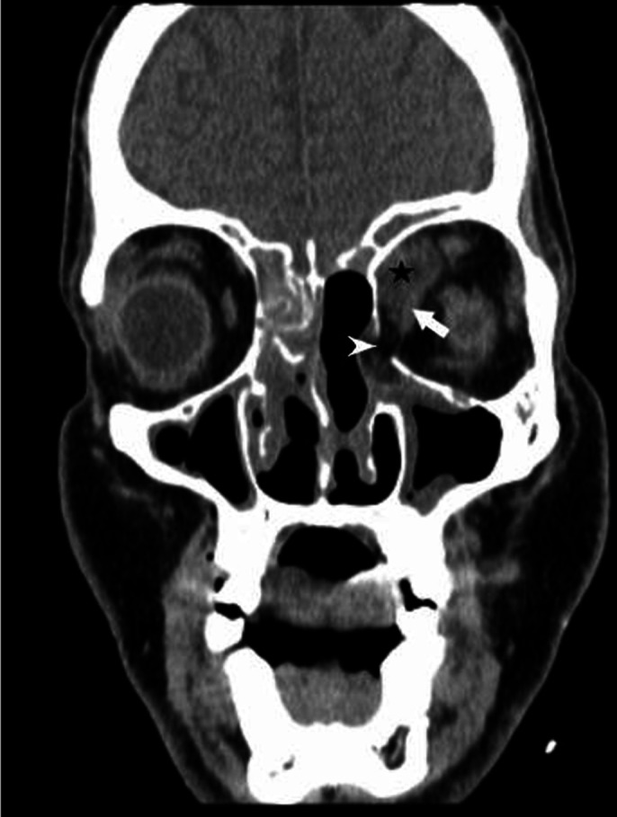
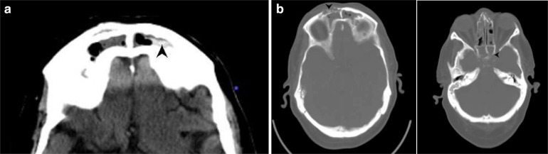
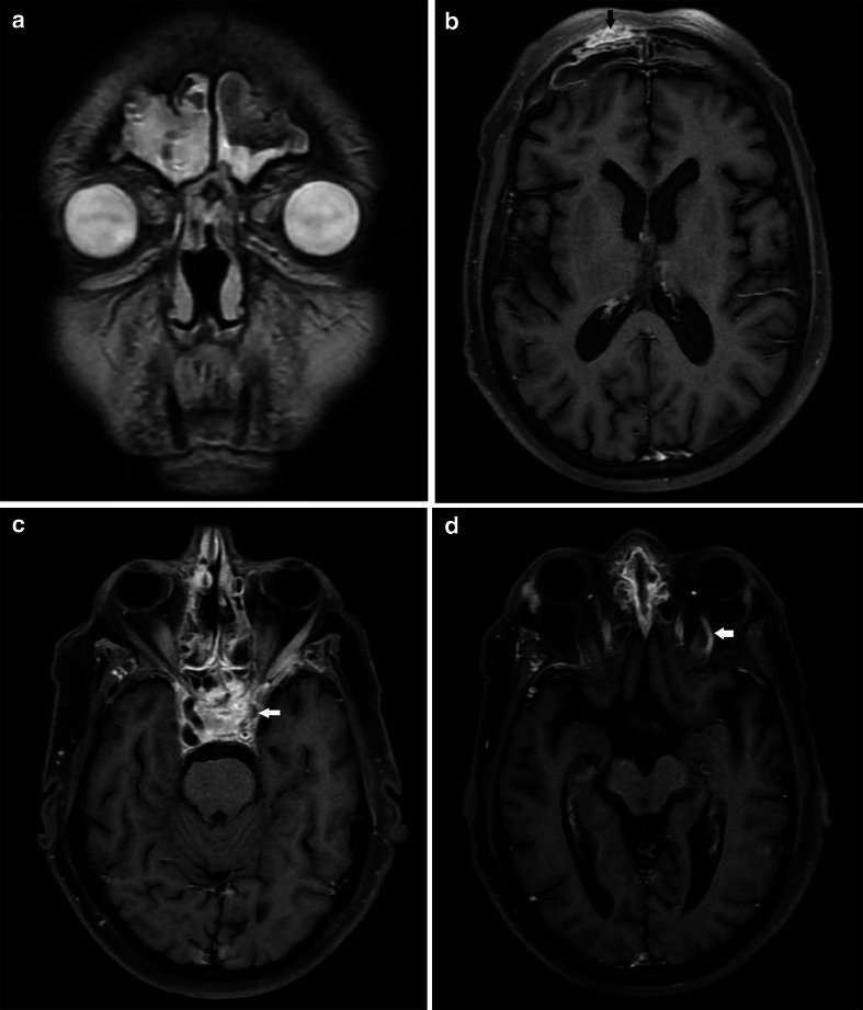
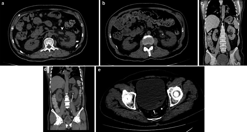
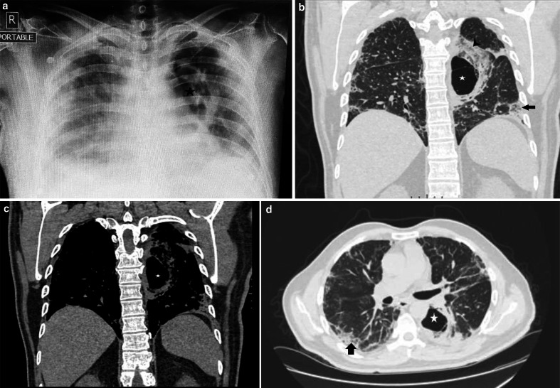
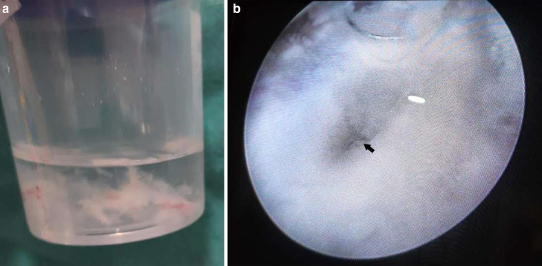
Mucormycosis, commonly known as the "black fungus" is recently emerging as a deadly complication in COVID patients in the Indian subcontinent. A growing number of cases are being reported from all over the country, with a majority of the patients either undergoing treatment or having recovered from COVID. Here, we report three cases of multisystem mucormycosis in COVID positive patients showing, rhino-orbital, cerebral, pulmonary, and genitourinary involvement. The first is a case of a 41-year-old male patient who during his treatment developed left periorbital swelling with ecchymosis and headache. CT and CE-MRI of the paranasal sinuses and brain revealed features of pan fungal sinusitis and subsequent invasion into the left orbit. The second case is of a 52-year-old male patient who after complaining of a severe left-sided hemicranial headache was diagnosed with cavernous sinus thrombosis. The third is of a 57-year-old male patient who presented with left flank pain and dysuria. HRCT (High-resolution CT) chest revealed a thick-walled cavitary lesion, and NCCT KUB (Non-contrast CT of Kidneys, ureters, and bladder) revealed left-sided pyelonephritis. A cystoscopic and microbiological evaluation revealed fungal growth. In all three patients, a biopsy from the involved area revealed broad aseptate filamentous fungal hyphae suggestive of mucormycosis, which was confirmed on culture. These are all unusual cases and physicians should be aware of the possibility of secondary invasive fungal infections in patients with COVID-19 infection.

摘要

毛霉菌病,通常被称为“黑真菌”,最近在印度次大陆成为新冠患者中一种致命的并发症。全国各地报告的病例越来越多,大多数患者正在接受治疗或已从新冠中康复。在此,我们报告3例新冠阳性患者发生多系统毛霉菌病的病例,表现为鼻眶、脑、肺和泌尿生殖系统受累。第一例是一名41岁男性患者,在治疗期间出现左眶周肿胀伴瘀斑和头痛。鼻窦和脑部的CT及增强MRI显示全真菌性鼻窦炎的特征,随后侵犯至左眼眶。第二例是一名52岁男性患者,在主诉严重左侧偏头痛后被诊断为海绵窦血栓形成。第三例是一名57岁男性患者,表现为左侧腰痛和排尿困难。胸部高分辨率CT(HRCT)显示一个厚壁空洞性病变,肾脏、输尿管和膀胱非增强CT(NCCT KUB)显示左侧肾盂肾炎。膀胱镜检查和微生物学评估显示有真菌生长。在所有3例患者中,受累部位的活检显示有宽的无隔丝状真菌菌丝,提示毛霉菌病,培养结果证实了这一点。这些都是不寻常的病例,医生应意识到新冠病毒感染患者发生继发性侵袭性真菌感染的可能性。

https://cdn.ncbi.nlm.nih.gov/pmc/blobs/00eb/8906162/de2c4561cce7/bjrcr.20210111.g010.jpg
https://cdn.ncbi.nlm.nih.gov/pmc/blobs/00eb/8906162/1c6795600a17/bjrcr.20210111.g001.jpg
https://cdn.ncbi.nlm.nih.gov/pmc/blobs/00eb/8906162/480d7a5682de/bjrcr.20210111.g002.jpg
https://cdn.ncbi.nlm.nih.gov/pmc/blobs/00eb/8906162/1cead2082767/bjrcr.20210111.g003.jpg
https://cdn.ncbi.nlm.nih.gov/pmc/blobs/00eb/8906162/ba7010445979/bjrcr.20210111.g004.jpg
https://cdn.ncbi.nlm.nih.gov/pmc/blobs/00eb/8906162/e23827a83b2a/bjrcr.20210111.g005.jpg
https://cdn.ncbi.nlm.nih.gov/pmc/blobs/00eb/8906162/98ce54b33916/bjrcr.20210111.g006.jpg
https://cdn.ncbi.nlm.nih.gov/pmc/blobs/00eb/8906162/a3da41a9be85/bjrcr.20210111.g007.jpg
https://cdn.ncbi.nlm.nih.gov/pmc/blobs/00eb/8906162/bdb97d1173d1/bjrcr.20210111.g008.jpg
https://cdn.ncbi.nlm.nih.gov/pmc/blobs/00eb/8906162/46a4e5dcc2c7/bjrcr.20210111.g009.jpg
https://cdn.ncbi.nlm.nih.gov/pmc/blobs/00eb/8906162/de2c4561cce7/bjrcr.20210111.g010.jpg

相似文献

1
Radiological spectrum of invasive mucormycosis in COVID-19.新型冠状病毒肺炎侵袭性毛霉病的放射学表现谱
BJR Case Rep. 2022 Mar 9;7(6):20210111. doi: 10.1259/bjrcr.20210111. eCollection 2022 Mar.
2
Rhino-Orbital Mucormycosis Associated With COVID-19.与新型冠状病毒肺炎相关的鼻眶毛霉病
Cureus. 2020 Sep 30;12(9):e10726. doi: 10.7759/cureus.10726.
3
Rhino-Orbital-Cerebral Mucormycosis: Battle with the Deadly Enemy.鼻眶脑毛霉菌病:与致命敌人的斗争
Indian J Otolaryngol Head Neck Surg. 2020 Mar;72(1):104-111. doi: 10.1007/s12070-019-01774-z. Epub 2019 Dec 5.
4
Study of Profile of Mucormycosis During the Second Wave of COVID-19 in a Tertiary Care Hospital.三级护理医院中新冠疫情第二波期间毛霉菌病概况研究
Cureus. 2022 Jan 9;14(1):e21054. doi: 10.7759/cureus.21054. eCollection 2022 Jan.
5
Rhino-Orbital-Cerebral Mucormycosis in a Young Diabetic Patient with COVID-19 in Ethiopia: A Case Report.埃塞俄比亚一名患有新冠肺炎的年轻糖尿病患者的鼻眶脑毛霉菌病:病例报告
Int Med Case Rep J. 2022 May 19;15:251-257. doi: 10.2147/IMCRJ.S364591. eCollection 2022.
6
Rhino-orbital mucormycosis in COVID-19 (Corona Virus Disease): A case report.新型冠状病毒肺炎(COVID-19)合并鼻眶毛霉菌病:一例报告
J Family Med Prim Care. 2022 Jun;11(6):3321-3325. doi: 10.4103/jfmpc.jfmpc_1194_21. Epub 2022 Jun 30.
7
A Case of Post-COVID-19 Rhino-Cerebral Mucormycosis in an Immunocompromised Patient.一名免疫功能低下患者的新冠后鼻脑型毛霉菌病病例
Cureus. 2023 Jul 29;15(7):e42652. doi: 10.7759/cureus.42652. eCollection 2023 Jul.
8
Rhino-Orbital-Cerebral Mucormycosis: A Rare Complication of Uncontrolled Diabetes.鼻眶脑毛霉菌病:糖尿病控制不佳的一种罕见并发症。
Case Rep Surg. 2022 Oct 5;2022:6535588. doi: 10.1155/2022/6535588. eCollection 2022.
9
Imaging Patterns of Rhino-Orbital-Cerebral Mucormycosis in Immunocompromised Patients : When to Suspect Complicated Mucormycosis.免疫功能低下患者鼻眶脑毛霉菌病的影像学表现:何时怀疑为复杂性毛霉菌病
Clin Neuroradiol. 2017 Dec;27(4):469-475. doi: 10.1007/s00062-017-0629-1. Epub 2017 Oct 12.
10
Covid-19-Associated Mucormycosis: Histopathology of the Deadly Fungal Infection.新型冠状病毒肺炎相关毛霉菌病:致命真菌感染的组织病理学
Int Arch Otorhinolaryngol. 2024 Jan 24;28(2):e240-e246. doi: 10.1055/s-0043-1776729. eCollection 2024 Apr.

引用本文的文献

1
Mucormycosis diagnosis revisited: Current and emerging diagnostic methodologies for the invasive fungal infection (Review).毛霉病诊断再探讨:侵袭性真菌感染的当前及新兴诊断方法(综述)
Exp Ther Med. 2022 Dec 5;25(1):47. doi: 10.3892/etm.2022.11746. eCollection 2023 Jan.

本文引用的文献

1
Mucormycosis in COVID-19: A systematic review of cases reported worldwide and in India.COVID-19 相关毛霉菌病:全球及印度病例报告的系统综述。
Diabetes Metab Syndr. 2021 Jul-Aug;15(4):102146. doi: 10.1016/j.dsx.2021.05.019. Epub 2021 May 21.
2
When Uncontrolled Diabetes Mellitus and Severe COVID-19 Converge: The Perfect Storm for Mucormycosis.当未控制的糖尿病与重症 COVID-19 相遇:毛霉菌病的完美风暴。
J Fungi (Basel). 2021 Apr 15;7(4):298. doi: 10.3390/jof7040298.
3
COVID-19 triggering mucormycosis in a susceptible patient: a new phenomenon in the developing world?
COVID-19 诱发易感患者发生毛霉菌病:发展中国家的一种新现象?
BMJ Case Rep. 2021 Apr 27;14(4):e241663. doi: 10.1136/bcr-2021-241663.
4
Epidemiology and Diagnosis of Mucormycosis: An Update.毛霉病的流行病学与诊断:最新进展
J Fungi (Basel). 2020 Nov 2;6(4):265. doi: 10.3390/jof6040265.
5
Rhino-Orbital Mucormycosis Associated With COVID-19.与新型冠状病毒肺炎相关的鼻眶毛霉病
Cureus. 2020 Sep 30;12(9):e10726. doi: 10.7759/cureus.10726.
6
Fungal Co-infections Associated with Global COVID-19 Pandemic: A Clinical and Diagnostic Perspective from China.真菌合并感染与全球 COVID-19 大流行相关:来自中国的临床和诊断视角。
Mycopathologia. 2020 Aug;185(4):599-606. doi: 10.1007/s11046-020-00462-9. Epub 2020 Jul 31.
7
Pulmonary Mucormycosis: Risk Factors, Radiologic Findings, and Pathologic Correlation.肺毛霉菌病:危险因素、影像学表现与病理学相关性。
Radiographics. 2020 May-Jun;40(3):656-666. doi: 10.1148/rg.2020190156. Epub 2020 Mar 20.
8
MR imaging of cavernous sinus thrombosis.海绵窦血栓形成的磁共振成像
Eur J Radiol Open. 2020 Mar 3;7:100226. doi: 10.1016/j.ejro.2020.100226. eCollection 2020.
9
The epidemiology and clinical manifestations of mucormycosis: a systematic review and meta-analysis of case reports.毛霉病的流行病学和临床表现:病例报告的系统评价和荟萃分析。
Clin Microbiol Infect. 2019 Jan;25(1):26-34. doi: 10.1016/j.cmi.2018.07.011. Epub 2018 Jul 21.
10
Pulmonary Mucormycosis: Radiologic Features at Presentation and Over Time.肺毛霉菌病:发病时和随时间推移的放射学特征。
AJR Am J Roentgenol. 2018 Apr;210(4):742-747. doi: 10.2214/AJR.17.18792. Epub 2018 Feb 22.