Bhatia Harsimran, Kaur Ravinder, Bedi Raveena
Department of Radiodiagnosis Government Medical College and Hospital, Sector 32, Chandigarh, 160030, India.
Eur J Radiol Open. 2020 Mar 3;7:100226. doi: 10.1016/j.ejro.2020.100226. eCollection 2020.
To determine the role of Contrast enhanced MRI (CEMRI) in the evaluation of Cavernous sinus thrombosis (CST).
The study included 7 patients with an imaging diagnosis of cavernous sinus thrombosis. A retrospective analysis of Contrast enhanced MRI of 9 affected cavernous sinuses and a control group of 7 patients (14 cavernous sinuses) was conducted. Various qualitative and quantitative parameters were then compared.
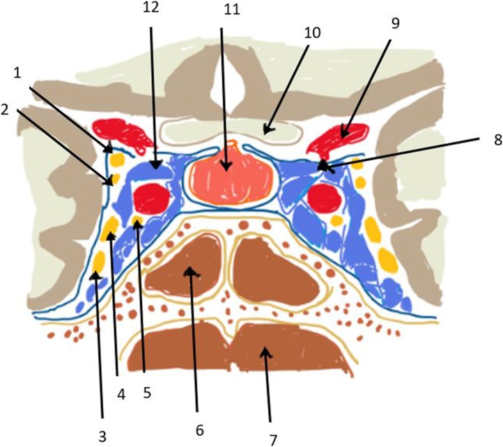
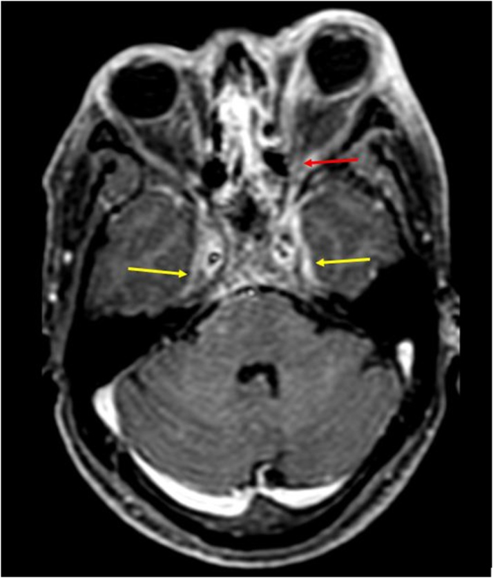
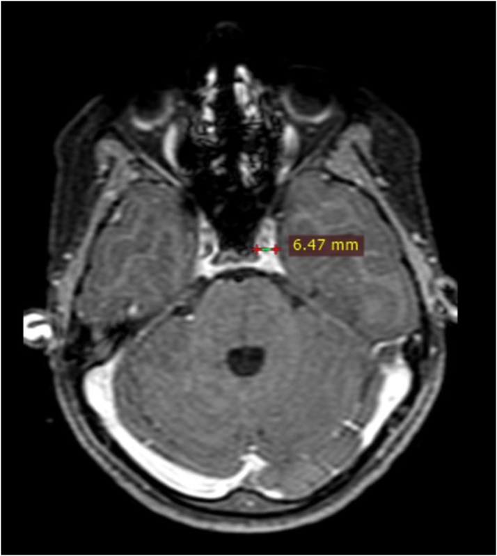
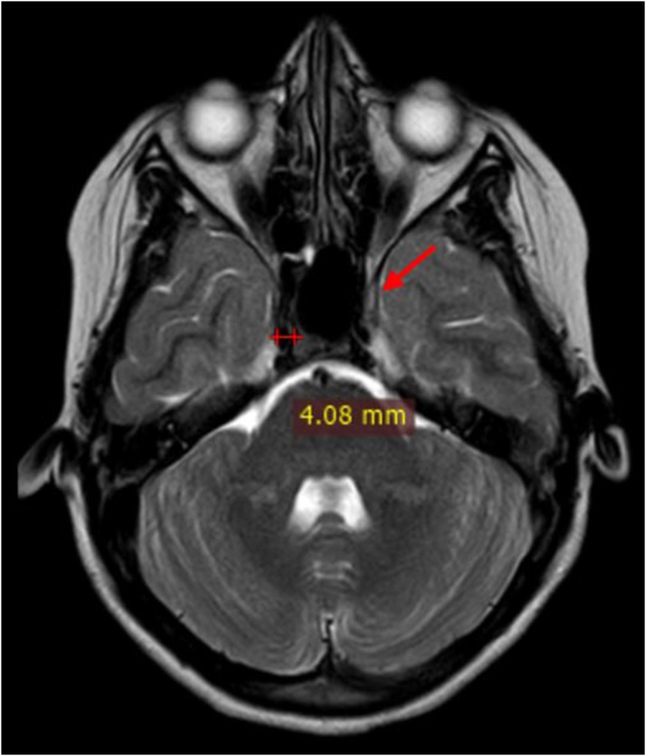
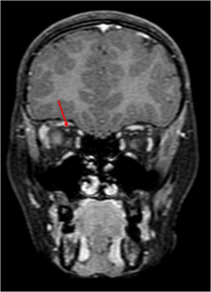
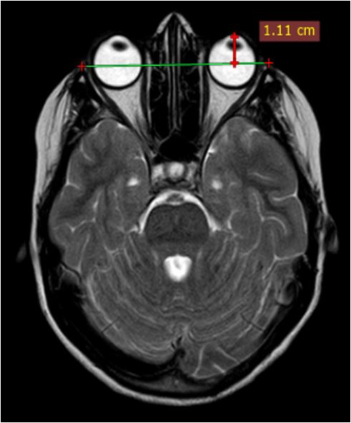
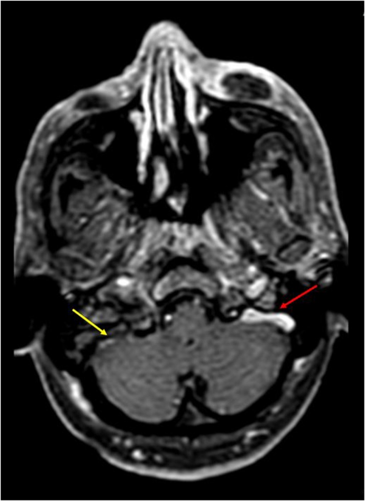
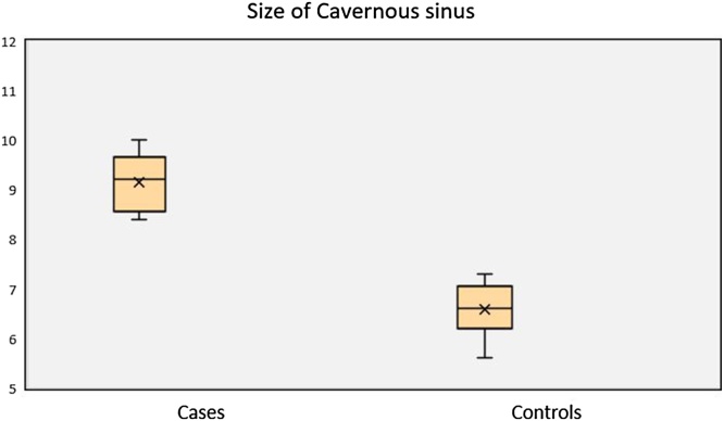
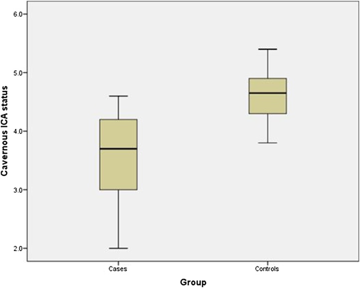
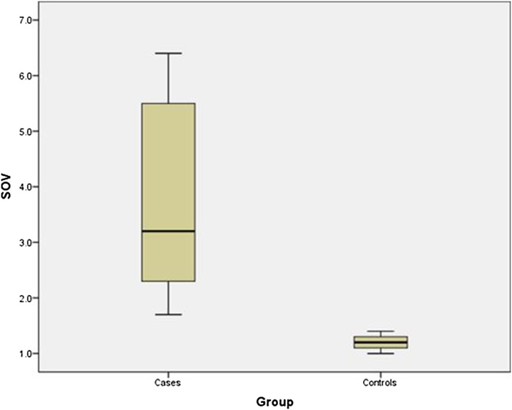
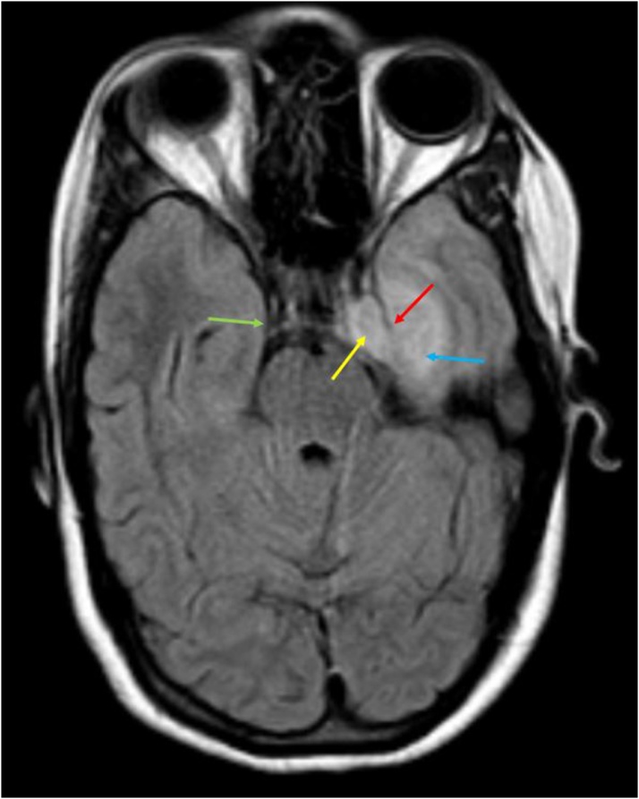
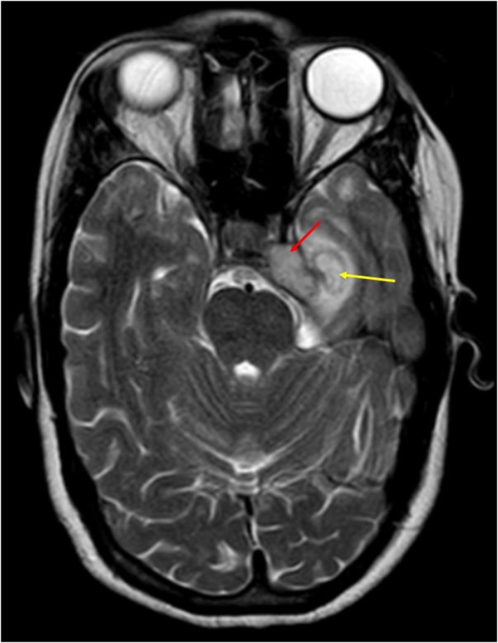
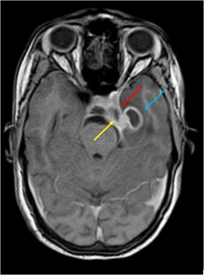
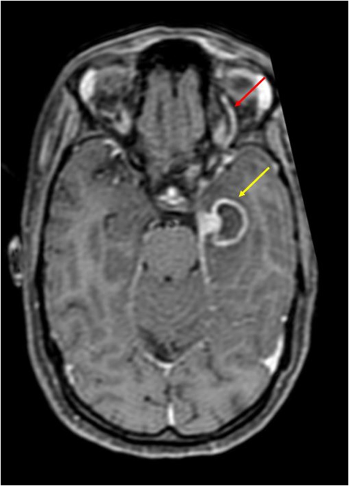
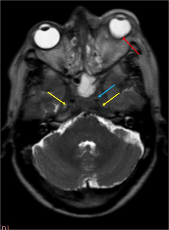
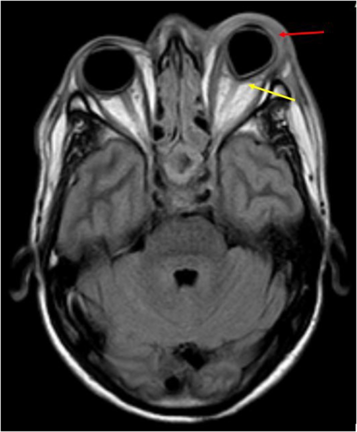
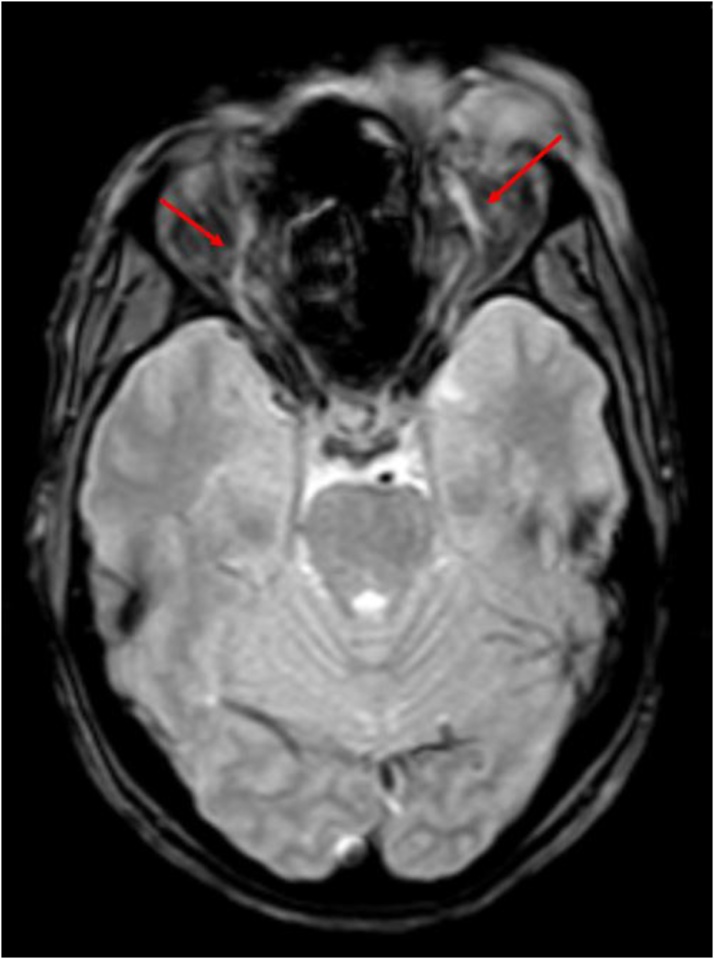
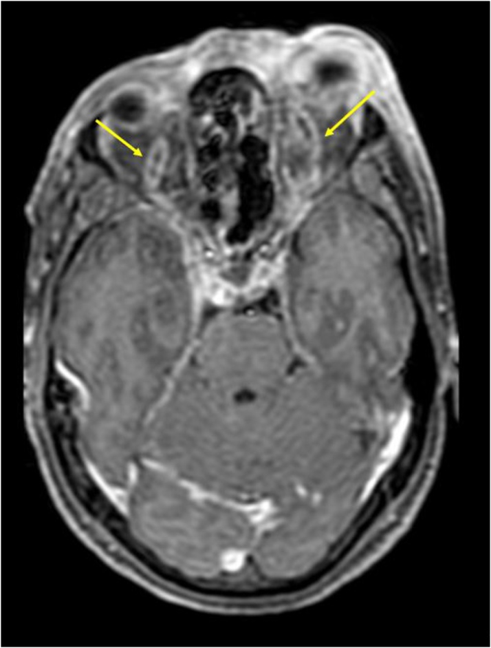
In the patient group, the mean Cavernous sinus (CS) diameter, Cavernous Internal Carotid Artery (ICA) diameter and Superior Ophthalmic Vein (SOV) diameter were 9.14 ± 0.56 mm, 3.5 mm ± 0.9 mm and 3.8 mm ± 1.79 mm respectively. While in the control group, the mean CS diameter, ICA diameter and SOV dimeter were 6.58 ± 0.54 mm, 4.6 mm ± 0.44 mm and 1.1 mm ± 0.11 mm respectively. The differences in the CS size, ICA and SOV diameters was statistically significant. (p < 0.05). Cut off points of ≥ 10 mm for CS diameter, ≥ 2.9 mm for SOV dilation, and ≤ 4.2 mm for ICA flow void diameter were estimated using receiver operating characteristic curves. Various other qualitative parameters, like bulging lateral walls of the sinus, heterogenous signal intensity with filling defects on post contrast images, abnormal dural enhancement along the lateral wall of the sinus and orbital apex involvement were more frequently observed in the CST group, in comparison to the control group.
CEMRI plays an invaluable role not only in the diagnosis of cavernous sinus thrombosis, but also in evaluating the extent of disease and its associated complications. The quantitative and qualitative parameters described here, provide more objectivity and accuracy in diagnosis of CST, thus, aiding prompt diagnosis and early treatment.
确定对比增强磁共振成像(CEMRI)在海绵窦血栓形成(CST)评估中的作用。
该研究纳入了7例经影像学诊断为海绵窦血栓形成的患者。对9个受累海绵窦的对比增强MRI进行回顾性分析,并与7例患者(14个海绵窦)的对照组进行对比。然后比较各种定性和定量参数。
在患者组中,海绵窦(CS)平均直径、海绵窦内颈动脉(ICA)直径和眼上静脉(SOV)直径分别为9.14±0.56mm、3.5mm±0.9mm和3.8mm±1.79mm。而在对照组中,CS平均直径、ICA直径和SOV直径分别为6.58±0.54mm、4.6mm±0.44mm和1.1mm±0.11mm。CS大小、ICA和SOV直径的差异具有统计学意义(p<0.05)。使用受试者工作特征曲线估计CS直径≥10mm、SOV扩张≥2.9mm和ICA血流空洞直径≤4.2mm的截断点。与对照组相比,CST组更常观察到各种其他定性参数,如窦外侧壁膨出、对比剂后图像上有充盈缺损的不均匀信号强度、沿窦外侧壁的硬脑膜异常强化以及眶尖受累。
CEMRI不仅在海绵窦血栓形成的诊断中发挥着重要作用,而且在评估疾病范围及其相关并发症方面也具有重要作用。这里描述的定量和定性参数为CST的诊断提供了更高的客观性和准确性,从而有助于及时诊断和早期治疗。