Department of Orthopedics, The Second Hospital of Hebei Medical University, No. 215, Hepingxi Road, Shijiazhuang, 050000, Hebei Province, China.
J Orthop Surg Res. 2022 Mar 18;17(1):172. doi: 10.1186/s13018-022-02985-x.
Repair of peripheral nerve defect presents a considerable challenge for reconstructive surgeons. The aim of this study is to develop a brain-derived neurotrophic factor (BDNF) from poly(D,L-lactide-co-glycolide) (PLGA) microspheres for the treatment of the peripheral nerve defect.
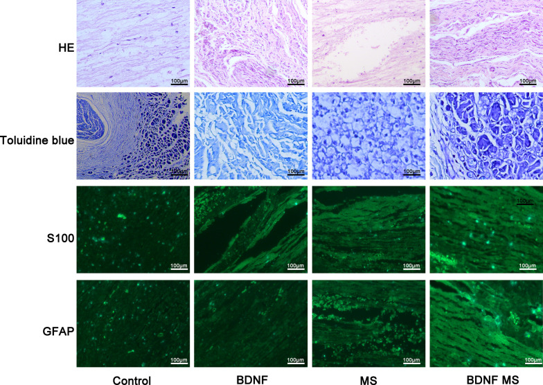
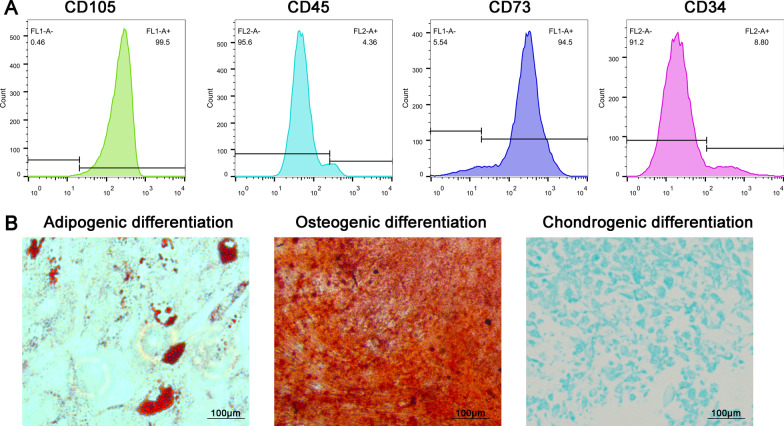
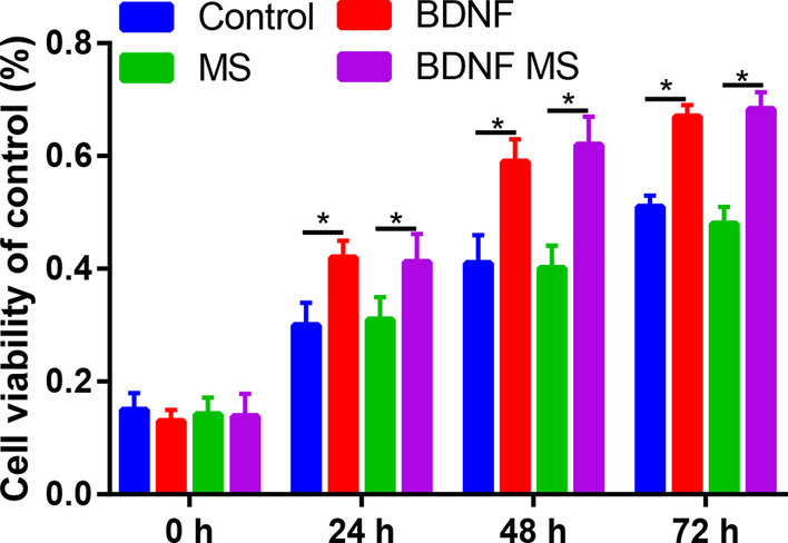
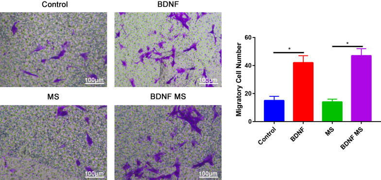
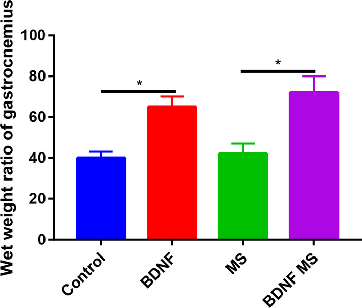
BDNF microspheres were prepared by using an oil-in-water emulsification-solvent evaporation method. The morphology, particle size, encapsulation efficiency, drug loading and sustained release performance of microspheres was observed and calculated. Adipose mesenchymal stem cells (ADSCs) were isolated and expanded. ADSCs were divided into four groups: control, BDNF, blank microsphere and BDNF microsphere groups. Cell count kit-8 (CCK-8) assays were used to assess cell proliferation. Cell migration was determined by Transwell assays. Twenty-eight male Sprague-Dawley rats underwent transection damage model on the right sciatic nerve. The wet weight ratio of the gastrocnemius muscle was calculated by comparing the weight of the gastrocnemius muscle from the operated side to that of the normal side. Neuroelectrophysiological testing was performed to assess nerve function recovery. Nerve regeneration was evaluated by histological analysis and immunohistochemical staining.
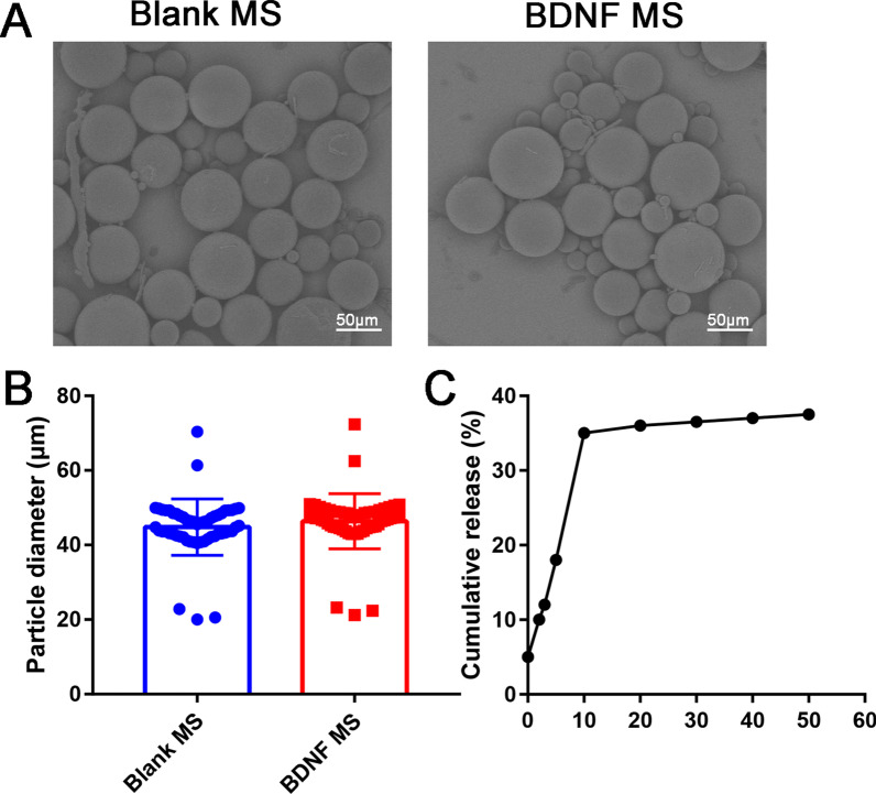
The microspheres were spherical and had uniform size (46.38 ± 1.00 μm), high encapsulation efficiency and high loading capacity. In vitro release studies showed that BDNF-loaded microspheres had good sustained release characteristics. The duration of BDNF release was extended to more than 50 days. BDNF or BDNF microsphere promote the proliferation and migration of ADSCs than control group (P < 0.05). Compared with control group, BDNF significantly decreased the nerve conduction velocity (NCV) and compound amplitude (AMP) (P < 0.05). The nerve fibers in the BDNF microsphere group were closely arranged and uniformly distributed than control group.
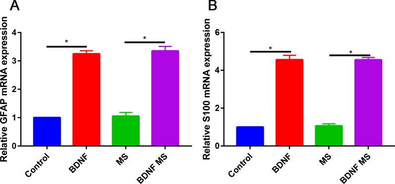
BDNF/PLGA sustained-release microsphere could promote the migration of ADSCs and promoted neural differentiation of ADSCs. Moreover, BDNF/PLGA sustained-release microsphere ameliorated nerve conduction velocity and prevented neuralgic amyotrophy.
周围神经缺损的修复对重建外科医生来说是一个相当大的挑战。本研究旨在开发一种脑源性神经营养因子(BDNF)聚(D,L-丙交酯-共-乙交酯)(PLGA)微球用于治疗周围神经缺损。
采用油包水乳化-溶剂挥发法制备 BDNF 微球。观察和计算微球的形态、粒径、包封率、载药量和缓释性能。分离并扩增脂肪间充质干细胞(ADSCs)。将 ADSCs 分为 4 组:对照组、BDNF 组、空白微球组和 BDNF 微球组。细胞计数试剂盒-8(CCK-8)法检测细胞增殖。Transwell 法检测细胞迁移。28 只雄性 Sprague-Dawley 大鼠右侧坐骨神经横断损伤模型。通过比较手术侧和正常侧腓肠肌的重量,计算腓肠肌湿重比。神经电生理检测评估神经功能恢复情况。通过组织学分析和免疫组织化学染色评估神经再生情况。
微球呈球形,粒径均匀(46.38±1.00μm),包封率高,载药量高。体外释放研究表明,BDNF 载药微球具有良好的缓释特性。BDNF 的释放持续时间延长至 50 天以上。与对照组相比,BDNF 或 BDNF 微球促进 ADSCs 的增殖和迁移(P<0.05)。与对照组相比,BDNF 显著降低神经传导速度(NCV)和复合幅度(AMP)(P<0.05)。BDNF 微球组的神经纤维排列紧密,分布均匀。
BDNF/PLGA 缓释微球能促进 ADSCs 的迁移,促进 ADSCs 的神经分化。此外,BDNF/PLGA 缓释微球改善神经传导速度,预防神经源性肌萎缩。