Yang Le, Li Manzhong, Zhan Yu, Feng Xuefeng, Lu Yun, Li Mingcong, Zhuang Yuming, Lei Jianfeng, Zhao Hui
School of Traditional Chinese Medicine, Capital Medical University, Beijing, China.
Beijing Key Lab of TCM Collateral Disease Theory Research, Beijing, China.
Front Neurol. 2022 Mar 2;13:834329. doi: 10.3389/fneur.2022.834329. eCollection 2022.
Identifying the alterations of the cerebral gray and white matter is an important prerequisite for developing potential pharmacological therapy for stroke. This study aimed to assess the changes of gray and white matter after permanent middle cerebral artery occlusion (pMCAO) in rats using magnetic resonance imaging (MRI), and to correlate them with the behavior performance.
Rats were subjected to pMCAO or sham surgery and reared for 30 days. Motor and cognitive function of the rats were examined by gait and Morris water maze (MWM) tests, respectively. Multimodal MRI was conducted to examine the functional and structural changes of the gray and white matter followed with luxol fast blue (LFB) staining.
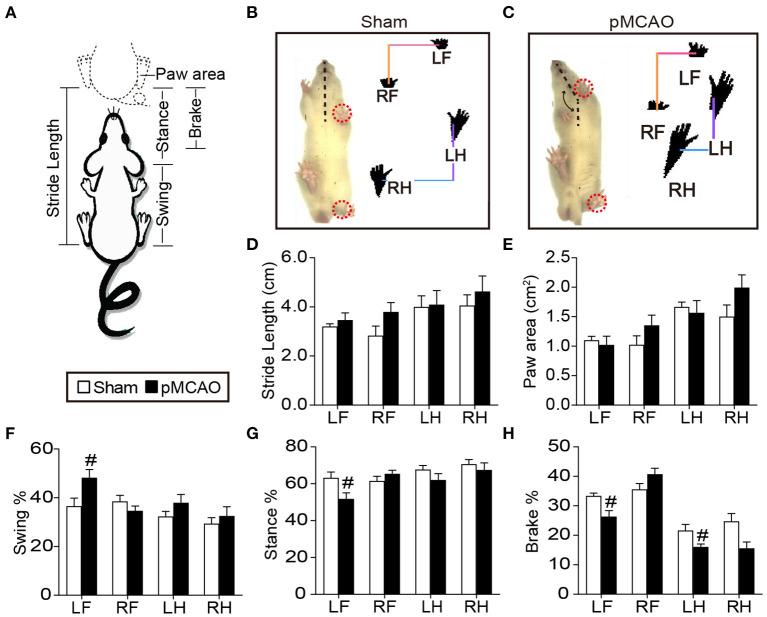
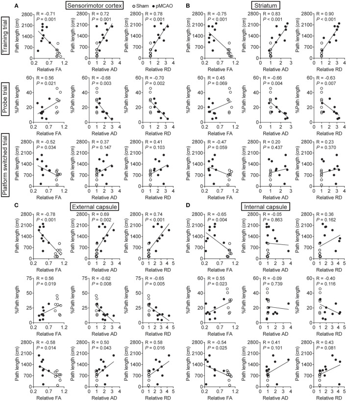
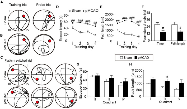
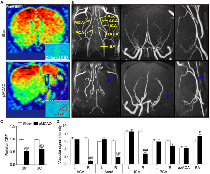
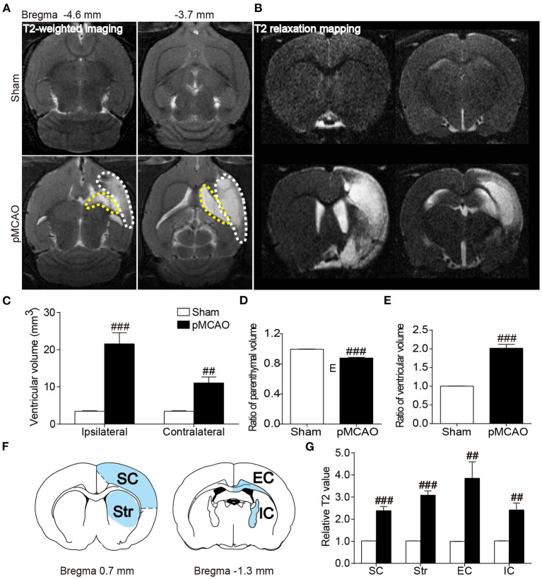
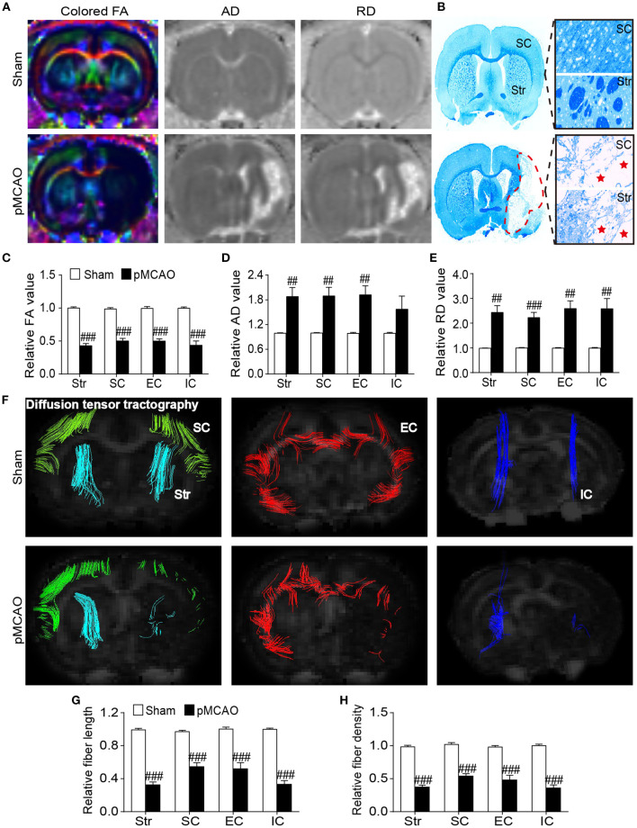
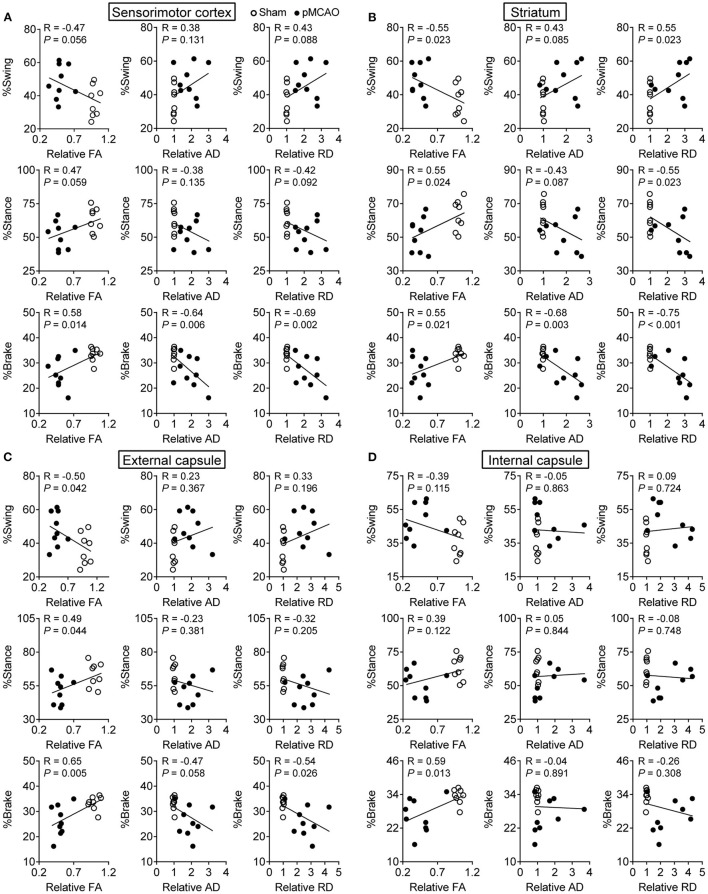
The gait and MWM tests revealed significant motor and cognitive dysfunction in pMCAO rats, respectively. Magnetic resonance angiography presented abnormal intracranial arteries in pMCAO rats with reduced signal intensity of the anterior cerebral artery, anterior communicating cerebral artery, internal carotid artery, and increased basilar artery vessel signal compared with sham rats. Arterial spin labeling confirmed the decreased cerebral blood flow in the infarcted sensorimotor cortex and striatum. Structural T2-weighted imaging and T2 mapping showed brain atrophy and elevation of T2 value in the gray (sensorimotor cortex, striatum) and white (external capsule, internal capsule) matter of pMCAO rats. The results from diffusion tensor imaging (DTI) corresponded well with LFB staining showing reduced relative FA accompanied with increased relative AD and RD in the gray and white matter of pMCAO rats compared with sham rats. Fiber tracking derived from DTI further observed significantly reduced fiber density and length in the corresponding brain regions of pMCAO rats compared with sham rats. Specially, the DTI parameters (especially FA) in the relevant gray matter and white matter significantly correlated with the behavior performance in the gait and MWM tests.
Collectively, the gray and white matter damages could be non-invasively monitored in pMCAO rats by multimodal MRI. DTI-derived parameters, particularly the FA, might be a good imaging index to stage gray and white matter damages associated with post-stroke motor and cognitive impairments.
识别脑灰质和白质的改变是开发中风潜在药物治疗的重要前提。本研究旨在使用磁共振成像(MRI)评估大鼠永久性大脑中动脉闭塞(pMCAO)后灰质和白质的变化,并将其与行为表现相关联。
将大鼠进行pMCAO或假手术,并饲养30天。分别通过步态和莫里斯水迷宫(MWM)试验检查大鼠的运动和认知功能。进行多模态MRI以检查灰质和白质的功能和结构变化,随后进行luxol快速蓝(LFB)染色。
步态和MWM试验分别显示pMCAO大鼠存在明显的运动和认知功能障碍。磁共振血管造影显示pMCAO大鼠颅内动脉异常,与假手术大鼠相比,大脑前动脉、大脑前交通动脉、颈内动脉信号强度降低,基底动脉血管信号增加。动脉自旋标记证实梗死感觉运动皮层和纹状体的脑血流量减少。结构T2加权成像和T2映射显示pMCAO大鼠的灰质(感觉运动皮层、纹状体)和白质(外囊、内囊)出现脑萎缩和T2值升高。扩散张量成像(DTI)结果与LFB染色结果相符,显示与假手术大鼠相比,pMCAO大鼠的灰质和白质中相对FA降低,相对AD和RD增加。从DTI得出的纤维追踪进一步观察到,与假手术大鼠相比,pMCAO大鼠相应脑区的纤维密度和长度明显降低。特别地,相关灰质和白质中的DTI参数(尤其是FA)与步态和MWM试验中的行为表现显著相关。
总体而言,多模态MRI可对pMCAO大鼠的灰质和白质损伤进行无创监测。DTI得出的参数,尤其是FA,可能是评估与中风后运动和认知障碍相关的灰质和白质损伤分期的良好成像指标。