• 文献检索
  • 文档翻译
  • 深度研究
  • 学术资讯
  • Suppr Zotero 插件Zotero 插件
  • 邀请有礼
  • 套餐&价格
  • 历史记录
应用&插件
Suppr Zotero 插件Zotero 插件浏览器插件Mac 客户端Windows 客户端微信小程序
定价
高级版会员购买积分包购买API积分包
服务
文献检索文档翻译深度研究API 文档MCP 服务
关于我们
关于 Suppr公司介绍联系我们用户协议隐私条款
关注我们

Suppr 超能文献

核心技术专利:CN118964589B侵权必究
粤ICP备2023148730 号-1Suppr @ 2026

文献检索

告别复杂PubMed语法,用中文像聊天一样搜索,搜遍4000万医学文献。AI智能推荐,让科研检索更轻松。

立即免费搜索

文件翻译

保留排版,准确专业,支持PDF/Word/PPT等文件格式,支持 12+语言互译。

免费翻译文档

深度研究

AI帮你快速写综述,25分钟生成高质量综述,智能提取关键信息,辅助科研写作。

立即免费体验

MRI-DTI 评估的白质和灰质完整性可以作为慢性神经创伤结构和功能改变的无创可靠指标。

White and gray matter integrity evaluated by MRI-DTI can serve as noninvasive and reliable indicators of structural and functional alterations in chronic neurotrauma.

机构信息

Department of Pediatrics, Chi Mei Medical Center, Tainan, 710, Taiwan.

Department of Biotechnology and Food Technology, Southern Taiwan University of Science and Technology, Tainan, 710, Taiwan.

出版信息

Sci Rep. 2024 Mar 27;14(1):7244. doi: 10.1038/s41598-024-57706-7.

DOI:10.1038/s41598-024-57706-7
PMID:38538745
原文链接:https://pmc.ncbi.nlm.nih.gov/articles/PMC10973494/
Abstract

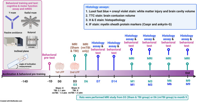
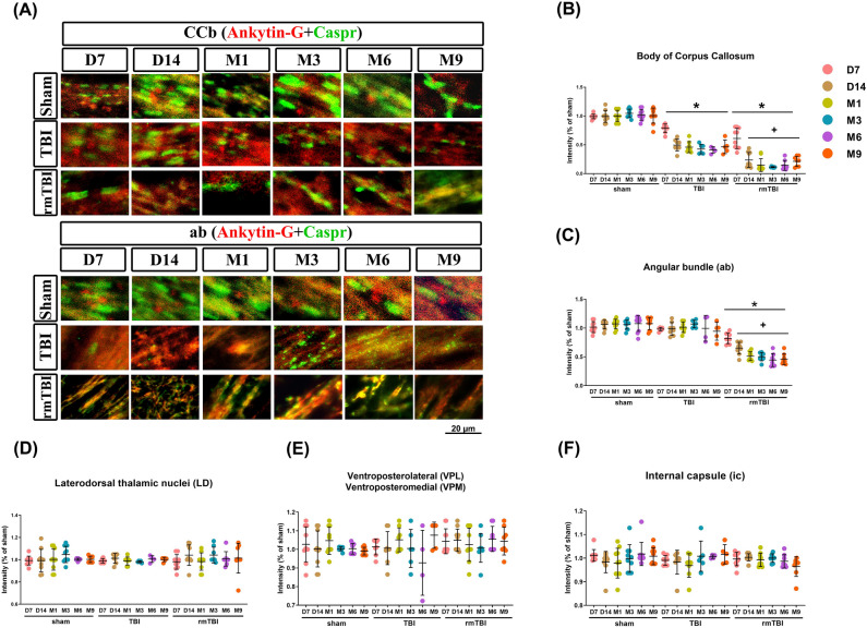
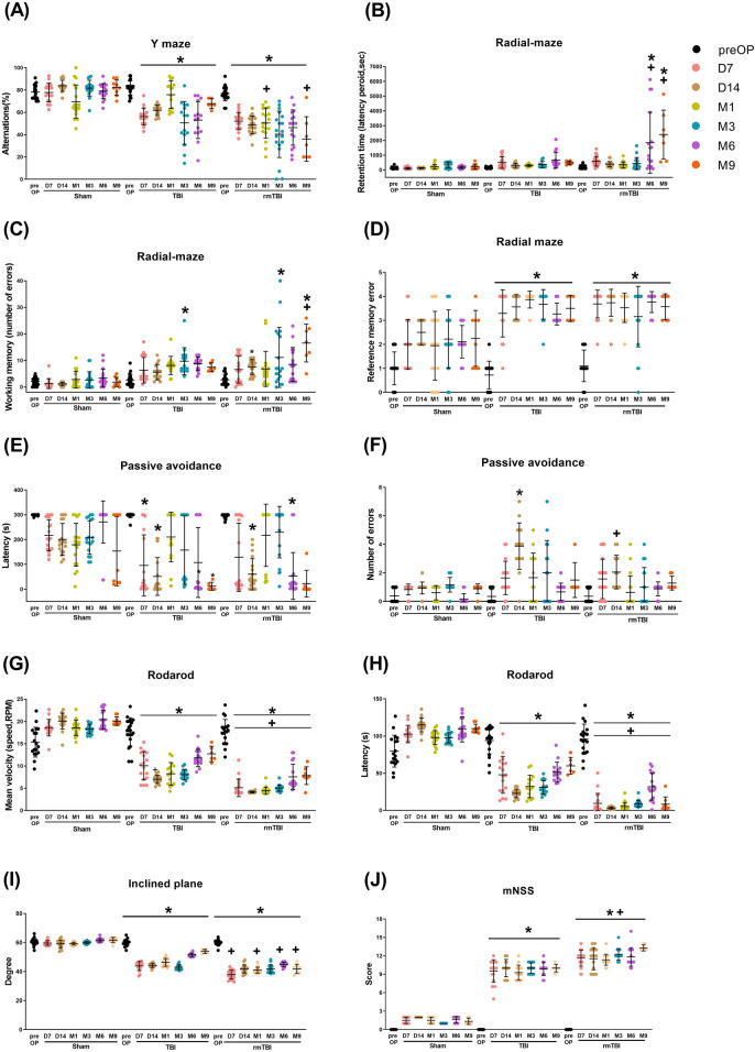
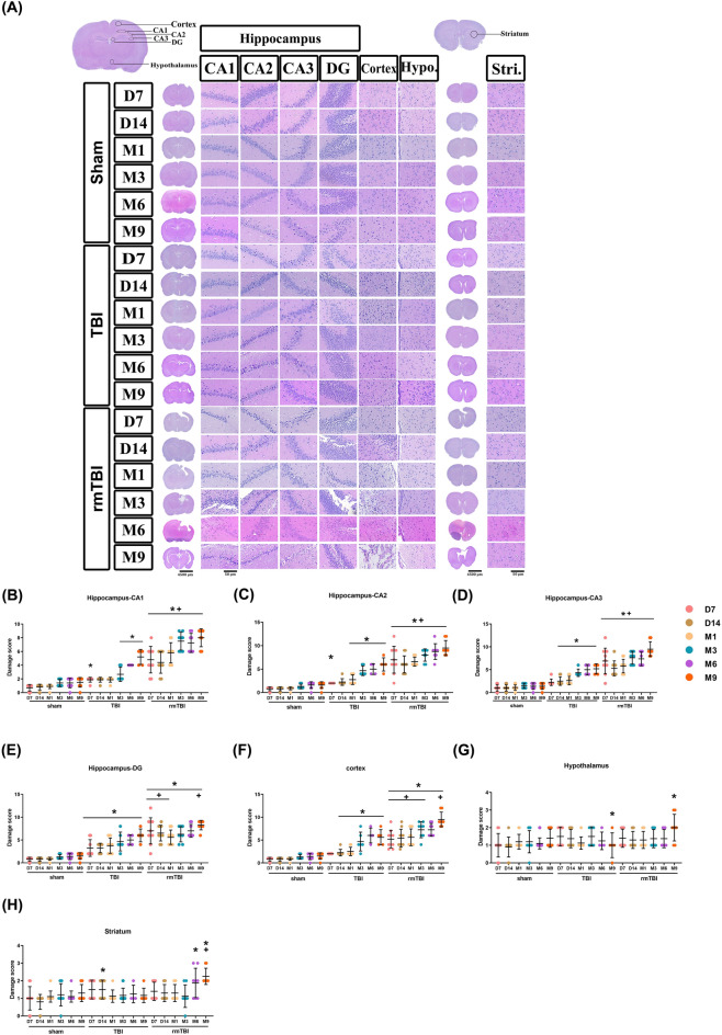
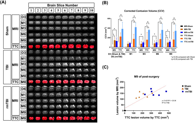
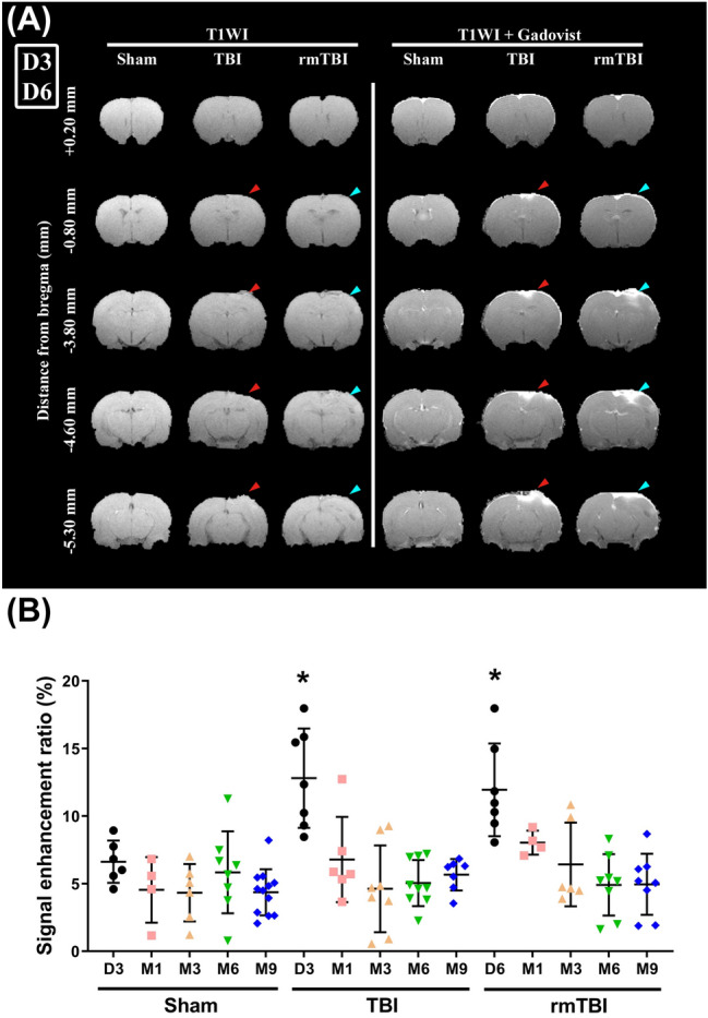
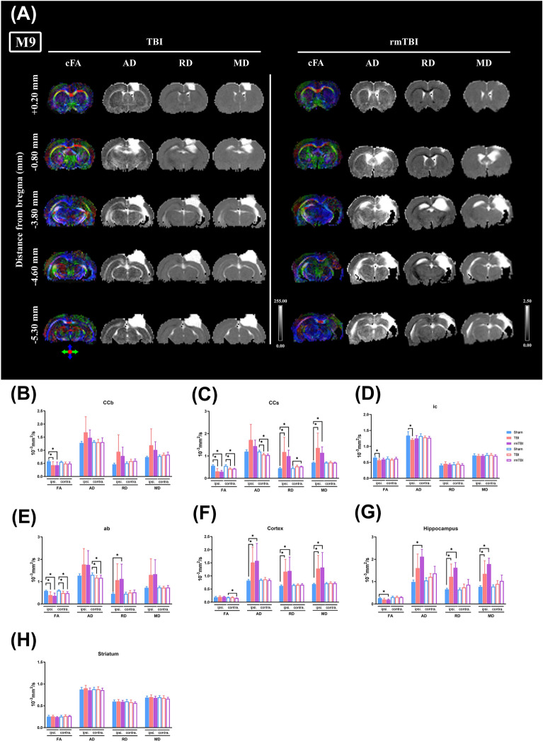
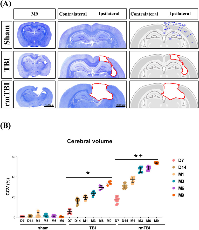
We aimed to evaluate whether white and gray matter microstructure changes observed with magnetic resonance imaging (MRI)-based diffusion tensor imaging (DTI) can be used to reflect the progression of chronic brain trauma. The MRI-DTI parameters, neuropathologic changes, and behavioral performance of adult male Wistar rats that underwent moderate (2.1 atm on day "0") or repeated mild (1.5 atm on days "0" and "2") traumatic brain injury (TBI or rmTBI) or sham operation were evaluated at 7 days, 14 days, and 1-9 months after surgery. Neurobehavioral tests showed that TBI causes long-term motor, cognitive and neurological deficits, whereas rmTBI results in more significant deficits in these paradigms. Both histology and MRI show that rmTBI causes more significant changes in brain lesion volumes than TBI. In vivo DTI further reveals that TBI and rmTBI cause persistent microstructural changes in white matter tracts (such as the body of the corpus callosum, splenium of corpus callus, internal capsule and/or angular bundle) of both two hemispheres. Luxol fast blue measurements reveal similar myelin loss (as well as reduction in white matter thickness) in ipsilateral and contralateral hemispheres as observed by DTI analysis in injured rats. These data indicate that the disintegration of microstructural changes in white and gray matter parameters analyzed by MRI-DTI can serve as noninvasive and reliable markers of structural and functional level alterations in chronic TBI.

摘要

我们旨在评估磁共振成像(MRI)-弥散张量成像(DTI)观察到的白质和灰质微观结构变化是否可用于反映慢性脑创伤的进展。评估了成年雄性 Wistar 大鼠的 MRI-DTI 参数、神经病理学变化和行为表现,这些大鼠接受了中度(第“0”天 2.1 大气压)或重复轻度(第“0”和“2”天 1.5 大气压)创伤性脑损伤(TBI 或 rmTBI)或假手术,分别在手术后 7 天、14 天和 1-9 个月进行。神经行为学测试表明,TBI 导致长期的运动、认知和神经功能缺陷,而 rmTBI 在这些范式中导致更显著的缺陷。组织学和 MRI 均显示,rmTBI 引起的脑损伤体积变化比 TBI 更显著。体内 DTI 进一步显示,TBI 和 rmTBI 导致两个半球的白质束(如胼胝体体部、胼胝体压部、内囊和/或角束)的微观结构变化持续存在。卢索快速蓝测量显示,在损伤大鼠的 DTI 分析中观察到同侧和对侧半球的髓鞘丢失(以及白质厚度减少)相似。这些数据表明,MRI-DTI 分析的白质和灰质参数的微观结构变化的解体可以作为慢性 TBI 结构和功能水平改变的无创和可靠标志物。

https://cdn.ncbi.nlm.nih.gov/pmc/blobs/30f2/10973494/528f73e1c4e3/41598_2024_57706_Fig10_HTML.jpg
https://cdn.ncbi.nlm.nih.gov/pmc/blobs/30f2/10973494/797d92a9b22c/41598_2024_57706_Fig1_HTML.jpg
https://cdn.ncbi.nlm.nih.gov/pmc/blobs/30f2/10973494/0603b38008ea/41598_2024_57706_Fig2_HTML.jpg
https://cdn.ncbi.nlm.nih.gov/pmc/blobs/30f2/10973494/97ddceacf72b/41598_2024_57706_Fig3_HTML.jpg
https://cdn.ncbi.nlm.nih.gov/pmc/blobs/30f2/10973494/01f583f8fbb4/41598_2024_57706_Fig4_HTML.jpg
https://cdn.ncbi.nlm.nih.gov/pmc/blobs/30f2/10973494/165ba7b7c26b/41598_2024_57706_Fig5_HTML.jpg
https://cdn.ncbi.nlm.nih.gov/pmc/blobs/30f2/10973494/5460d75b3ed5/41598_2024_57706_Fig6_HTML.jpg
https://cdn.ncbi.nlm.nih.gov/pmc/blobs/30f2/10973494/aeabeb295ebf/41598_2024_57706_Fig7_HTML.jpg
https://cdn.ncbi.nlm.nih.gov/pmc/blobs/30f2/10973494/a7a4800ca889/41598_2024_57706_Fig8_HTML.jpg
https://cdn.ncbi.nlm.nih.gov/pmc/blobs/30f2/10973494/14e507497942/41598_2024_57706_Fig9_HTML.jpg
https://cdn.ncbi.nlm.nih.gov/pmc/blobs/30f2/10973494/528f73e1c4e3/41598_2024_57706_Fig10_HTML.jpg

相似文献

1
White and gray matter integrity evaluated by MRI-DTI can serve as noninvasive and reliable indicators of structural and functional alterations in chronic neurotrauma.MRI-DTI 评估的白质和灰质完整性可以作为慢性神经创伤结构和功能改变的无创可靠指标。
Sci Rep. 2024 Mar 27;14(1):7244. doi: 10.1038/s41598-024-57706-7.
2
Evaluating spatiotemporal microstructural alterations following diffuse traumatic brain injury.评估弥漫性创伤性脑损伤后的时空微观结构改变。
Neuroimage Clin. 2020;25:102136. doi: 10.1016/j.nicl.2019.102136. Epub 2019 Dec 14.
3
Acute Blood Levels of Neurofilament Light Indicate One-Year White Matter Pathology and Functional Impairment in Repetitive Mild Traumatic Brain Injured Mice.急性血液神经丝轻链水平提示重复性轻度创伤性脑损伤小鼠一年的白质病理学和功能损伤。
J Neurotrauma. 2023 Jun;40(11-12):1144-1163. doi: 10.1089/neu.2022.0252. Epub 2023 Mar 13.
4
Common Patterns of Regional Brain Injury Detectable by Diffusion Tensor Imaging in Otherwise Normal-Appearing White Matter in Patients with Early Moderate to Severe Traumatic Brain Injury.早期中重度创伤性脑损伤患者,弥散张量成像可发现正常外观的脑白质中存在的常见区域性脑损伤模式。
J Neurotrauma. 2018 Mar 1;35(5):739-749. doi: 10.1089/neu.2016.4944. Epub 2018 Feb 9.
5
Imaging Markers for the Characterization of Gray and White Matter Changes from Acute to Chronic Stages after Experimental Traumatic Brain Injury.实验性颅脑损伤后从急性期到慢性期的灰、白质变化的影像学标志物。
J Neurotrauma. 2021 Jun 15;38(12):1642-1653. doi: 10.1089/neu.2020.7151. Epub 2021 Jan 11.
6
Multimodal magnetic resonance imaging after experimental moderate and severe traumatic brain injury: A longitudinal correlative assessment of structural and cerebral blood flow changes.实验性中重度创伤性脑损伤后的多模态磁共振成像:结构和脑血流变化的纵向相关评估。
PLoS One. 2023 Aug 7;18(8):e0289786. doi: 10.1371/journal.pone.0289786. eCollection 2023.
7
Repeated mild blast exposure in young adult rats results in dynamic and persistent microstructural changes in the brain.反复轻度爆震暴露于年轻成年大鼠导致大脑中动态和持久的微观结构变化。
Neuroimage Clin. 2018 Jan 11;18:60-73. doi: 10.1016/j.nicl.2018.01.007. eCollection 2018.
8
Radiological-pathological correlation of diffusion tensor and magnetization transfer imaging in a closed head traumatic brain injury model.闭合性颅脑创伤模型中扩散张量成像与磁化传递成像的放射学-病理学相关性
Ann Neurol. 2016 Jun;79(6):907-20. doi: 10.1002/ana.24641. Epub 2016 Apr 18.
9
Changes in Apparent Fiber Density and Track-Weighted Imaging Metrics in White Matter following Experimental Traumatic Brain Injury.实验性创伤性脑损伤后白质中表观纤维密度和轨迹加权成像指标的变化
J Neurotrauma. 2017 Jul 1;34(13):2109-2118. doi: 10.1089/neu.2016.4730. Epub 2017 Apr 13.
10
Diffusion Magnetic Resonance Imaging Unveils the Spatiotemporal Microstructural Gray Matter Changes following Injury in the Rodent Brain.扩散磁共振成像揭示了啮齿动物脑损伤后时空微观结构灰质的变化。
J Neurotrauma. 2019 Apr 15;36(8):1306-1317. doi: 10.1089/neu.2018.5972. Epub 2018 Dec 20.

引用本文的文献

1
The Collapse of Brain Clearance: Glymphatic-Venous Failure, Aquaporin-4 Breakdown, and AI-Empowered Precision Neurotherapeutics in Intracranial Hypertension.脑清除功能的崩溃:颅内高压中的淋巴-静脉功能衰竭、水通道蛋白4的破坏以及人工智能助力的精准神经治疗
Int J Mol Sci. 2025 Jul 25;26(15):7223. doi: 10.3390/ijms26157223.
2
Bundle-specific tractography approach for identifying white matter microstructural changes following traumatic brain injury in rats: An EpiBioS4Rx study.用于识别大鼠创伤性脑损伤后白质微观结构变化的束特异性纤维束成像方法:一项EpiBioS4Rx研究。
Imaging Neurosci (Camb). 2025 Feb 18;3. doi: 10.1162/imag_a_00471. eCollection 2025.
3

本文引用的文献

1
White Matter Organization and Cortical Thickness Differ Among Active Duty Service Members With Chronic Mild, Moderate, and Severe Traumatic Brain Injury.慢性轻度、中度和重度创伤性脑损伤的现役军人的白质组织和皮质厚度存在差异。
J Neurotrauma. 2024 Apr;41(7-8):818-835. doi: 10.1089/neu.2023.0336. Epub 2023 Nov 15.
2
Multimodal magnetic resonance imaging after experimental moderate and severe traumatic brain injury: A longitudinal correlative assessment of structural and cerebral blood flow changes.实验性中重度创伤性脑损伤后的多模态磁共振成像:结构和脑血流变化的纵向相关评估。
PLoS One. 2023 Aug 7;18(8):e0289786. doi: 10.1371/journal.pone.0289786. eCollection 2023.
3
Diffusion Tensor Imaging Findings in Cerebral Sensorimotor Areas in Patients After Spinal Cord Injury Correlate With Neurophysiological Deficits.
脊髓损伤患者大脑感觉运动区的扩散张量成像结果与神经生理缺陷相关。
Neurorehabil Neural Repair. 2025 Jun 19;39(9):15459683251345434. doi: 10.1177/15459683251345434.
4
White matter microstructure abnormalities in thalassemia patients using TBSS analysis based on DTI.基于扩散张量成像(DTI)的体素-基于的形态学分析(TBSS)对地中海贫血患者白质微观结构异常的研究
Sci Rep. 2025 May 23;15(1):17929. doi: 10.1038/s41598-025-99505-8.
5
Closed-loop rehabilitation of upper-limb dyskinesia after stroke: from natural motion to neuronal microfluidics.中风后上肢运动障碍的闭环康复:从自然运动到神经微流体
J Neuroeng Rehabil. 2025 Apr 19;22(1):87. doi: 10.1186/s12984-025-01617-9.
6
Traumatic brain injury: Bridging pathophysiological insights and precision treatment strategies.创伤性脑损伤:连接病理生理见解与精准治疗策略
Neural Regen Res. 2026 Mar 1;21(3):887-907. doi: 10.4103/NRR.NRR-D-24-01398. Epub 2025 Mar 25.
7
Preliminary feasibility study on DTI to assess the early brain injury in germinal matrix-intraventricular hemorrhage rats.利用扩散张量成像评估脑室周围-脑室内出血大鼠早期脑损伤的初步可行性研究。
Sci Rep. 2025 Mar 21;15(1):9839. doi: 10.1038/s41598-025-94934-x.
8
Independent associations of sleep and physical activity with cognition are mediated by hippocampal microstructure in middle-aged and older adults.睡眠和身体活动与认知的独立关联由中老年人的海马体微观结构介导。
Neurobiol Aging. 2025 Mar;147:22-31. doi: 10.1016/j.neurobiolaging.2024.11.011. Epub 2024 Dec 5.
9
Assessing biological self-organization patterns using statistical complexity characteristics: a tool for diffusion tensor imaging analysis.利用统计复杂性特征评估生物自组织模式:一种扩散张量成像分析工具。
MAGMA. 2024 Jul 28. doi: 10.1007/s10334-024-01185-4.
Diffusion MRI approaches for investigating microstructural complexity in a rat model of traumatic brain injury.
扩散 MRI 方法研究创伤性脑损伤大鼠模型中的微观结构复杂性。
Sci Rep. 2023 Feb 8;13(1):2219. doi: 10.1038/s41598-023-29010-3.
4
Chronic traumatic encephalopathy: Diagnostic updates and advances.慢性创伤性脑病:诊断的更新与进展
AIMS Neurosci. 2022 Dec 19;9(4):519-535. doi: 10.3934/Neuroscience.2022030. eCollection 2022.
5
A multiscale tissue assessment in a rat model of mild traumatic brain injury.大鼠轻度创伤性脑损伤模型的多尺度组织评估。
J Neuropathol Exp Neurol. 2022 Dec 19;82(1):71-83. doi: 10.1093/jnen/nlac100.
6
Combining Multiple Indices of Diffusion Tensor Imaging Can Better Differentiate Patients with Traumatic Brain Injury from Healthy Subjects.结合扩散张量成像的多个指标可以更好地区分创伤性脑损伤患者与健康受试者。
Neuropsychiatr Dis Treat. 2022 Aug 23;18:1801-1814. doi: 10.2147/NDT.S354265. eCollection 2022.
7
White matter pathology in alzheimer's transgenic mice with chronic exposure to low-level ambient fine particulate matter.慢性暴露于低水平环境细颗粒物的阿尔茨海默病转基因小鼠的脑白质病变。
Part Fibre Toxicol. 2022 Jun 30;19(1):44. doi: 10.1186/s12989-022-00485-8.
8
Microstructural Tissue Changes in a Rat Model of Mild Traumatic Brain Injury.轻度创伤性脑损伤大鼠模型中的微观组织变化
Front Neurosci. 2021 Nov 26;15:746214. doi: 10.3389/fnins.2021.746214. eCollection 2021.
9
Long-term follow-up of neurodegenerative phenomenon in severe traumatic brain injury using MRI.利用 MRI 对严重创伤性脑损伤的神经退行性现象进行长期随访。
Ann Phys Rehabil Med. 2022 Nov;65(6):101599. doi: 10.1016/j.rehab.2021.101599. Epub 2022 Feb 15.
10
Contrast enhanced magnetic resonance imaging highlights neurovasculature changes following experimental traumatic brain injury in the rat.对比增强磁共振成像突出显示大鼠实验性创伤性脑损伤后的神经血管变化。
Sci Rep. 2020 Dec 4;10(1):21252. doi: 10.1038/s41598-020-77975-2.