Brain Research Center, Third Military Medical University, Chongqing, 400038, China.
State Key Laboratory of Trauma, Burns, and Combined Injury, Third Military Medical University, Chongqing, 400038, China.
Nat Commun. 2022 Mar 22;13(1):1531. doi: 10.1038/s41467-022-29229-0.
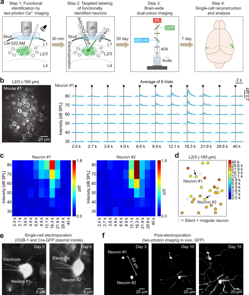
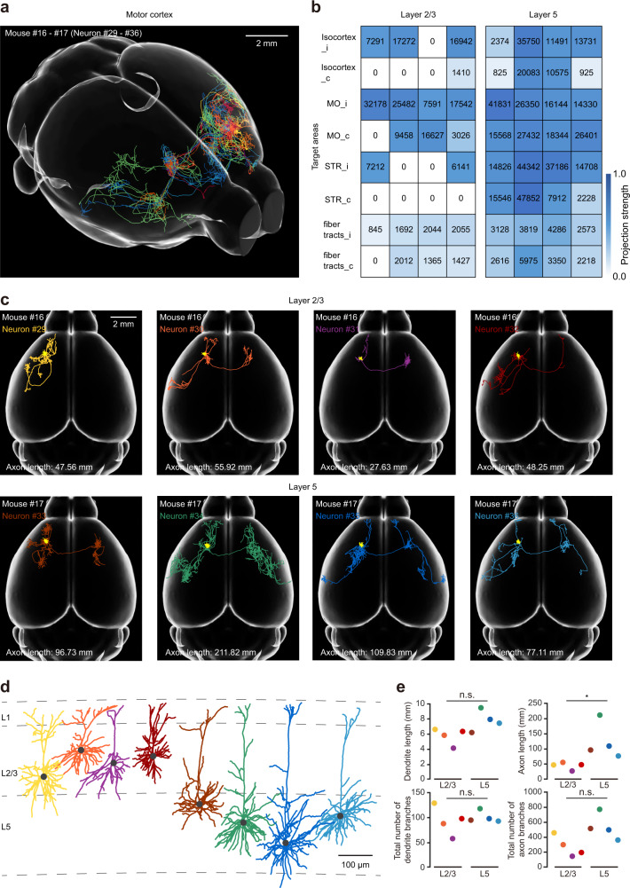
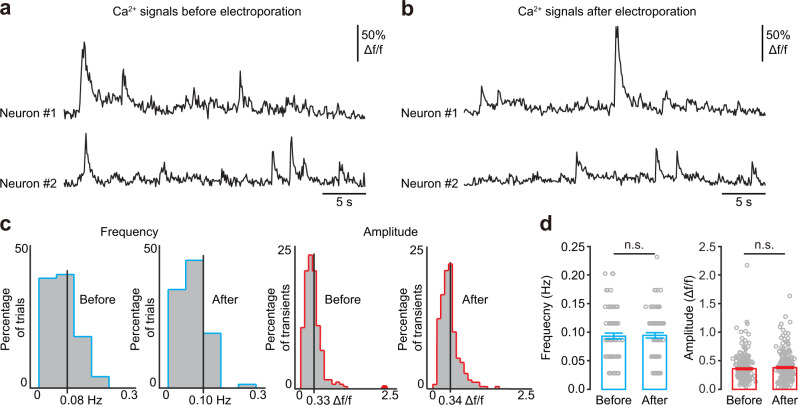
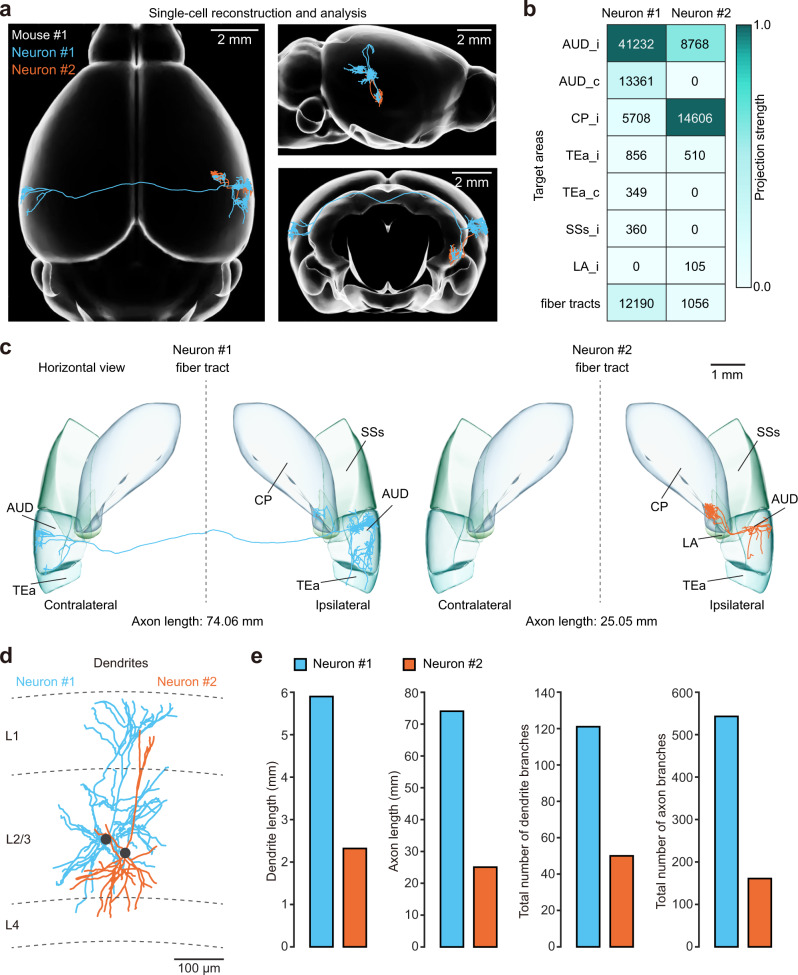
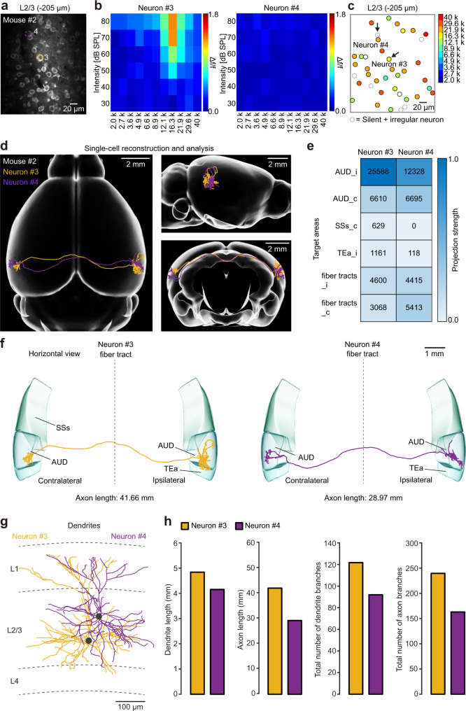
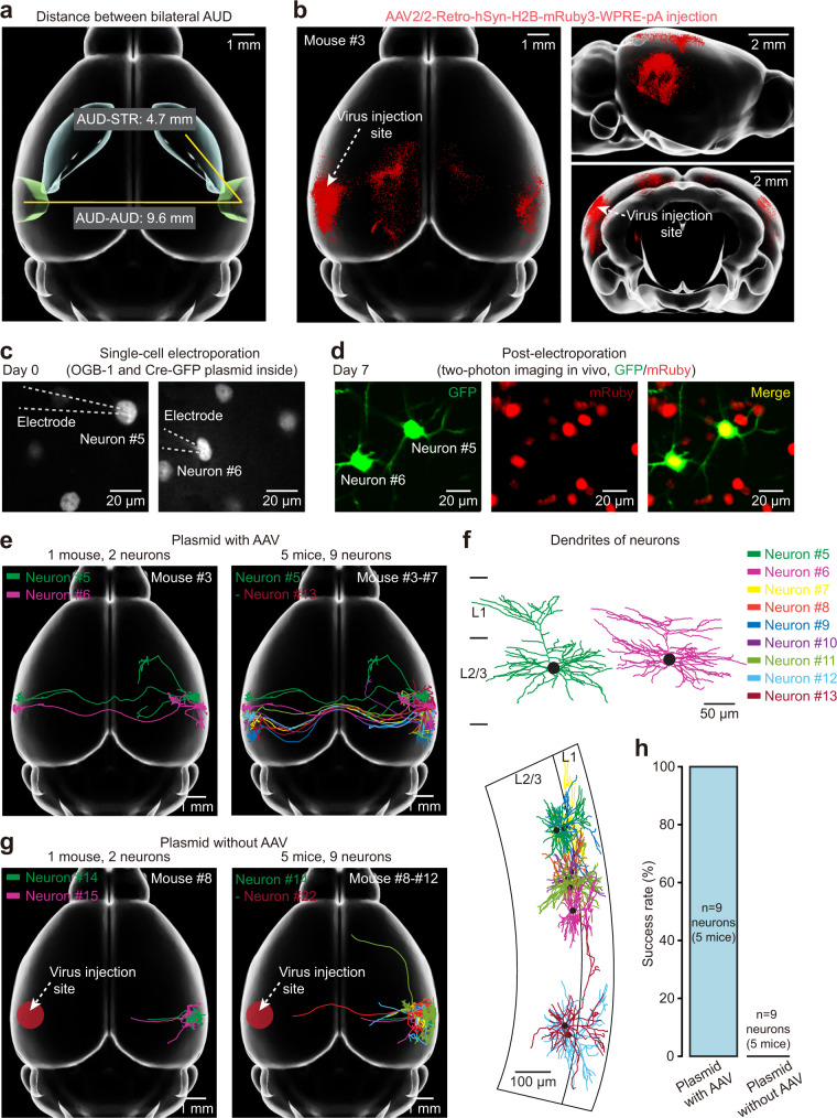
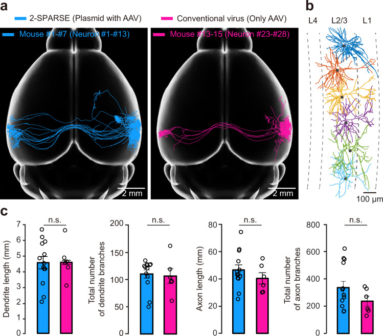
Reconstructing axonal projections of single neurons at the whole-brain level is currently a converging goal of the neuroscience community that is fundamental for understanding the logic of information flow in the brain. Thousands of single neurons from different brain regions have recently been morphologically reconstructed, but the corresponding physiological functional features of these reconstructed neurons are unclear. By combining two-photon Ca imaging with targeted single-cell plasmid electroporation, we reconstruct the brain-wide morphologies of single neurons that are defined by a sound-evoked response map in the auditory cortices (AUDs) of awake mice. Long-range interhemispheric projections can be reliably labelled via co-injection with an adeno-associated virus, which enables enhanced expression of indicator protein in the targeted neurons. Here we show that this method avoids the randomness and ambiguity of conventional methods of neuronal morphological reconstruction, offering an avenue for developing a precise one-to-one map of neuronal projection patterns and physiological functional features.
在全脑水平上重建单个神经元的轴突投射,是当前神经科学界的一个共同目标,对于理解大脑中信息流的逻辑至关重要。最近,已经对来自不同脑区的数千个单个神经元进行了形态重建,但这些重建神经元的相应生理功能特征尚不清楚。通过结合双光子 Ca 成像和靶向单细胞质粒电穿孔,我们重建了在清醒小鼠听觉皮层(AUD)中由声刺激反应图谱定义的单个神经元的全脑形态。通过共注射腺相关病毒,可以可靠地标记长程半球间投射,这使得靶向神经元中指示剂蛋白的表达增强。本文中我们展示了这种方法避免了传统神经元形态重建方法的随机性和模糊性,为开发精确的神经元投射模式和生理功能特征的一一对应图谱提供了途径。