Istituto di Ricerche Farmacologiche Mario Negri IRCCS, Bergamo, Italy.
Unit of Nephrology and Dialysis, Azienda Socio Sanitaria Territoriale (ASST) Papa Giovanni XXIII, Bergamo, Italy.
Front Immunol. 2022 Mar 7;13:827146. doi: 10.3389/fimmu.2022.827146. eCollection 2022.
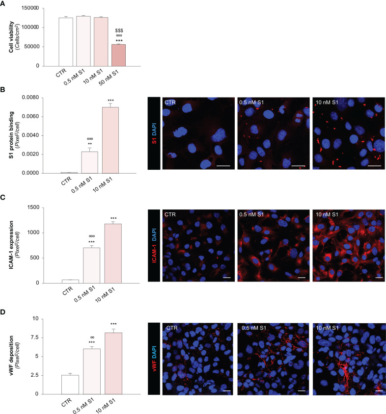
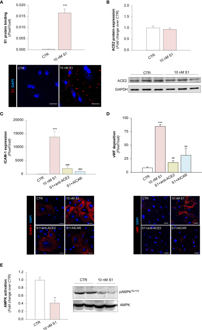
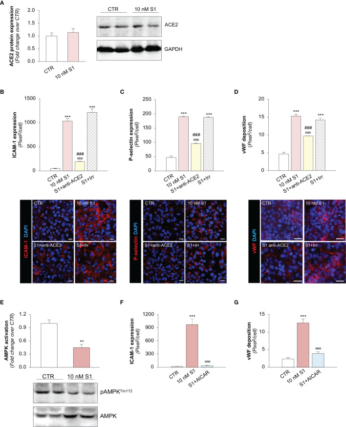
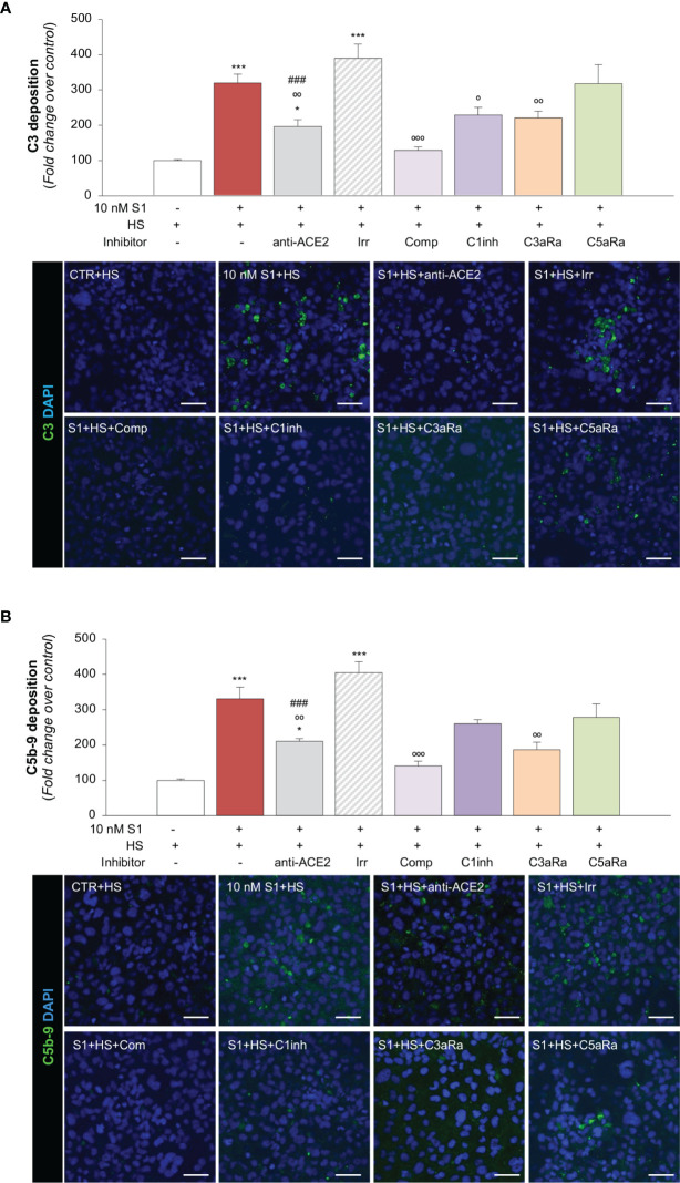
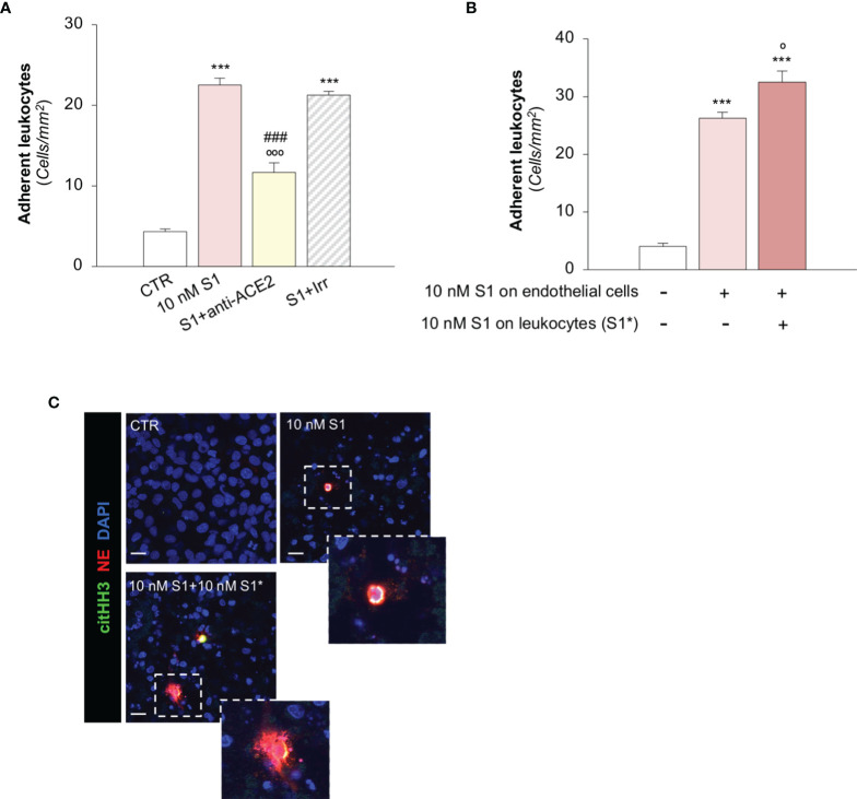
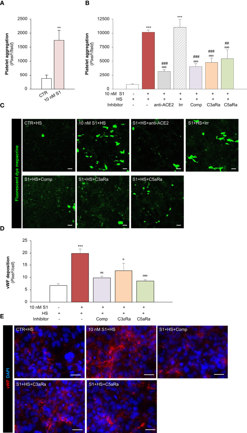
Microvascular thrombosis is associated with multiorgan failure and mortality in coronavirus disease 2019 (COVID-19). Although thrombotic complications may be ascribed to the ability of SARS-CoV-2 to infect and replicate in endothelial cells, it has been poorly investigated whether, in the complexity of viral infection in the human host, specific viral elements alone can induce endothelial damage. Detection of circulating spike protein in the sera of severe COVID-19 patients was evaluated by ELISA. experiments were performed on human microvascular endothelial cells from the derma and lung exposed to SARS-CoV-2-derived spike protein 1 (S1). The expression of adhesive molecules was studied by immunofluorescence and leukocyte adhesion and platelet aggregation were assessed under flow conditions. Angiotensin converting enzyme 2 (ACE2) and AMPK expression were investigated by Western Blot analysis. In addition, S1-treated endothelial cells were incubated with anti-ACE2 blocking antibody, AMPK agonist, or complement inhibitors. Our results show that significant levels of spike protein were found in the 30.4% of severe COVID-19 patients. , the activation of endothelial cells with S1 protein, ACE2, impaired AMPK signalling, leading to robust leukocyte recruitment due to increased adhesive molecule expression and thrombomodulin loss. This S1-induced pro-inflammatory phenotype led to exuberant C3 and C5b-9 deposition on endothelial cells, along with C3a and C5a generation that further amplified S1-induced complement activation. Functional blockade of ACE2 or complement inhibition halted S1-induced platelet aggregates by limiting von Willebrand factor and P-selectin exocytosis and expression on endothelial cells. Overall, we demonstrate that SARS-CoV-2-derived S1 is sufficient in itself to propagate inflammatory and thrombogenic processes in the microvasculature, amplified by the complement system, recapitulating the thromboembolic complications of COVID-19.
微血管血栓形成与 2019 年冠状病毒病(COVID-19)的多器官衰竭和死亡率有关。虽然血栓形成并发症可能归因于 SARS-CoV-2 感染和在血管内皮细胞中复制的能力,但在人类宿主中病毒感染的复杂性中,是否仅特定的病毒成分就可以诱导内皮损伤,这一点还没有得到很好的研究。通过 ELISA 评估严重 COVID-19 患者血清中循环刺突蛋白的检测。在暴露于 SARS-CoV-2 衍生的刺突蛋白 1(S1)的人皮肤和肺微血管内皮细胞上进行了 实验。通过免疫荧光研究粘附分子的表达,并在流动条件下评估白细胞黏附和血小板聚集。通过 Western Blot 分析研究血管紧张素转换酶 2(ACE2)和 AMPK 表达。此外,用抗 ACE2 阻断抗体、AMPK 激动剂或补体抑制剂孵育 S1 处理的内皮细胞。我们的结果表明,在 30.4%的严重 COVID-19 患者中发现了显著水平的刺突蛋白。 ,S1 蛋白激活内皮细胞, ACE2,AMPK 信号受损,导致粘附分子表达增加和血栓调节蛋白丢失,导致白细胞募集增加。这种 S1 诱导的促炎表型导致丰富的 C3 和 C5b-9 在血管内皮细胞上沉积,以及 C3a 和 C5a 的产生,进一步放大了 S1 诱导的补体激活。ACE2 或补体抑制的功能阻断通过限制血管内皮细胞上 von Willebrand 因子和 P-选择素的胞吐作用和表达,阻止了 S1 诱导的血小板聚集。总的来说,我们证明 SARS-CoV-2 衍生的 S1 本身足以在微血管中传播炎症和血栓形成过程,补体系统放大了这一过程,再现了 COVID-19 的血栓栓塞并发症。