• 文献检索
  • 文档翻译
  • 深度研究
  • 学术资讯
  • Suppr Zotero 插件Zotero 插件
  • 邀请有礼
  • 套餐&价格
  • 历史记录
应用&插件
Suppr Zotero 插件Zotero 插件浏览器插件Mac 客户端Windows 客户端微信小程序
定价
高级版会员购买积分包购买API积分包
服务
文献检索文档翻译深度研究API 文档MCP 服务
关于我们
关于 Suppr公司介绍联系我们用户协议隐私条款
关注我们

Suppr 超能文献

核心技术专利:CN118964589B侵权必究
粤ICP备2023148730 号-1Suppr @ 2026

文献检索

告别复杂PubMed语法,用中文像聊天一样搜索,搜遍4000万医学文献。AI智能推荐,让科研检索更轻松。

立即免费搜索

文件翻译

保留排版,准确专业,支持PDF/Word/PPT等文件格式,支持 12+语言互译。

免费翻译文档

深度研究

AI帮你快速写综述,25分钟生成高质量综述,智能提取关键信息,辅助科研写作。

立即免费体验

局部补体在 SARS-CoV-2 感染中导致肺血管内皮细胞的致病激活。

Local Complement Contributes to Pathogenic Activation of Lung Endothelial Cells in SARS-CoV-2 Infection.

机构信息

Cardiovascular Pulmonary Research Laboratories, Department of Pediatrics and Medicine.

Department of Immunology and Microbiology, and.

出版信息

Am J Respir Cell Mol Biol. 2023 Aug;69(2):210-219. doi: 10.1165/rcmb.2022-0373OC.

DOI:10.1165/rcmb.2022-0373OC
PMID:37071849
原文链接:https://pmc.ncbi.nlm.nih.gov/articles/PMC10399142/
Abstract

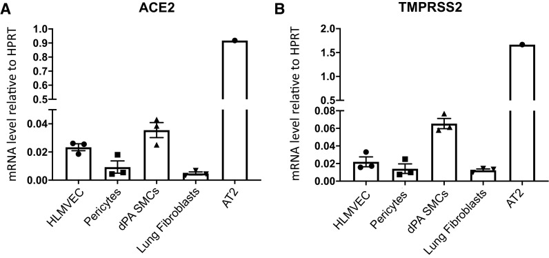
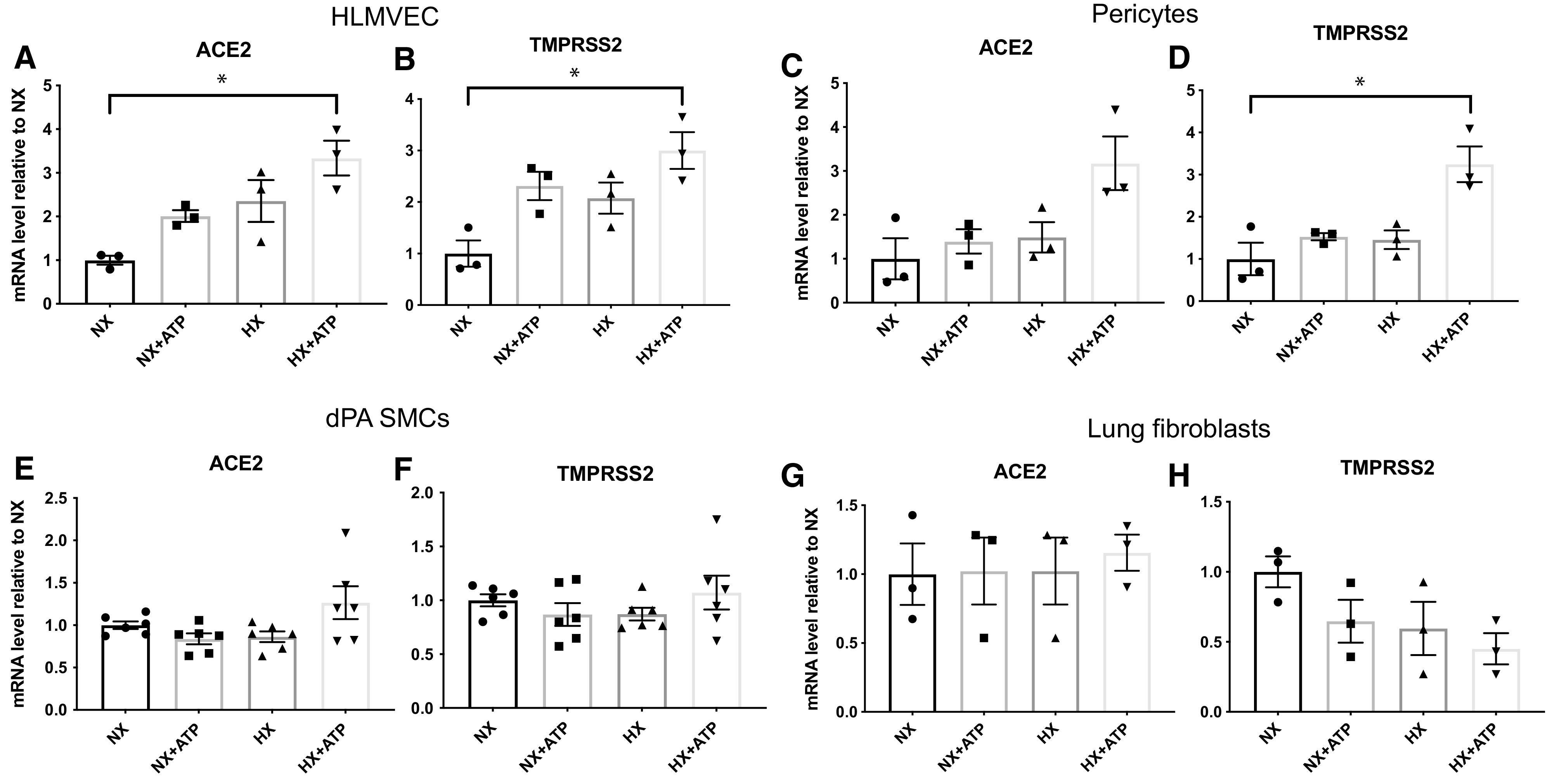
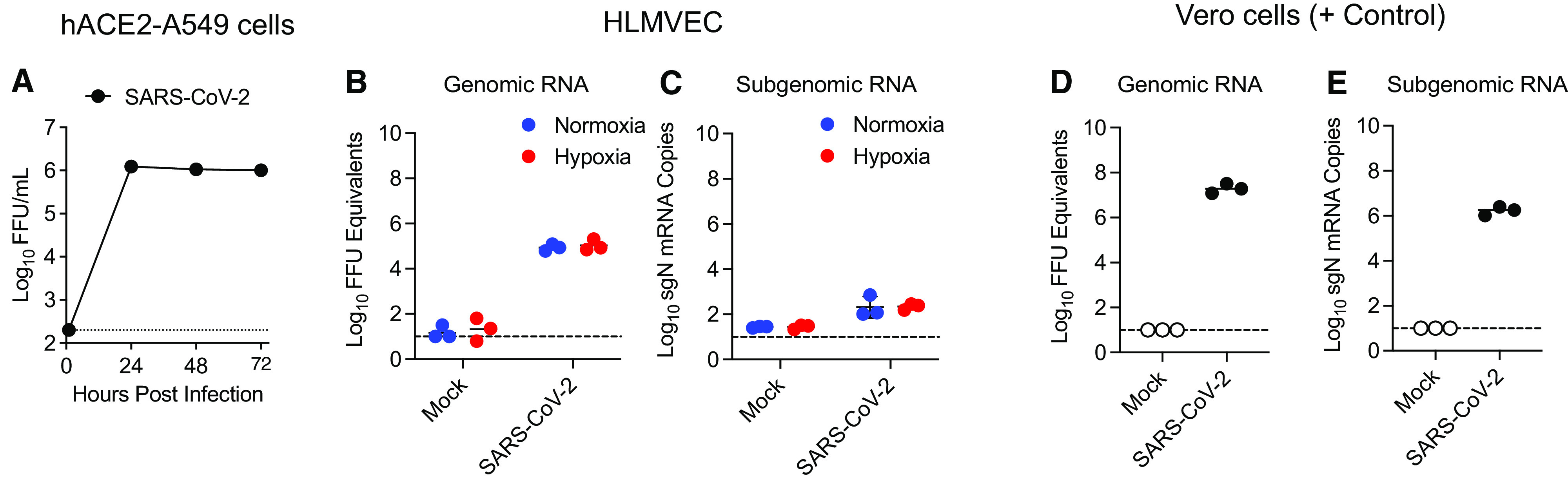
Endothelial dysfunction and inflammation contribute to the vascular pathology of coronavirus disease (COVID-19). However, emerging evidence does not support direct infection of endothelial or other vascular wall cells, and thus inflammation may be better explained as a secondary response to epithelial cell infection. In this study, we sought to determine whether lung endothelial or other resident vascular cells are susceptible to productive severe acute respiratory syndrome coronavirus 2 (SARS-CoV-2) infection and how local complement activation contributes to endothelial dysfunction and inflammation in response to hypoxia and SARS-CoV-2-infected lung alveolar epithelial cells. We found that ACE2 (angiotensin-converting enzyme 2) and TMPRSS2 (transmembrane serine protease 2) mRNA expression in lung vascular cells, including primary human lung microvascular endothelial cells (HLMVECs), pericytes, smooth muscle cells, and fibroblasts, was 20- to 90-fold lower compared with primary human alveolar epithelial type II cells. Consistently, we found that HLMVECs and other resident vascular cells were not susceptible to productive SARS-CoV-2 infection under either normoxic or hypoxic conditions. However, viral uptake without replication (abortive infection) was observed in HLMVECs when exposed to conditioned medium from SARS-CoV-2-infected human ACE2 stably transfected A549 epithelial cells. Furthermore, we demonstrated that exposure of HLMVECs to conditioned medium from SARS-CoV-2-infected human ACE2 stably transfected A549 epithelial cells and hypoxia resulted in upregulation of inflammatory factors such as ICAM-1 (intercellular adhesion molecule 1), VCAM-1 (vascular cell adhesion molecule 1), and IL-6 (interleukin 6) as well as complement components such as C3 (complement C3), C3AR1 (complement C3a receptor 1), C1QA (complement C1q A chain), and CFB (complement factor B). Taken together, our data support a model in which lung endothelial and vascular dysfunction during COVID-19 involves the activation of complement and inflammatory signaling and does not involve productive viral infection of endothelial cells.

摘要

内皮功能障碍和炎症是导致冠状病毒病 (COVID-19) 血管病变的原因。然而,新出现的证据并不支持内皮细胞或其他血管壁细胞的直接感染,因此炎症可能更好地解释为对上皮细胞感染的继发反应。在这项研究中,我们试图确定肺内皮细胞或其他常驻血管细胞是否容易受到严重急性呼吸综合征冠状病毒 2(SARS-CoV-2)的有效感染,以及局部补体激活如何导致对缺氧和 SARS-CoV-2 感染的肺肺泡上皮细胞的内皮功能障碍和炎症反应。我们发现,肺血管细胞(包括原代人肺微血管内皮细胞 [HLMVEC]、周细胞、平滑肌细胞和成纤维细胞)中的 ACE2(血管紧张素转换酶 2)和 TMPRSS2(跨膜丝氨酸蛋白酶 2)mRNA 表达水平比原代人肺泡上皮细胞 II 型细胞低 20-90 倍。一致地,我们发现,在常氧或低氧条件下,HLMVEC 和其他常驻血管细胞均不易受到有效 SARS-CoV-2 感染。然而,当暴露于 SARS-CoV-2 感染的人 ACE2 稳定转染 A549 上皮细胞的条件培养基时,HLMVEC 中观察到无复制的病毒摄取(流产感染)。此外,我们证明,当 HLMVEC 暴露于 SARS-CoV-2 感染的人 ACE2 稳定转染 A549 上皮细胞的条件培养基和缺氧时,细胞间黏附分子 1(ICAM-1)、血管细胞黏附分子 1(VCAM-1)和白细胞介素 6(IL-6)等炎症因子以及补体 C3(补体 C3)、补体 C3a 受体 1(C3AR1)、补体 C1q A 链(C1QA)和补体因子 B(CFB)等补体成分的表达上调。综上所述,我们的数据支持这样一种模型,即在 COVID-19 期间,肺内皮和血管功能障碍涉及补体和炎症信号的激活,而不涉及内皮细胞的有效病毒感染。

https://cdn.ncbi.nlm.nih.gov/pmc/blobs/7f11/10399142/00a9128f6a57/rcmb.2022-0373OCf6.jpg
https://cdn.ncbi.nlm.nih.gov/pmc/blobs/7f11/10399142/4679a17c4934/rcmb.2022-0373OCf1.jpg
https://cdn.ncbi.nlm.nih.gov/pmc/blobs/7f11/10399142/dafee9112c57/rcmb.2022-0373OCf2.jpg
https://cdn.ncbi.nlm.nih.gov/pmc/blobs/7f11/10399142/d370548a52fa/rcmb.2022-0373OCf3.jpg
https://cdn.ncbi.nlm.nih.gov/pmc/blobs/7f11/10399142/ee06cfc42590/rcmb.2022-0373OCf4.jpg
https://cdn.ncbi.nlm.nih.gov/pmc/blobs/7f11/10399142/94e27c071004/rcmb.2022-0373OCf5.jpg
https://cdn.ncbi.nlm.nih.gov/pmc/blobs/7f11/10399142/00a9128f6a57/rcmb.2022-0373OCf6.jpg

相似文献

1
Local Complement Contributes to Pathogenic Activation of Lung Endothelial Cells in SARS-CoV-2 Infection.局部补体在 SARS-CoV-2 感染中导致肺血管内皮细胞的致病激活。
Am J Respir Cell Mol Biol. 2023 Aug;69(2):210-219. doi: 10.1165/rcmb.2022-0373OC.
2
SARS-CoV-2 Spike Protein Destabilizes Microvascular Homeostasis.SARS-CoV-2 刺突蛋白破坏微血管内稳态。
Microbiol Spectr. 2021 Dec 22;9(3):e0073521. doi: 10.1128/Spectrum.00735-21.
3
SARS-CoV-2 deregulates the vascular and immune functions of brain pericytes via Spike protein.SARS-CoV-2 通过 Spike 蛋白使脑周细胞的血管和免疫功能失调。
Neurobiol Dis. 2021 Dec;161:105561. doi: 10.1016/j.nbd.2021.105561. Epub 2021 Nov 13.
4
Complement Activation-Independent Attenuation of SARS-CoV-2 Infection by C1q and C4b-Binding Protein.补体激活非依赖性的 C1q 和 C4b 结合蛋白对 SARS-CoV-2 感染的抑制作用
Viruses. 2023 May 29;15(6):1269. doi: 10.3390/v15061269.
5
Docked severe acute respiratory syndrome coronavirus 2 proteins within the cutaneous and subcutaneous microvasculature and their role in the pathogenesis of severe coronavirus disease 2019.在皮肤和皮下微血管中发现了严重急性呼吸综合征冠状病毒 2 蛋白及其在严重 2019 冠状病毒病发病机制中的作用。
Hum Pathol. 2020 Dec;106:106-116. doi: 10.1016/j.humpath.2020.10.002. Epub 2020 Oct 12.
6
SARS-CoV-2 Spike Protein Induces Paracrine Senescence and Leukocyte Adhesion in Endothelial Cells.SARS-CoV-2 刺突蛋白诱导内皮细胞旁分泌衰老和白细胞黏附。
J Virol. 2021 Aug 10;95(17):e0079421. doi: 10.1128/JVI.00794-21.
7
Recombinant ACE2 Expression Is Required for SARS-CoV-2 To Infect Primary Human Endothelial Cells and Induce Inflammatory and Procoagulative Responses.重组 ACE2 表达是 SARS-CoV-2 感染原代人血管内皮细胞并诱导炎症和促凝反应所必需的。
mBio. 2020 Dec 11;11(6):e03185-20. doi: 10.1128/mBio.03185-20.
8
SARS-CoV-2 Spike Protein 1 Activates Microvascular Endothelial Cells and Complement System Leading to Platelet Aggregation.SARS-CoV-2 刺突蛋白 1 激活微血管内皮细胞和补体系统导致血小板聚集。
Front Immunol. 2022 Mar 7;13:827146. doi: 10.3389/fimmu.2022.827146. eCollection 2022.
9
SARS-CoV-2 induced vascular endothelial dysfunction: direct or indirect effects?SARS-CoV-2 诱导的血管内皮功能障碍:直接还是间接作用?
Cardiovasc Res. 2024 Feb 27;120(1):34-43. doi: 10.1093/cvr/cvad191.
10
Flow-Mediated Susceptibility and Molecular Response of Cerebral Endothelia to SARS-CoV-2 Infection.血流介导易损性与脑内皮对 SARS-CoV-2 感染的分子反应。
Stroke. 2021 Jan;52(1):260-270. doi: 10.1161/STROKEAHA.120.032764. Epub 2020 Nov 9.

引用本文的文献

1
Emerging role of complement in COVID-19 and other respiratory virus diseases.补体系统在 COVID-19 和其他呼吸道病毒疾病中的新作用。
Cell Mol Life Sci. 2024 Feb 18;81(1):94. doi: 10.1007/s00018-024-05157-8.
2
SARS-CoV-2 Lurking and Lingering in the Depths of Lungs.严重急性呼吸综合征冠状病毒2潜伏并 lingering 在肺部深处。(注:原英文中“lingering”这个词有误,正确拼写应该是“lingering”,意思是“徘徊、逗留”,这里结合语境意译为“留存”更合适) 正确译文:严重急性呼吸综合征冠状病毒2潜伏并留存于肺部深处。
Am J Respir Crit Care Med. 2024 Apr 1;209(7):779-780. doi: 10.1164/rccm.202401-0067ED.

本文引用的文献

1
Novel Insight into Pulmonary Fibrosis and Long COVID.对肺纤维化和长期新冠的新见解。
Am J Respir Crit Care Med. 2023 Apr 15;207(8):1105-1107. doi: 10.1164/rccm.202212-2314LE.
2
COVID-19 and the Vasculature: Current Aspects and Long-Term Consequences.新冠病毒与血管系统:当前状况及长期后果
Front Cell Dev Biol. 2022 Feb 15;10:824851. doi: 10.3389/fcell.2022.824851. eCollection 2022.
3
Infection of Brain Pericytes Underlying Neuropathology of COVID-19 Patients.COVID-19 患者神经病理学相关脑周细胞感染。
Int J Mol Sci. 2021 Oct 27;22(21):11622. doi: 10.3390/ijms222111622.
4
Endothelial cells are not productively infected by SARS-CoV-2.内皮细胞不会被严重急性呼吸综合征冠状病毒2(SARS-CoV-2)有效感染。
Clin Transl Immunology. 2021 Oct 24;10(10):e1350. doi: 10.1002/cti2.1350. eCollection 2021.
5
Lipid nanoparticle formulation of niclosamide (nano NCM) effectively inhibits SARS-CoV-2 replication .氯硝柳胺脂质纳米颗粒制剂(纳米NCM)可有效抑制新型冠状病毒(SARS-CoV-2)的复制。
Precis Nanomed. 2021 Apr 17;4(1):724-737. doi: 10.33218/001c.18813. Epub 2021 Jan 17.
6
Mechanisms of SARS-CoV-2 entry into cells.SARS-CoV-2 进入细胞的机制。
Nat Rev Mol Cell Biol. 2022 Jan;23(1):3-20. doi: 10.1038/s41580-021-00418-x. Epub 2021 Oct 5.
7
Increased susceptibility of human endothelial cells to infections by SARS-CoV-2 variants.人类血管内皮细胞对 SARS-CoV-2 变异株感染易感性增加。
Basic Res Cardiol. 2021 Jul 5;116(1):42. doi: 10.1007/s00395-021-00882-8.
8
An evidence that SARS-Cov-2/COVID-19 spike protein (SP) damages hematopoietic stem/progenitor cells in the mechanism of pyroptosis in Nlrp3 inflammasome-dependent manner.有证据表明,严重急性呼吸综合征冠状病毒2/冠状病毒病(SARS-CoV-2/COVID-19)刺突蛋白(SP)以Nlrp3炎性小体依赖性方式通过细胞焦亡机制损害造血干细胞/祖细胞。
Leukemia. 2021 Oct;35(10):3026-3029. doi: 10.1038/s41375-021-01332-z. Epub 2021 Jun 23.
9
Mechanisms of SARS-CoV-2-induced lung vascular disease: potential role of complement.严重急性呼吸综合征冠状病毒2(SARS-CoV-2)诱导的肺血管疾病机制:补体的潜在作用
Pulm Circ. 2021 May 18;11(2):20458940211015799. doi: 10.1177/20458940211015799. eCollection 2021 Apr-Jun.
10
COVID-19 tissue atlases reveal SARS-CoV-2 pathology and cellular targets.COVID-19 组织图谱揭示了 SARS-CoV-2 的病理学和细胞靶标。
Nature. 2021 Jul;595(7865):107-113. doi: 10.1038/s41586-021-03570-8. Epub 2021 Apr 29.