Department of Radiology, Michigan Medicine, University of Michigan, Ann Arbor, Michigan, United States of America.
Department of Biomedical Engineering, Michigan Medicine, University of Michigan, Ann Arbor, Michigan, United States of America.
PLoS One. 2021 Mar 24;16(3):e0248902. doi: 10.1371/journal.pone.0248902. eCollection 2021.
Radiologic evidence of air trapping (AT) on expiratory computed tomography (CT) scans is associated with early pulmonary dysfunction in patients with cystic fibrosis (CF). However, standard techniques for quantitative assessment of AT are highly variable, resulting in limited efficacy for monitoring disease progression.
To investigate the effectiveness of a convolutional neural network (CNN) model for quantifying and monitoring AT, and to compare it with other quantitative AT measures obtained from threshold-based techniques.
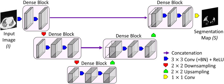
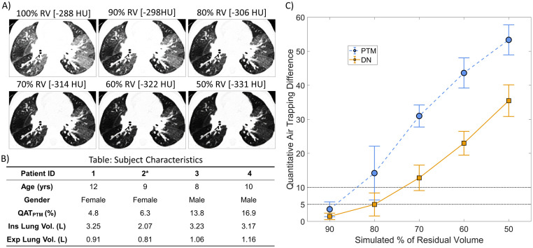
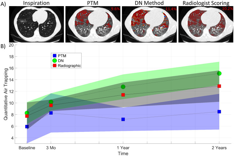
Paired volumetric whole lung inspiratory and expiratory CT scans were obtained at four time points (0, 3, 12 and 24 months) on 36 subjects with mild CF lung disease. A densely connected CNN (DN) was trained using AT segmentation maps generated from a personalized threshold-based method (PTM). Quantitative AT (QAT) values, presented as the relative volume of AT over the lungs, from the DN approach were compared to QAT values from the PTM method. Radiographic assessment, spirometric measures, and clinical scores were correlated to the DN QAT values using a linear mixed effects model.
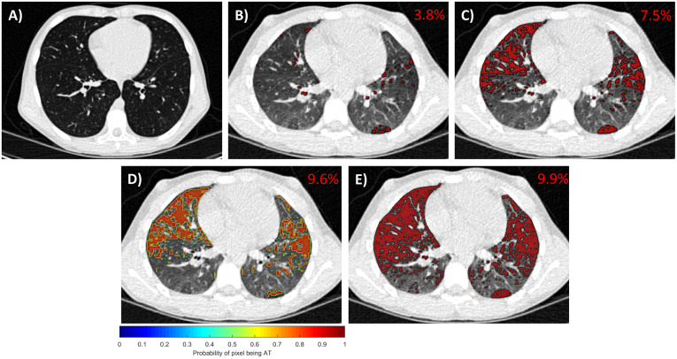
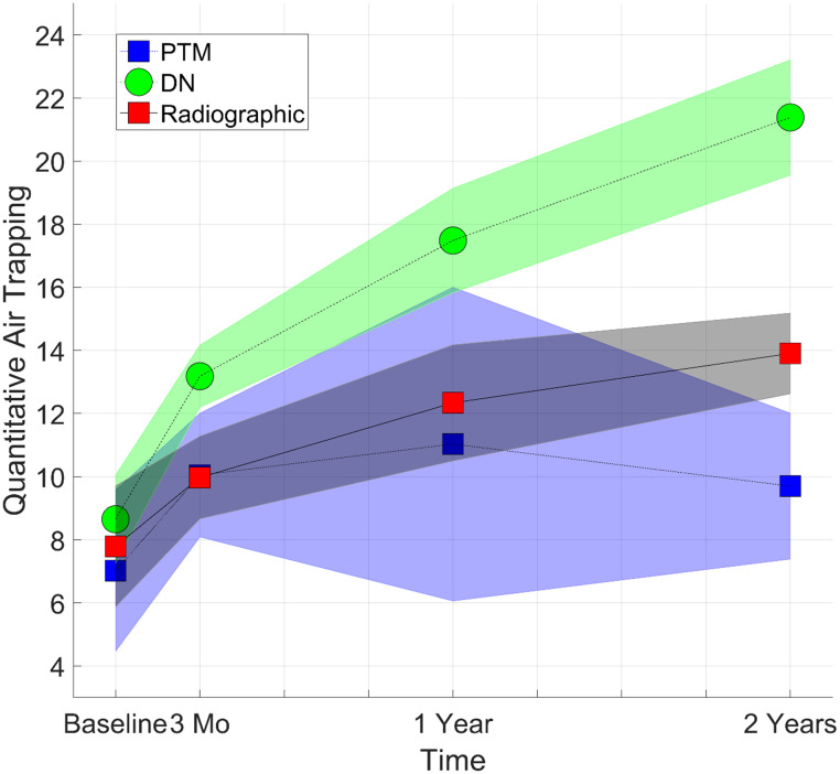
QAT values from the DN were found to increase from 8.65% ± 1.38% to 21.38% ± 1.82%, respectively, over a two-year period. Comparison of CNN model results to intensity-based measures demonstrated a systematic drop in the Dice coefficient over time (decreased from 0.86 ± 0.03 to 0.45 ± 0.04). The trends observed in DN QAT values were consistent with clinical scores for AT, bronchiectasis, and mucus plugging. In addition, the DN approach was found to be less susceptible to variations in expiratory deflation levels than the threshold-based approach.
The CNN model effectively delineated AT on expiratory CT scans, which provides an automated and objective approach for assessing and monitoring AT in CF patients.
呼气 CT 扫描中的空气滞留(AT)的放射学证据与囊性纤维化(CF)患者的早期肺功能障碍有关。然而,用于定量评估 AT 的标准技术具有高度可变性,导致监测疾病进展的效果有限。
研究卷积神经网络(CNN)模型量化和监测 AT 的有效性,并将其与基于阈值的技术获得的其他定量 AT 测量方法进行比较。
对 36 例轻度 CF 肺部疾病患者的 4 个时间点(0、3、12 和 24 个月)的容积全肺吸气和呼气 CT 扫描进行了配对。使用个性化基于阈值的方法(PTM)生成的 AT 分割图训练了一个密集连接的 CNN(DN)。从 DN 方法得到的定量 AT(QAT)值,以肺内 AT 的相对体积表示,与来自 PTM 方法的 QAT 值进行了比较。使用线性混合效应模型将影像学评估、肺量计测量和临床评分与 DN QAT 值相关联。
DN 的 QAT 值从 8.65%±1.38%增加到 21.38%±1.82%,在两年内增加。将 CNN 模型结果与基于强度的测量值进行比较,发现 Dice 系数随时间呈系统性下降(从 0.86±0.03 下降到 0.45±0.04)。DN QAT 值的趋势与 AT、支气管扩张和黏液栓的临床评分一致。此外,与基于阈值的方法相比,DN 方法对呼气排空水平的变化不太敏感。
CNN 模型有效地描绘了呼气 CT 扫描中的 AT,为评估和监测 CF 患者的 AT 提供了一种自动化和客观的方法。