• 文献检索
  • 文档翻译
  • 深度研究
  • 学术资讯
  • Suppr Zotero 插件Zotero 插件
  • 邀请有礼
  • 套餐&价格
  • 历史记录
应用&插件
Suppr Zotero 插件Zotero 插件浏览器插件Mac 客户端Windows 客户端微信小程序
定价
高级版会员购买积分包购买API积分包
服务
文献检索文档翻译深度研究API 文档MCP 服务
关于我们
关于 Suppr公司介绍联系我们用户协议隐私条款
关注我们

Suppr 超能文献

核心技术专利:CN118964589B侵权必究
粤ICP备2023148730 号-1Suppr @ 2026

文献检索

告别复杂PubMed语法,用中文像聊天一样搜索,搜遍4000万医学文献。AI智能推荐,让科研检索更轻松。

立即免费搜索

文件翻译

保留排版,准确专业,支持PDF/Word/PPT等文件格式,支持 12+语言互译。

免费翻译文档

深度研究

AI帮你快速写综述,25分钟生成高质量综述,智能提取关键信息,辅助科研写作。

立即免费体验

局限性硬皮病患者的临床和实验室特征及 UVA-1 光疗的反应:体内和体外皮肤模型。

Clinical and laboratory characterization of patients with localized scleroderma and response to UVA-1 phototherapy: In vivo and in vitro skin models.

机构信息

Dermatology Unit and Skin Bank Unit, Department of Clinical Surgical and Neuro-sciences, University of Siena, Siena, Italy.

Laboratory of Molecular Microbiology and Biotechnology, Department of Medical Biotechnologies, University of Siena, Siena, Italy.

出版信息

Photodermatol Photoimmunol Photomed. 2022 Nov;38(6):531-540. doi: 10.1111/phpp.12786. Epub 2022 Apr 3.

DOI:10.1111/phpp.12786
PMID:35324032
原文链接:https://pmc.ncbi.nlm.nih.gov/articles/PMC9790552/
Abstract

BACKGROUND/PURPOSE: Localized scleroderma (LS) is a rare disease leading to progressive hardening and induration of the skin and subcutaneous tissues. LS is responsive to UVA-1 phototherapy, though its exact mechanism of action dermal fibrosis is yet to be fully elucidated. We aimed to investigate the molecular changes induced by UVA-1 rays in human primary fibroblasts cultures.

METHODS

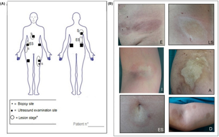
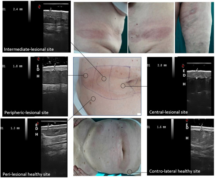
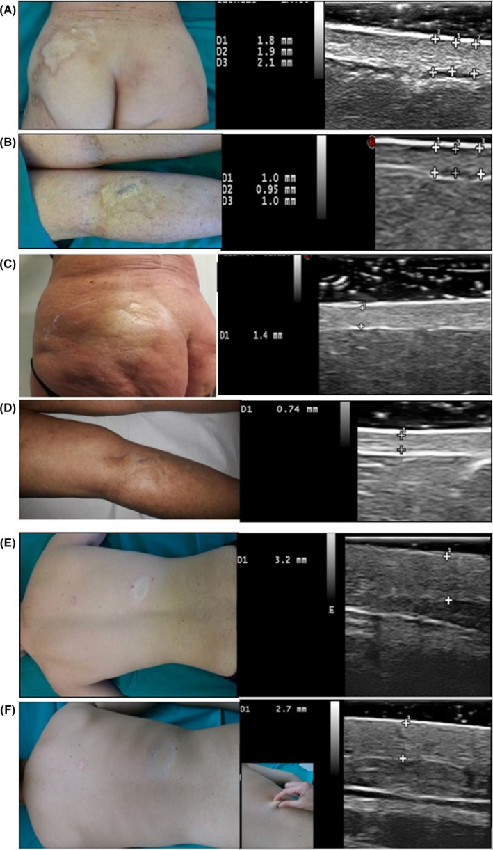
A total of 16 LS patients were treated with medium-dose UVA-1 phototherapy. At baseline, during and after therapy, Localized Scleroderma Assessment Tool, Dermatology Life Quality Index and lesions' staging and mapping were performed along with high-frequency ultrasound (HFUS) examination for dermal thickness assessment. Gene expression analysis for 23 mRNA transcripts, in vitro UVA-1 irradiation and viability tests were realized on lesional fibroblasts' primary cultures, before and 3 months after therapy.

RESULTS

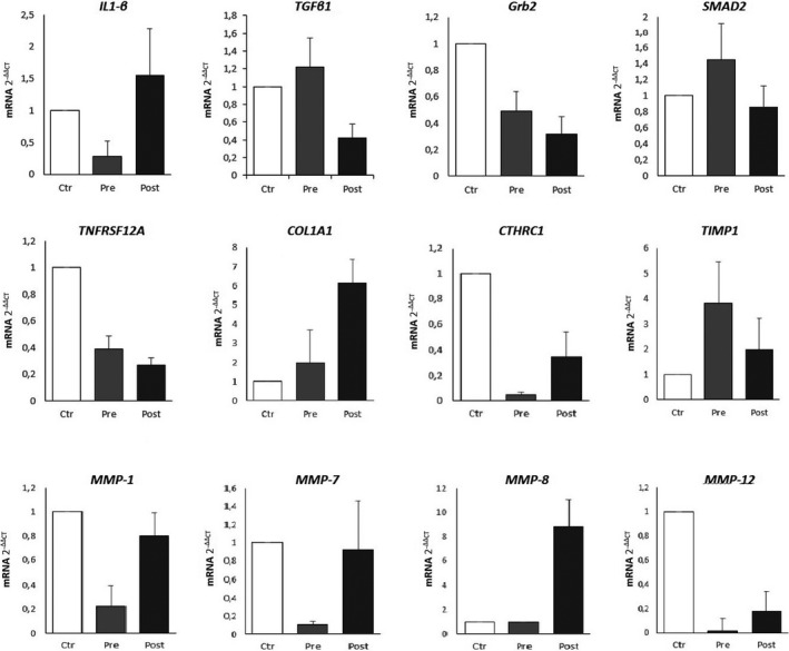
The dermal thickness, the LoSCAT and the DLQI progressively decreased starting from the last phototherapy session up to the 6 and 9 month follow-ups (-57% and -60%, respectively). Molecular gene analysis (rt-PCR) revealed that UVA-1 phototherapy exerts multiple effects: the activation of specific anti-fibrotic pathways (e.g., overexpression of CTHRC1 and metalloproteases 1, 2, 7, 8, 9, 12, suppression of TIMP-1), the downregulation of peculiar pro-fibrotic pathways (e.g., downregulation of TGF-ß, TGF-ßrII, Grb2, SMAD 2/3, TNRSF12A, CTGF) through a significant overexpression of IL-1ß; the stabilization of collagen synthesis acting on genes COL1A1, COL3A1, COL8A1, COL10A1, COL12A1.

CONCLUSION

UVA-1 phototherapy adds significant benefits in local tissue remodeling, rebalancing the alteration between pro-fibrotic and anti-fibrotic pathways; these changes can be well monitored by HFUS.

摘要

背景/目的:局限性硬皮病(LS)是一种罕见的疾病,会导致皮肤和皮下组织逐渐变硬和硬化。LS 对 UVA-1 光疗有反应,尽管其确切的作用机制——真皮纤维化尚未完全阐明。我们旨在研究 UVA-1 射线在人原代成纤维细胞培养物中诱导的分子变化。

方法

共有 16 名 LS 患者接受中剂量 UVA-1 光疗。在基线、治疗期间和治疗后,进行局部硬皮病评估工具、皮肤病生活质量指数和病变分期和绘图,以及高频超声(HFUS)检查评估真皮厚度。对病变成纤维细胞原代培养物进行 23 种 mRNA 转录物的基因表达分析,在治疗前和治疗后 3 个月进行体外 UVA-1 照射和活力测试。

结果

从最后一次光疗开始,直到 6 个月和 9 个月的随访,真皮厚度、LoSCAT 和 DLQI 逐渐下降(分别为-57%和-60%)。分子基因分析(rt-PCR)显示,UVA-1 光疗具有多种作用:激活特定的抗纤维化途径(例如,CTHRC1 和金属蛋白酶 1、2、7、8、9、12 的过度表达,TIMP-1 的抑制),下调特定的促纤维化途径(例如,TGF-ß、TGF-ßrII、Grb2、SMAD 2/3、TNRSF12A、CTGF 的下调),通过 IL-1ß 的显著过度表达;通过对 COL1A1、COL3A1、COL8A1、COL10A1、COL12A1 基因的作用稳定胶原合成。

结论

UVA-1 光疗在局部组织重塑方面有显著的益处,重新平衡了促纤维化和抗纤维化途径之间的变化;这些变化可以通过 HFUS 很好地监测。

https://cdn.ncbi.nlm.nih.gov/pmc/blobs/5741/9790552/8131e87e2b6a/PHPP-38-531-g001.jpg
https://cdn.ncbi.nlm.nih.gov/pmc/blobs/5741/9790552/633eb73c4e4b/PHPP-38-531-g003.jpg
https://cdn.ncbi.nlm.nih.gov/pmc/blobs/5741/9790552/b9c0c2dc850e/PHPP-38-531-g002.jpg
https://cdn.ncbi.nlm.nih.gov/pmc/blobs/5741/9790552/5695b58aa160/PHPP-38-531-g004.jpg
https://cdn.ncbi.nlm.nih.gov/pmc/blobs/5741/9790552/8131e87e2b6a/PHPP-38-531-g001.jpg

相似文献

1
Clinical and laboratory characterization of patients with localized scleroderma and response to UVA-1 phototherapy: In vivo and in vitro skin models.局限性硬皮病患者的临床和实验室特征及 UVA-1 光疗的反应:体内和体外皮肤模型。
Photodermatol Photoimmunol Photomed. 2022 Nov;38(6):531-540. doi: 10.1111/phpp.12786. Epub 2022 Apr 3.
2
UVA-1 phototherapy as adjuvant treatment for eosinophilic fasciitis: in vitro and in vivo functional characterization.UVA-1 光疗作为嗜酸性筋膜炎的辅助治疗:体外和体内功能特征。
Int J Dermatol. 2022 Jun;61(6):718-726. doi: 10.1111/ijd.16003. Epub 2021 Dec 8.
3
Ultraviolet A1 phototherapy decreases inhibitory SMAD7 gene expression in localized scleroderma.紫外线A1光疗可降低局限性硬皮病中抑制性SMAD7基因的表达。
Arch Dermatol Res. 2006 Nov;298(6):265-72. doi: 10.1007/s00403-006-0695-8. Epub 2006 Sep 19.
4
Successful ultraviolet A1 phototherapy in the treatment of localized scleroderma: a retrospective and prospective study.成功应用长波紫外线 A1 光疗治疗局限性硬皮病:一项回顾性和前瞻性研究。
Br J Dermatol. 2010 Feb 1;162(2):445-7. doi: 10.1111/j.1365-2133.2009.09438.x. Epub 2009 Aug 8.
5
Ultraviolet A1-induced downregulation of human beta-defensins and interleukin-6 and interleukin-8 correlates with clinical improvement in localized scleroderma.紫外线A1诱导的人β-防御素、白细胞介素-6和白细胞介素-8下调与局限性硬皮病的临床改善相关。
Br J Dermatol. 2006 Sep;155(3):600-7. doi: 10.1111/j.1365-2133.2006.07391.x.
6
Biological effects of a new ultraviolet A prototype based on light-emitting diodes on the treatment of localized scleroderma.基于发光二极管的新型紫外线 A 原型在局部硬皮病治疗中的生物学效应。
Exp Dermatol. 2020 Dec;29(12):1199-1208. doi: 10.1111/exd.14135. Epub 2020 Jul 20.
7
Suggested mechanisms of action of UVA phototherapy in morphea: a molecular study.UVA光疗治疗硬斑病的作用机制探讨:一项分子研究
Photodermatol Photoimmunol Photomed. 2004 Apr;20(2):93-100. doi: 10.1111/j.1600-0781.2004.00080.x.
8
Ultraviolet A phototherapy for sclerotic skin diseases: a systematic review.紫外线A光疗治疗硬化性皮肤病:一项系统评价。
J Am Acad Dermatol. 2008 Dec;59(6):1017-30. doi: 10.1016/j.jaad.2008.07.042. Epub 2008 Oct 2.
9
UVA irradiation induced heme oxygenase-1: a novel phototherapy for morphea.紫外线A照射诱导血红素加氧酶-1:一种治疗硬斑病的新型光疗法。
Photochem Photobiol. 2015 Jan-Feb;91(1):210-20. doi: 10.1111/php.12342. Epub 2014 Nov 8.
10
UVA1 irradiation attenuates collagen production via Ficz/AhR/MAPK signaling activation in scleroderma.UVA1 照射通过 Ficz/AhR/MAPK 信号通路激活减轻硬皮病中的胶原产生。
Int Immunopharmacol. 2023 Mar;116:109764. doi: 10.1016/j.intimp.2023.109764. Epub 2023 Jan 25.

引用本文的文献

1
Efficacy and Satisfaction of Low Doses UVA1 Phototherapy: A Spanish Experience from a Single Centre.低剂量UVA1光疗的疗效与满意度:来自单一中心的西班牙经验
Life (Basel). 2023 Feb 28;13(3):669. doi: 10.3390/life13030669.

本文引用的文献

1
UVA-1 phototherapy as adjuvant treatment for eosinophilic fasciitis: in vitro and in vivo functional characterization.UVA-1 光疗作为嗜酸性筋膜炎的辅助治疗:体外和体内功能特征。
Int J Dermatol. 2022 Jun;61(6):718-726. doi: 10.1111/ijd.16003. Epub 2021 Dec 8.
2
Associations between Disease Activity/Severity and Damage and Health-Related Quality of Life in Adult Patients with Localized Scleroderma-A Comparison of LoSCAT and Visual Analogue Scales.局限性硬皮病成年患者疾病活动/严重程度与损伤及健康相关生活质量的关联——LoSCAT与视觉模拟量表的比较
J Clin Med. 2020 Mar 11;9(3):756. doi: 10.3390/jcm9030756.
3
MicroRNA in localized scleroderma: a review of literature.
局限性硬皮病中的 microRNA:文献综述。
Arch Dermatol Res. 2020 Jul;312(5):317-324. doi: 10.1007/s00403-019-01991-0. Epub 2019 Oct 21.
4
Interventions for morphea.硬斑病的干预措施。
Cochrane Database Syst Rev. 2019 Jul 16;7(7):CD005027. doi: 10.1002/14651858.CD005027.pub5.
5
Using the Localized Scleroderma Cutaneous Assessment Tool (LoSCAT) to classify morphoea by severity and identify clinically significant change.使用局部硬皮病皮肤评估工具(LoSCAT)对硬斑病进行严重程度分类,并识别具有临床意义的变化。
Br J Dermatol. 2020 Feb;182(2):398-404. doi: 10.1111/bjd.18097. Epub 2019 Aug 14.
6
Morphea: Current concepts.硬斑病:当前概念
Clin Dermatol. 2018 Jul-Aug;36(4):475-486. doi: 10.1016/j.clindermatol.2018.04.005. Epub 2018 Apr 10.
7
Diagnostic criteria, severity classification and guidelines of localized scleroderma.局限性硬皮病的诊断标准、严重程度分类及治疗指南。
J Dermatol. 2018 Jul;45(7):755-780. doi: 10.1111/1346-8138.14161. Epub 2018 Apr 23.
8
Regarding "Transcriptional and Cytokine Profiles Identify CXCL9 as a Biomarker of Disease Activity in Morphea".关于“转录组和细胞因子谱鉴定CXCL9为硬皮病疾病活动的生物标志物”。
J Invest Dermatol. 2018 May;138(5):1212-1215. doi: 10.1016/j.jid.2017.11.032. Epub 2017 Dec 12.
9
European Dermatology Forum S1-guideline on the diagnosis and treatment of sclerosing diseases of the skin, Part 1: localized scleroderma, systemic sclerosis and overlap syndromes.欧洲皮肤病论坛 S1 指南:皮肤硬化病的诊断和治疗,第 1 部分:局限性硬皮病、系统性硬皮病和重叠综合征。
J Eur Acad Dermatol Venereol. 2017 Sep;31(9):1401-1424. doi: 10.1111/jdv.14458. Epub 2017 Aug 9.
10
Morphea and Eosinophilic Fasciitis: An Update.硬斑病和嗜酸性筋膜炎:最新进展
Am J Clin Dermatol. 2017 Aug;18(4):491-512. doi: 10.1007/s40257-017-0269-x.