van Vliet-Pérez Sharline M, van de Berg Nick J, Manni Francesca, Lai Marco, Rijstenberg Lucia, Hendriks Benno H W, Dankelman Jenny, Ewing-Graham Patricia C, Nieuwenhuyzen-de Boer Gatske M, van Beekhuizen Heleen J
Department of Biomechanical Engineering, Delft University of Technology, 2628 CD Delft, The Netherlands.
Department of Radiotherapy, Erasmus MC Cancer Institute, University Medical Center Rotterdam, 3015 GD Rotterdam, The Netherlands.
Cancers (Basel). 2022 Mar 10;14(6):1422. doi: 10.3390/cancers14061422.
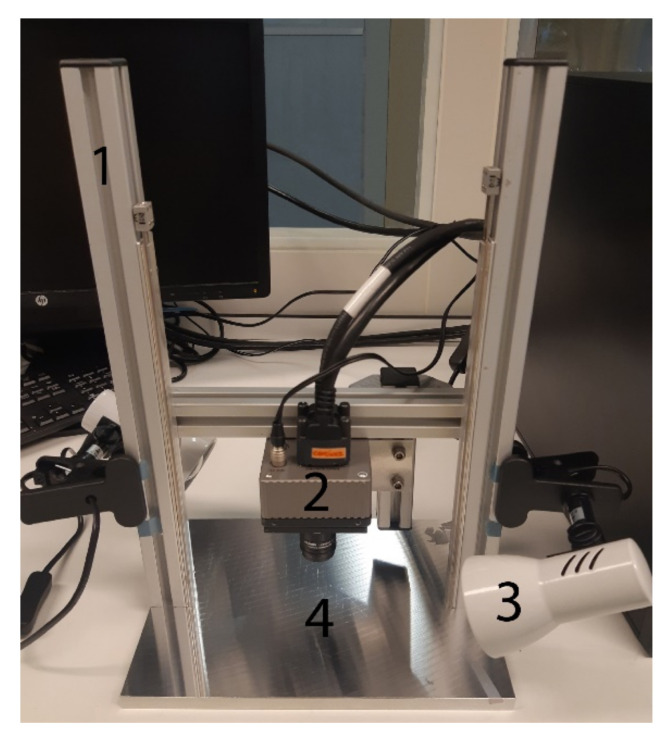
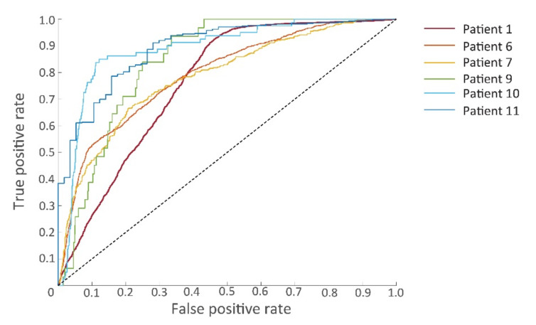
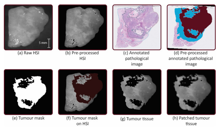
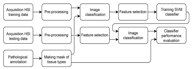
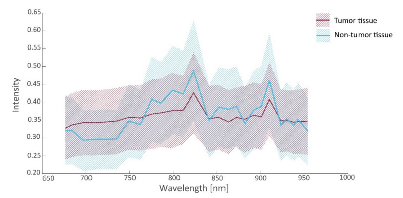
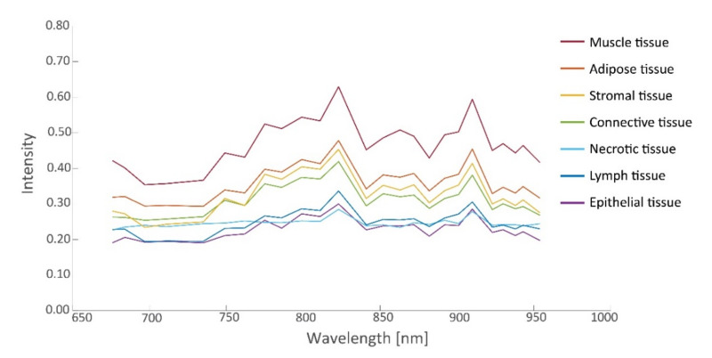
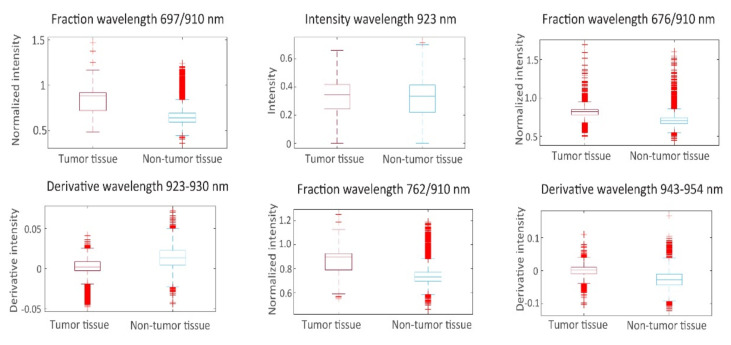
The most important prognostic factor for the survival of advanced-stage epithelial ovarian cancer (EOC) is the completeness of cytoreductive surgery (CRS). Therefore, an intraoperative technique to detect microscopic tumors would be of great value. The aim of this pilot study is to assess the feasibility of near-infrared hyperspectral imaging (HSI) for EOC detection in ex vivo tissue samples. Images were collected during CRS in 11 patients in the wavelength range of 665−975 nm, and processed by calibration, normalization, and noise filtering. A linear support vector machine (SVM) was employed to classify healthy and tumorous tissue (defined as >50% tumor cells). Classifier performance was evaluated using leave-one-out cross-validation. Images of 26 tissue samples from 10 patients were included, containing 26,446 data points that were matched to histopathology. Tumorous tissue could be classified with an area under the curve of 0.83, a sensitivity of 0.81, a specificity of 0.70, and Matthew’s correlation coefficient of 0.41. This study paves the way to in vivo and intraoperative use of HSI during CRS. Hyperspectral imaging can scan a whole tissue surface in a fast and non-contact way. Our pilot study demonstrates that HSI and SVM learning can be used to discriminate EOC from surrounding tissue.
晚期上皮性卵巢癌(EOC)生存的最重要预后因素是肿瘤细胞减灭术(CRS)的彻底性。因此,一种检测微小肿瘤的术中技术将具有重要价值。这项初步研究的目的是评估近红外高光谱成像(HSI)在体外组织样本中检测EOC的可行性。在11例患者的CRS过程中,于665−975 nm波长范围内采集图像,并通过校准、归一化和噪声滤波进行处理。采用线性支持向量机(SVM)对健康组织和肿瘤组织(定义为肿瘤细胞>50%)进行分类。使用留一法交叉验证评估分类器性能。纳入了来自10例患者的26个组织样本的图像,包含与组织病理学匹配的26446个数据点。肿瘤组织分类的曲线下面积为0.83,灵敏度为0.81,特异性为0.70,马修斯相关系数为0.41。本研究为CRS期间HSI的体内和术中应用铺平了道路。高光谱成像可以快速、非接触地扫描整个组织表面。我们的初步研究表明,HSI和SVM学习可用于区分EOC与周围组织。