Bychkov Maxim L, Kirichenko Artem V, Mikhaylova Irina N, Paramonov Alexander S, Yastremsky Evgeny V, Kirpichnikov Mikhail P, Shulepko Mikhail A, Lyukmanova Ekaterina N
Shemyakin-Ovchinnikov Institute of Bioorganic Chemistry, Russian Academy of Sciences, 119997 Moscow, Russia.
Moscow Institute of Physics and Technology, State University, 141701 Dolgoprudny, Russia.
Biomedicines. 2022 Mar 12;10(3):660. doi: 10.3390/biomedicines10030660.
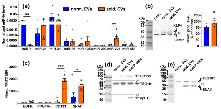
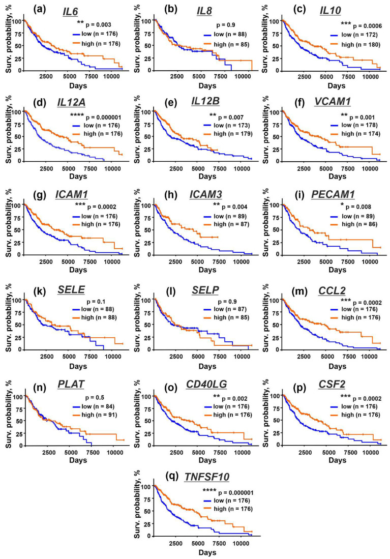
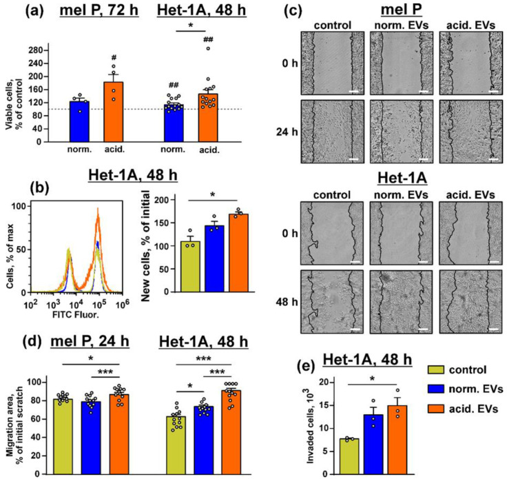
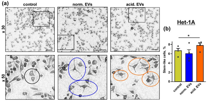
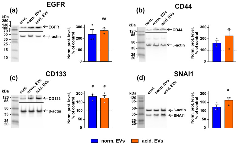
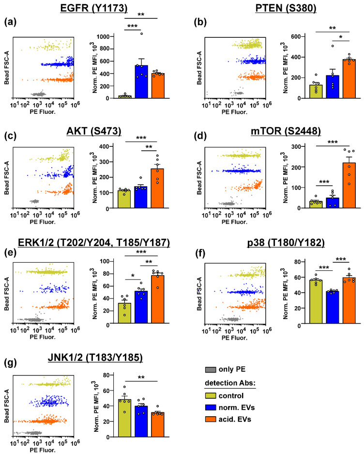
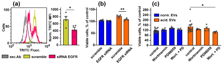
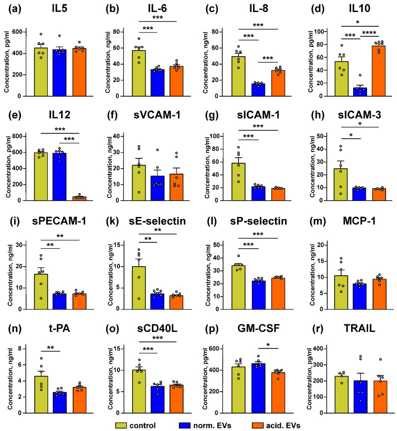
Metastatic melanoma is a highly malignant tumor. Melanoma cells release extracellular vesicles (EVs), which contribute to the growth, metastasis, and malignancy of neighboring cells by transfer of tumor-promoting miRNAs, mRNA, and proteins. Melanoma microenvironment acidification promotes tumor progression and determines EVs' properties. We studied the influence of EVs derived from metastatic melanoma cells cultivated at acidic (6.5) and normal (7.4) pH on the morphology and homeostasis of normal keratinocytes. Acidification of metastatic melanoma environment made EVs more prooncogenic with increased expression of prooncogenic mi221 RNA, stemless factor CD133, and pro-migration factor SNAI1, as well as with downregulated antitumor mir7 RNA. Incubation with EVs stimulated growth and migration both of metastatic melanoma cells and keratinocytes and changed the morphology of keratinocytes to stem-like phenotype, which was confirmed by increased expression of the stemness factors KLF and CD133. Activation of the AKT/mTOR and ERK signaling pathways and increased expression of epidermal growth factor receptor EGFR and SNAI1 were detected in keratinocytes upon incubation with EVs. Moreover, EVs reduced the production of different cytokines (IL6, IL10, and IL12) and adhesion factors (sICAM-1, sICAM-3, sPecam-1, and sCD40L) usually secreted by keratinocytes to control melanoma progression. Bioinformatic analysis revealed the correlation between decreased expression of these secreted factors and worse survival prognosis for patients with metastatic melanoma. Altogether, our data mean that metastatic melanoma EVs are important players in the transformation of normal keratinocytes.
转移性黑色素瘤是一种高度恶性的肿瘤。黑色素瘤细胞释放细胞外囊泡(EVs),这些囊泡通过转移促肿瘤的微小RNA(miRNAs)、信使核糖核酸(mRNA)和蛋白质,促进邻近细胞的生长、转移和恶性转化。黑色素瘤微环境酸化促进肿瘤进展并决定细胞外囊泡的特性。我们研究了在酸性(6.5)和正常(7.4)pH条件下培养的转移性黑色素瘤细胞来源的细胞外囊泡对正常角质形成细胞形态和稳态的影响。转移性黑色素瘤环境的酸化使细胞外囊泡更具促肿瘤性,促肿瘤mi221 RNA、无柄因子CD133和促迁移因子SNAI1的表达增加,同时抗肿瘤mir7 RNA表达下调。与细胞外囊泡共孵育刺激了转移性黑色素瘤细胞和角质形成细胞的生长和迁移,并将角质形成细胞的形态改变为干细胞样表型,这通过干细胞因子KLF和CD133表达增加得到证实。在与细胞外囊泡共孵育的角质形成细胞中检测到AKT/mTOR和ERK信号通路的激活以及表皮生长因子受体EGFR和SNAI1表达增加。此外,细胞外囊泡减少了角质形成细胞通常分泌的用于控制黑色素瘤进展的不同细胞因子(IL6、IL10和IL12)和黏附因子(可溶性细胞间黏附分子-1[sICAM-1]、可溶性细胞间黏附分子-3[sICAM-3]、可溶性血小板内皮细胞黏附分子-1[sPecam-1]和可溶性CD40配体[sCD40L])的产生。生物信息学分析揭示了这些分泌因子表达降低与转移性黑色素瘤患者较差的生存预后之间的相关性。总之,我们的数据表明转移性黑色素瘤细胞外囊泡是正常角质形成细胞转化的重要参与者。