Zhu Yihua, Fried Daniel
Department of Preventive and Restorative Dental Sciences, University of California, San Francisco, 707 Parnassus Ave., San Francisco, CA 94143, USA.
Diagnostics (Basel). 2022 Feb 26;12(3):597. doi: 10.3390/diagnostics12030597.
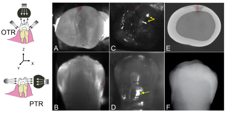
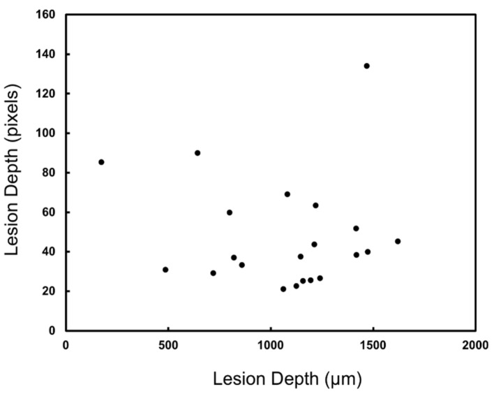
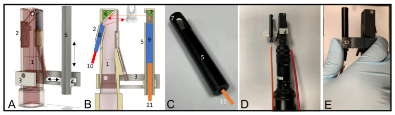
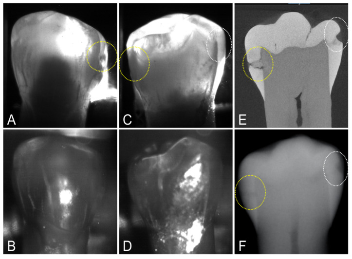
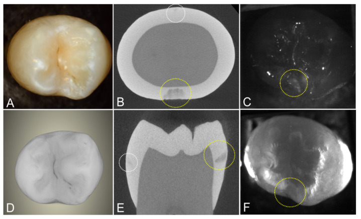
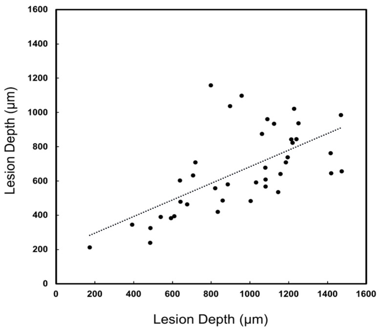
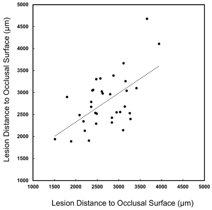
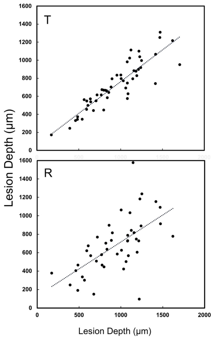
The aim of this study was to compare the diagnostic performance of dual short-wavelength infrared (SWIR) transillumination and reflectance multispectral imaging devices for imaging interproximal lesions with radiography using extracted teeth that had been imaged with micro-computed tomography (microCT). Thirty-six extracted teeth with 67 lesions on the proximal surfaces were imaged using a newly fabricated SWIR multispectral proximal transillumination and reflectance imaging device along with an existing SWIR multispectral occlusal transillumination and reflectance device. The ability of SWIR imaging and radiography to detect lesions and accurately assess lesion dimensions were compared using microCT as a standard. Occlusal and proximal transillumination and occlusal reflectance performed best for imaging interproximal lesions while proximal reflectance was not useful for imaging interproximal lesions from tooth buccal and lingual surfaces. There was high correlation of the lesion dimensions measured in occlusal and proximal transillumination images compared to microCT and moderate correlation in occlusal reflectance images. The correlation between the lesion depth measured in radiographs and the lesion depth measured with microCT was not significant. This study demonstrates that SWIR occlusal and proximal transillumination and SWIR occlusal reflectance images are useful for imaging interproximal lesions and they provide more accurate measurements of lesion severity.
本研究的目的是使用已通过微型计算机断层扫描(microCT)成像的离体牙,比较双短波长红外(SWIR)透照和反射多光谱成像设备与X线摄影对邻面病变成像的诊断性能。使用新制作的SWIR多光谱邻面透照和反射成像设备以及现有的SWIR多光谱咬合面透照和反射设备,对36颗在近中面有67处病变的离体牙进行成像。以microCT作为标准,比较SWIR成像和X线摄影检测病变以及准确评估病变大小的能力。咬合面和邻面透照以及咬合面反射在邻面病变成像方面表现最佳,而邻面反射对于从牙齿颊面和舌面成像邻面病变并无用处。与microCT相比,咬合面和邻面透照图像中测量的病变大小具有高度相关性,咬合面反射图像中具有中度相关性。X线片中测量的病变深度与microCT测量的病变深度之间的相关性不显著。本研究表明,SWIR咬合面和邻面透照以及SWIR咬合面反射图像对于邻面病变成像很有用,并且它们能更准确地测量病变严重程度。