Farahat Ibrahim Shawky, Sharafeldeen Ahmed, Elsharkawy Mohamed, Soliman Ahmed, Mahmoud Ali, Ghazal Mohammed, Taher Fatma, Bilal Maha, Abdel Razek Ahmed Abdel Khalek, Aladrousy Waleed, Elmougy Samir, Tolba Ahmed Elsaid, El-Melegy Moumen, El-Baz Ayman
BioImaging Laboratory, Department of Bioengineering, University of Louisville, Louisville, KY 40292, USA.
Electrical, Computer, and Biomedical Engineering Department, College of Engineering, Abu Dhabi University, Abu Dhabi 59911, United Arab Emirates.
Diagnostics (Basel). 2022 Mar 12;12(3):696. doi: 10.3390/diagnostics12030696.
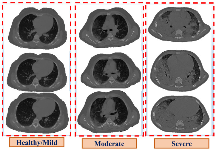
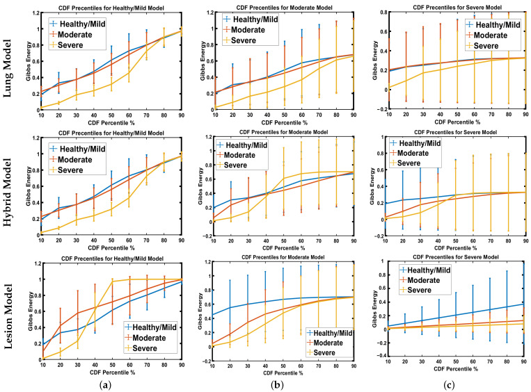
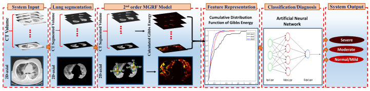
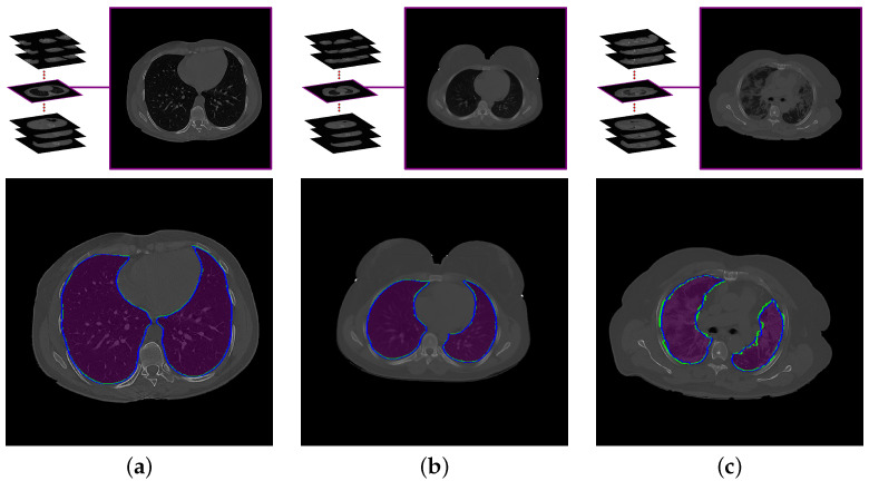
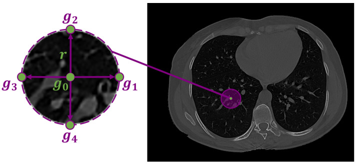
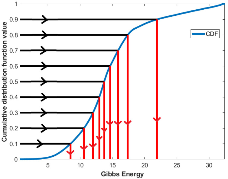
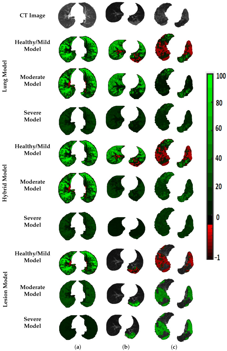
Early grading of coronavirus disease 2019 (COVID-19), as well as ventilator support machines, are prime ways to help the world fight this virus and reduce the mortality rate. To reduce the burden on physicians, we developed an automatic Computer-Aided Diagnostic (CAD) system to grade COVID-19 from Computed Tomography (CT) images. This system segments the lung region from chest CT scans using an unsupervised approach based on an appearance model, followed by 3D rotation invariant Markov-Gibbs Random Field (MGRF)-based morphological constraints. This system analyzes the segmented lung and generates precise, analytical imaging markers by estimating the MGRF-based analytical potentials. Three Gibbs energy markers were extracted from each CT scan by tuning the MGRF parameters on each lesion separately. The latter were healthy/mild, moderate, and severe lesions. To represent these markers more reliably, a Cumulative Distribution Function (CDF) was generated, then statistical markers were extracted from it, namely, 10th through 90th CDF percentiles with 10% increments. Subsequently, the three extracted markers were combined together and fed into a backpropagation neural network to make the diagnosis. The developed system was assessed on 76 COVID-19-infected patients using two metrics, namely, accuracy and Kappa. In this paper, the proposed system was trained and tested by three approaches. In the first approach, the MGRF model was trained and tested on the lungs. This approach achieved 95.83% accuracy and 93.39% kappa. In the second approach, we trained the MGRF model on the lesions and tested it on the lungs. This approach achieved 91.67% accuracy and 86.67% kappa. Finally, we trained and tested the MGRF model on lesions. It achieved 100% accuracy and 100% kappa. The results reported in this paper show the ability of the developed system to accurately grade COVID-19 lesions compared to other machine learning classifiers, such as k-Nearest Neighbor (KNN), decision tree, naïve Bayes, and random forest.
2019冠状病毒病(COVID-19)的早期分级以及呼吸机支持设备,是帮助全球对抗这种病毒并降低死亡率的主要方式。为减轻医生的负担,我们开发了一种自动计算机辅助诊断(CAD)系统,用于根据计算机断层扫描(CT)图像对COVID-19进行分级。该系统使用基于外观模型的无监督方法从胸部CT扫描中分割出肺区域,随后基于三维旋转不变马尔可夫-吉布斯随机场(MGRF)进行形态学约束。该系统分析分割后的肺,并通过估计基于MGRF的分析势来生成精确的分析成像标记。通过分别调整每个病变的MGRF参数,从每次CT扫描中提取三个吉布斯能量标记。后者分别为健康/轻度、中度和重度病变。为更可靠地表示这些标记,生成了累积分布函数(CDF),然后从中提取统计标记,即第10至第90百分位的CDF,增量为10%。随后,将提取的三个标记组合在一起并输入到反向传播神经网络中进行诊断。使用准确性和卡帕系数这两个指标,在76名COVID-19感染患者身上对开发的系统进行了评估。在本文中,所提出的系统通过三种方法进行训练和测试。在第一种方法中,MGRF模型在肺部进行训练和测试。这种方法的准确率达到95.83%,卡帕系数达到93.39%。在第二种方法中,我们在病变上训练MGRF模型,并在肺部进行测试。这种方法的准确率达到91.67%,卡帕系数达到86.67%。最后,我们在病变上训练和测试MGRF模型。其准确率达到100%,卡帕系数达到100%。本文报告的结果表明,与其他机器学习分类器(如k近邻(KNN)、决策树、朴素贝叶斯和随机森林)相比,开发的系统能够准确地对COVID-19病变进行分级。