• 文献检索
  • 文档翻译
  • 深度研究
  • 学术资讯
  • Suppr Zotero 插件Zotero 插件
  • 邀请有礼
  • 套餐&价格
  • 历史记录
应用&插件
Suppr Zotero 插件Zotero 插件浏览器插件Mac 客户端Windows 客户端微信小程序
定价
高级版会员购买积分包购买API积分包
服务
文献检索文档翻译深度研究API 文档MCP 服务
关于我们
关于 Suppr公司介绍联系我们用户协议隐私条款
关注我们

Suppr 超能文献

核心技术专利:CN118964589B侵权必究
粤ICP备2023148730 号-1Suppr @ 2026

文献检索

告别复杂PubMed语法,用中文像聊天一样搜索,搜遍4000万医学文献。AI智能推荐,让科研检索更轻松。

立即免费搜索

文件翻译

保留排版,准确专业,支持PDF/Word/PPT等文件格式,支持 12+语言互译。

免费翻译文档

深度研究

AI帮你快速写综述,25分钟生成高质量综述,智能提取关键信息,辅助科研写作。

立即免费体验

使用皮肤镜和高频超声检查评估基底细胞癌

Assessment of Basal Cell Carcinoma Using Dermoscopy and High Frequency Ultrasound Examination.

作者信息

Halip Ioana-Alina, Vâţă Dan, Statescu Laura, Salahoru Paul, Patraşcu Adriana Ionela, Temelie Olinici Doinita, Tarcau Bogdan, Popescu Ioana-Adriana, Mocanu Madalina, Constantin Anne-Marie, Crisan Maria, Brihan Ilarie, Nicolescu Alin Codrut, Gheuca-Solovastru Laura

机构信息

Department of Dermatology, "Grigore T. Popa" University of Medicine and Pharmacy, 700115 Iasi, Romania.

Dermatology Clinic, "St. Spiridon" County Emergency Clinical Hospital, 700115 Iasi, Romania.

出版信息

Diagnostics (Basel). 2022 Mar 18;12(3):735. doi: 10.3390/diagnostics12030735.

DOI:10.3390/diagnostics12030735
PMID:35328289
原文链接:https://pmc.ncbi.nlm.nih.gov/articles/PMC8947530/
Abstract

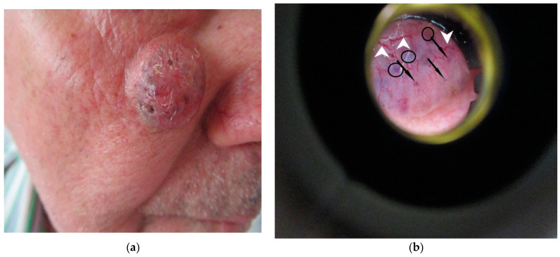
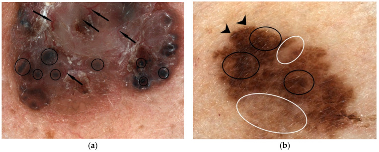
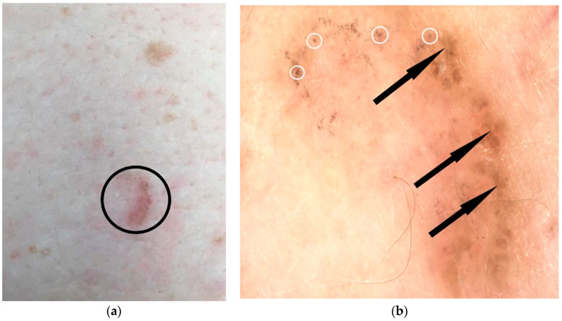
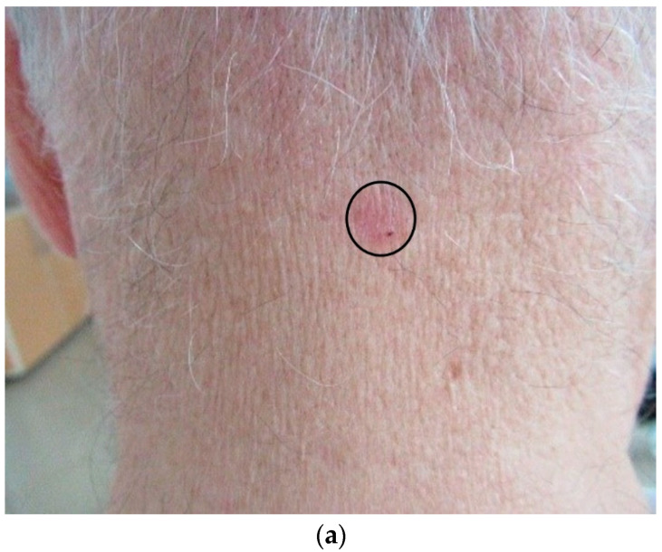
Basal cell carcinoma (BCC) is the most common form of cutaneous neoplasia in humans, and dermoscopy may provide valuable information for histopathological classification of BCC, which allows for the choice of non-invasive topical or surgical therapy. Similarly, dermoscopy may allow for the identification of incipient forms of BCC that cannot be detected in clinical examination. The importance of early diagnosis using the dermoscopy of superficial BCC forms is proven by the fact that despite their indolent clinical appearance, they can be included in high-risk BCC forms due to the rate of postoperative recurrence. Nodular pigmentary forms of BCCs present ovoid gray-blue nests or multiple gray-blue dots/globules associated with arborized vessels, sometimes undetectable on clinical examination. The management of BCC depends on this, as pigmentary forms have been shown to have a poor response to photodynamic therapy. High frequency ultrasound examination (HFUS) aids in the diagnosis of BCC with hypoechoic tumour masses, as well as in estimating tumour size (thickness and diameter), presurgical margin delineation, and surgical planning. The examination is also useful for determining the invasion of adjacent structures and for studying local recurrences. The use of dermoscopy in combination with HFUS allows for optimisation of the management of the oncological patient.

摘要

基底细胞癌(BCC)是人类皮肤肿瘤最常见的形式,皮肤镜检查可为BCC的组织病理学分类提供有价值的信息,这有助于选择非侵入性的局部或手术治疗方法。同样,皮肤镜检查可能有助于识别在临床检查中无法检测到的早期BCC形式。使用皮肤镜检查浅表BCC形式进行早期诊断的重要性已得到证实,尽管它们在临床上表现为惰性,但由于术后复发率,它们可被归类为高风险的BCC形式。结节性色素型BCC表现为椭圆形灰蓝色巢状结构或多个与树枝状血管相关的灰蓝色点/小球,有时在临床检查中无法检测到。BCC的治疗取决于此,因为色素型已被证明对光动力疗法反应不佳。高频超声检查(HFUS)有助于诊断具有低回声肿瘤块的BCC,以及估计肿瘤大小(厚度和直径)、术前边缘划定和手术规划。该检查对于确定相邻结构的侵犯情况和研究局部复发也很有用。皮肤镜检查与HFUS联合使用可优化肿瘤患者的管理。

https://cdn.ncbi.nlm.nih.gov/pmc/blobs/3a80/8947530/6c68a65922a0/diagnostics-12-00735-g004.jpg
https://cdn.ncbi.nlm.nih.gov/pmc/blobs/3a80/8947530/faf597317be7/diagnostics-12-00735-g001.jpg
https://cdn.ncbi.nlm.nih.gov/pmc/blobs/3a80/8947530/a838abc2bebb/diagnostics-12-00735-g002.jpg
https://cdn.ncbi.nlm.nih.gov/pmc/blobs/3a80/8947530/dca019292e53/diagnostics-12-00735-g003a.jpg
https://cdn.ncbi.nlm.nih.gov/pmc/blobs/3a80/8947530/6c68a65922a0/diagnostics-12-00735-g004.jpg

相似文献

1
Assessment of Basal Cell Carcinoma Using Dermoscopy and High Frequency Ultrasound Examination.使用皮肤镜和高频超声检查评估基底细胞癌
Diagnostics (Basel). 2022 Mar 18;12(3):735. doi: 10.3390/diagnostics12030735.
2
Dermoscopy uncovers clinically undetectable pigmentation in basal cell carcinoma.皮肤镜检查揭示了临床无法检测到的基底细胞癌中的色素沉着。
Br J Dermatol. 2014 Jan;170(1):192-5. doi: 10.1111/bjd.12634.
3
Dermoscopic features of basal cell carcinoma and its subtypes: A systematic review.基底细胞癌及其亚型的皮肤镜特征:系统综述。
J Am Acad Dermatol. 2021 Sep;85(3):653-664. doi: 10.1016/j.jaad.2019.11.008. Epub 2019 Nov 7.
4
Basal cell carcinoma thickness evaluated by high-frequency ultrasounds and correlation with dermoscopic features.高频超声评估基底细胞癌厚度与皮肤镜特征的相关性。
Ital J Dermatol Venerol. 2021 Oct;156(5):610-615. doi: 10.23736/S2784-8671.20.06576-1. Epub 2020 Sep 17.
5
Dermoscopic criteria and basal cell carcinoma.皮肤镜检查标准与基底细胞癌。
G Ital Dermatol Venereol. 2012 Apr;147(2):135-40.
6
High-frequency ultrasound: an essential non-invasive tool for the pre-therapeutic assessment of basal cell carcinoma.高频超声:基底细胞癌术前评估的一种不可或缺的无创工具。
Rom J Morphol Embryol. 2021 Apr-Jun;62(2):545-551. doi: 10.47162/RJME.62.2.21.
7
Dermatoscopy of basal cell carcinoma: morphologic variability of global and local features and accuracy of diagnosis.基底细胞癌的皮肤镜检查:整体和局部特征的形态变异性及诊断准确性。
J Am Acad Dermatol. 2010 Jan;62(1):67-75. doi: 10.1016/j.jaad.2009.05.035. Epub 2009 Oct 13.
8
Performance of Ultrasound for Identifying Morphological Characteristics and Thickness of Cutaneous Basal Cell Carcinoma: A Systematic Review.超声在识别皮肤基底细胞癌形态特征及厚度中的表现:系统评价。
Dermatology. 2022;238(4):692-710. doi: 10.1159/000520751. Epub 2022 Jan 13.
9
A Comparative Study of Pigmented and Non-pigmented Basal Cell Carcinoma in Reflectance Confocal Microscopy.反射共聚焦显微镜下色素性和非色素性基底细胞癌的对比研究。
In Vivo. 2021 Jan-Feb;35(1):423-427. doi: 10.21873/invivo.12274.
10
Dermoscopic features of small size pigmented basal cell carcinomas.小尺寸色素性基底细胞癌的皮肤镜特征
J Dermatol. 2016 May;43(5):543-6. doi: 10.1111/1346-8138.13173. Epub 2015 Oct 13.

引用本文的文献

1
Clinical Versus Dermoscopic Evaluation of Tumor Margins Prior to Surgical Excision-A Systematic Review.手术切除前肿瘤边缘的临床评估与皮肤镜评估——一项系统综述
J Clin Med. 2025 Aug 26;14(17):6014. doi: 10.3390/jcm14176014.
2
Advances in Skin Ultrasonography for Malignant and Benign Tumors of the Head and Neck: Current Insights and Future Directions.头颈部恶性和良性肿瘤的皮肤超声检查进展:当前见解与未来方向
J Clin Med. 2025 Mar 27;14(7):2298. doi: 10.3390/jcm14072298.
3
Advanced Doppler Ultrasound Insights: A Multicenter Prospective Study on Healthy Skin.

本文引用的文献

1
Unusual dermoscopic patterns of basal cell carcinoma mimicking melanoma.基底细胞癌酷似黑素瘤的不寻常皮肤镜表现。
Exp Dermatol. 2022 Jun;31(6):890-898. doi: 10.1111/exd.14533. Epub 2022 Feb 6.
2
Use of imaging techniques for melanocytic naevi and basal cell carcinoma in integrative analysis (Review).成像技术在综合分析中用于黑素细胞痣和基底细胞癌的应用(综述)
Exp Ther Med. 2020 Jul;20(1):78-86. doi: 10.3892/etm.2020.8620. Epub 2020 Mar 20.
3
Vascular patterns in basal cell carcinoma: Dermoscopic, confocal and histopathological perspectives.
先进的多普勒超声见解:一项关于健康皮肤的多中心前瞻性研究。
Diagnostics (Basel). 2025 Feb 26;15(5):569. doi: 10.3390/diagnostics15050569.
4
Basal Cell Carcinoma: Comprehensive Review with Emphasis on Scar Tissue Manifestation and Post-Vaccination Incidence.基底细胞癌:重点关注瘢痕组织表现及接种疫苗后发病率的综合综述
Biomedicines. 2024 Aug 6;12(8):1769. doi: 10.3390/biomedicines12081769.
5
Preliminary Experience in Ultra-High Frequency Ultrasound Assessment of Cutaneous Primary Lymphomas: An Innovative Classification.皮肤原发性淋巴瘤的超高频率超声评估初步经验:一种创新分类法
Cancers (Basel). 2024 Jul 4;16(13):2456. doi: 10.3390/cancers16132456.
6
[Diagnosis and treatment monitoring of basal cell carcinoma in one step: BCC one-stop shop : In vivo and ex vivo monitoring of basal cell carcinoma margins].[基底细胞癌的一步式诊断与治疗监测:基底细胞癌一站式服务:对基底细胞癌边缘进行体内和体外监测]
Dermatologie (Heidelb). 2024 Jul;75(7):587-591. doi: 10.1007/s00105-024-05378-9. Epub 2024 Jun 27.
7
Spatio-Temporal Pattern of Tuberculosis Distribution in Romania and Particulate Matter Pollution Associated With Risk of Infection.罗马尼亚结核病分布的时空模式以及与感染风险相关的颗粒物污染
Geohealth. 2024 Feb 6;8(2):e2023GH000972. doi: 10.1029/2023GH000972. eCollection 2024 Feb.
8
Multimodal Method for Differentiating Various Clinical Forms of Basal Cell Carcinoma and Benign Neoplasms In Vivo.用于在体内鉴别基底细胞癌和良性肿瘤的各种临床形式的多模态方法。
Diagnostics (Basel). 2024 Jan 17;14(2):202. doi: 10.3390/diagnostics14020202.
9
Optically Guided High-Frequency Ultrasound to Differentiate High-Risk Basal Cell Carcinoma Subtypes: A Single-Centre Prospective Study.光学引导高频超声鉴别高危基底细胞癌亚型:一项单中心前瞻性研究
J Clin Med. 2023 Nov 3;12(21):6910. doi: 10.3390/jcm12216910.
10
The Spatial-Temporal Dimension of Oncological Prevalence and Mortality in Romania.罗马尼亚肿瘤患病率和死亡率的时空维度
Geohealth. 2023 Oct 4;7(10):e2023GH000901. doi: 10.1029/2023GH000901. eCollection 2023 Oct.
基底细胞癌的血管形态:皮肤镜、共聚焦显微镜及组织病理学视角
Oncol Lett. 2019 May;17(5):4112-4125. doi: 10.3892/ol.2019.10070. Epub 2019 Feb 25.
4
Basosquamous carcinoma: Dermoscopic clues to diagnosis.基底鳞状细胞癌:诊断的皮肤镜线索
J Dermatol. 2017 Feb;44(2):127-134. doi: 10.1111/1346-8138.13563. Epub 2016 Aug 29.
5
Cancer statistics, 2016.癌症统计数据,2016 年。
CA Cancer J Clin. 2016 Jan-Feb;66(1):7-30. doi: 10.3322/caac.21332. Epub 2016 Jan 7.
6
Vascular structures in dermoscopy.皮肤镜检查中的血管结构
An Bras Dermatol. 2015 Jul-Aug;90(4):545-53. doi: 10.1590/abd1806-4841.20153452.
7
Trends in Basal Cell Carcinoma Incidence and Identification of High-Risk Subgroups, 1998-2012.1998-2012 年基底细胞癌发病率趋势及高危亚组鉴定。
JAMA Dermatol. 2015 Sep;151(9):976-81. doi: 10.1001/jamadermatol.2015.1188.
8
Incidence Estimate of Nonmelanoma Skin Cancer (Keratinocyte Carcinomas) in the U.S. Population, 2012.美国 2012 年人群中非黑色素瘤皮肤癌(角质形成细胞癌)的发病率估计。
JAMA Dermatol. 2015 Oct;151(10):1081-6. doi: 10.1001/jamadermatol.2015.1187.
9
Integrative analysis of cutaneous skin tumours using ultrasonogaphic criteria. Preliminary results.使用超声标准对皮肤肿瘤进行综合分析。初步结果。
Med Ultrason. 2014 Dec;16(4):285-90. doi: 10.11152/mu.201.3.2066.164.dcafb.
10
Ultrasound as predictor of histologic subtypes linked to recurrence in basal cell carcinoma of the skin.超声作为皮肤基底细胞癌组织学亚型与复发相关性的预测指标。
J Eur Acad Dermatol Venereol. 2015 Apr;29(4):702-7. doi: 10.1111/jdv.12660. Epub 2014 Sep 8.