• 文献检索
  • 文档翻译
  • 深度研究
  • 学术资讯
  • Suppr Zotero 插件Zotero 插件
  • 邀请有礼
  • 套餐&价格
  • 历史记录
应用&插件
Suppr Zotero 插件Zotero 插件浏览器插件Mac 客户端Windows 客户端微信小程序
定价
高级版会员购买积分包购买API积分包
服务
文献检索文档翻译深度研究API 文档MCP 服务
关于我们
关于 Suppr公司介绍联系我们用户协议隐私条款
关注我们

Suppr 超能文献

核心技术专利:CN118964589B侵权必究
粤ICP备2023148730 号-1Suppr @ 2026

文献检索

告别复杂PubMed语法,用中文像聊天一样搜索,搜遍4000万医学文献。AI智能推荐,让科研检索更轻松。

立即免费搜索

文件翻译

保留排版,准确专业,支持PDF/Word/PPT等文件格式,支持 12+语言互译。

免费翻译文档

深度研究

AI帮你快速写综述,25分钟生成高质量综述,智能提取关键信息,辅助科研写作。

立即免费体验

利用结节内和结节周围放射组学特征预测表现为磨玻璃密度结节的早期肺腺癌 Ki-67 标记指数水平。

Predicting Ki-67 labeling index level in early-stage lung adenocarcinomas manifesting as ground-glass opacity nodules using intra-nodular and peri-nodular radiomic features.

机构信息

Department of Respiratory Medicine, Chinese People's Liberation Army General Hospital, Beijing, China.

Department of Pulmonary and Critical Care Medicine, Zhongnan Hospital of Wuhan University, Wuhan, Hubei, China.

出版信息

Cancer Med. 2022 Nov;11(21):3982-3992. doi: 10.1002/cam4.4719. Epub 2022 Mar 24.

DOI:10.1002/cam4.4719
PMID:35332684
原文链接:https://pmc.ncbi.nlm.nih.gov/articles/PMC9636499/
Abstract

OBJECTIVES

To explore the diagnostic value of radiomics in differentiating between lung adenocarcinomas appearing as ground-glass opacity nodules (GGO) with high- and low Ki-67 expression levels.

MATERIALS AND METHODS

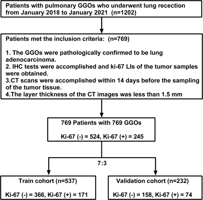
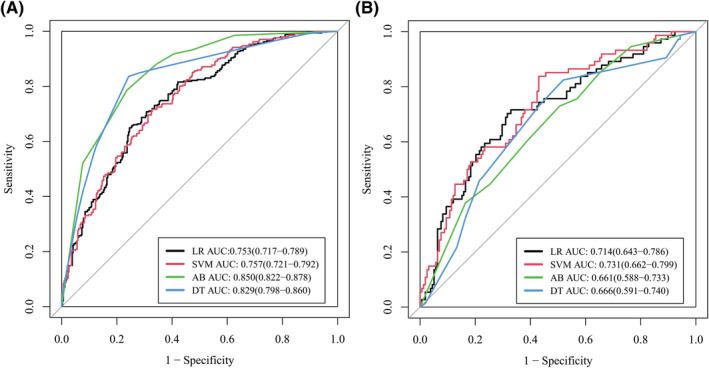
From January 2018 to January 2021, patients with pulmonary GGO who received lung resection were evaluated for potential enrollment. The included GGOs were then randomly divided into a training cohort and a validation cohort with a ratio of 7:3. Logistic regression (LR), decision tree (DT), support vector machines (SVM), and adaboost (AB) were applied for radiomic model construction. Area under the curve (AUC) of the receiver operating characteristic (ROC) curve was used to evaluate the diagnostic efficacy of the established models.

RESULTS

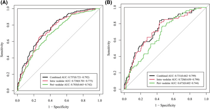
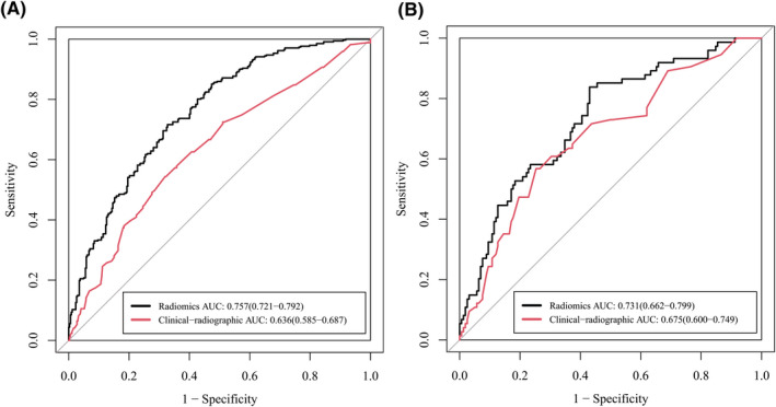
Seven hundred and sixty-nine patients with 769 GGOs were included in this study. Two hundred and forty-five GGOs were confirmed to be of high Ki-67 labeling index (LI). In the training cohort, gender, age, spiculation sign, pleural indentation sign, bubble sign, and maximum 2D diameter of the nodule were found to be significantly different between high- and low Ki-67 LI groups (p < 0.05), and spiculation sign and maximum 2D diameter of the nodule were further confirmed to be risk factors for Ki-67 LI. The radiomic model established using SVM exhibited an AUC of 0.731 in the validation cohort, which was higher than that of the clinical-radiographic model (AUC = 0.675). Moreover, radiomic model combining both intra- and peri-nodular features showed better diagnostic efficacy than using intra-nodular features alone (AUC = 0.731 and 0.720, respectively).

CONCLUSIONS

The established radiomic model exhibited good diagnostic efficacy in differentiating between lung adenocarcinoma GGOs with high and low Ki-67 LI, which was higher than the clinical-radiographic model. Peri-nodular radiomic features showed added benefits to the radiomic model. As a novel noninvasive method, radiomics have the potential to be applied in the preliminary classification of Ki-67 expression level in lung adenocarcinoma GGOs.

摘要

目的

探讨影像组学在区分 Ki-67 高表达和低表达的肺腺癌磨玻璃结节(GGO)中的诊断价值。

材料与方法

本研究纳入了 2018 年 1 月至 2021 年 1 月期间因肺 GGO 接受肺切除术的患者,并评估了其潜在的入组情况。将纳入的 GGO 随机分为训练队列和验证队列,比例为 7:3。应用逻辑回归(LR)、决策树(DT)、支持向量机(SVM)和自适应增强(AB)构建影像组学模型。采用受试者工作特征(ROC)曲线下面积(AUC)评估建立模型的诊断效能。

结果

本研究共纳入 769 例患者的 769 个 GGO。其中 245 个 GGO 的 Ki-67 标记指数(LI)较高。在训练队列中,高 Ki-67 LI 组和低 Ki-67 LI 组在性别、年龄、分叶征、胸膜凹陷征、泡征和结节最大二维直径方面存在显著差异(p<0.05),其中分叶征和结节最大二维直径被进一步确认为 Ki-67 LI 的危险因素。在验证队列中,SVM 构建的影像组学模型的 AUC 为 0.731,高于临床-影像学模型(AUC=0.675)。此外,同时结合结节内和结节周特征的影像组学模型比单独使用结节内特征的诊断效能更好(AUC 分别为 0.731 和 0.720)。

结论

本研究建立的影像组学模型在区分 Ki-67 高表达和低表达的肺腺癌 GGO 方面具有良好的诊断效能,高于临床-影像学模型。结节周影像组学特征为影像组学模型提供了附加价值。作为一种新的非侵入性方法,影像组学有望用于肺腺癌 GGO 中 Ki-67 表达水平的初步分类。

https://cdn.ncbi.nlm.nih.gov/pmc/blobs/ded4/9636499/6ff85ace5fa0/CAM4-11-3982-g001.jpg
https://cdn.ncbi.nlm.nih.gov/pmc/blobs/ded4/9636499/662c8c3c6967/CAM4-11-3982-g006.jpg
https://cdn.ncbi.nlm.nih.gov/pmc/blobs/ded4/9636499/7bb515557743/CAM4-11-3982-g004.jpg
https://cdn.ncbi.nlm.nih.gov/pmc/blobs/ded4/9636499/6c3d00adf139/CAM4-11-3982-g007.jpg
https://cdn.ncbi.nlm.nih.gov/pmc/blobs/ded4/9636499/999f37ba7adb/CAM4-11-3982-g003.jpg
https://cdn.ncbi.nlm.nih.gov/pmc/blobs/ded4/9636499/91b7d6e5e9fe/CAM4-11-3982-g002.jpg
https://cdn.ncbi.nlm.nih.gov/pmc/blobs/ded4/9636499/6ff85ace5fa0/CAM4-11-3982-g001.jpg

相似文献

1
Predicting Ki-67 labeling index level in early-stage lung adenocarcinomas manifesting as ground-glass opacity nodules using intra-nodular and peri-nodular radiomic features.利用结节内和结节周围放射组学特征预测表现为磨玻璃密度结节的早期肺腺癌 Ki-67 标记指数水平。
Cancer Med. 2022 Nov;11(21):3982-3992. doi: 10.1002/cam4.4719. Epub 2022 Mar 24.
2
A computerized tomography-based radiomic model for assessing the invasiveness of lung adenocarcinoma manifesting as ground-glass opacity nodules.基于计算机断层扫描的放射组学模型评估表现为磨玻璃密度结节的肺腺癌侵袭性。
Respir Res. 2022 Apr 16;23(1):96. doi: 10.1186/s12931-022-02016-7.
3
CT-Imaging Based Analysis of Invasive Lung Adenocarcinoma Presenting as Ground Glass Nodules Using Peri- and Intra-nodular Radiomic Features.基于CT成像,利用结节周围和结节内的影像组学特征对表现为磨玻璃结节的浸润性肺腺癌进行分析。
Front Oncol. 2020 May 27;10:838. doi: 10.3389/fonc.2020.00838. eCollection 2020.
4
Radiomics for lung adenocarcinoma manifesting as pure ground-glass nodules: invasive prediction.肺腺癌纯磨玻璃结节的影像组学分析:侵袭性预测。
Eur Radiol. 2020 Jul;30(7):3650-3659. doi: 10.1007/s00330-020-06776-y. Epub 2020 Mar 11.
5
A comparative study to evaluate CT-based semantic and radiomic features in preoperative diagnosis of invasive pulmonary adenocarcinomas manifesting as subsolid nodules.一项基于 CT 的语义和放射组学特征在术前诊断表现为亚实性结节的浸润性肺腺癌的对比研究。
Sci Rep. 2021 Jan 18;11(1):66. doi: 10.1038/s41598-020-79690-4.
6
Radiomics nomogram for preoperative differentiation of pulmonary mucinous adenocarcinoma from tuberculoma in solitary pulmonary solid nodules.基于影像组学的列线图模型术前鉴别肺内孤立实性结节中的黏液型肺腺癌与结核瘤。
BMC Cancer. 2023 Mar 21;23(1):261. doi: 10.1186/s12885-023-10734-4.
7
[Clinical value of a differentiation prediction model for invasive lung adenocarcinoma].[浸润性肺腺癌鉴别预测模型的临床价值]
Zhonghua Zhong Liu Za Zhi. 2022 Jul 23;44(7):767-775. doi: 10.3760/cma.j.cn112152-20200102-00002.
8
Predicting invasion in early-stage ground-glass opacity pulmonary adenocarcinoma: a radiomics-based machine learning approach.基于影像组学的机器学习方法预测早期磨玻璃密度肺腺癌的侵袭性。
BMC Med Imaging. 2024 Sep 13;24(1):240. doi: 10.1186/s12880-024-01421-2.
9
A comparative study for the evaluation of CT-based conventional, radiomic, combined conventional and radiomic, and delta-radiomic features, and the prediction of the invasiveness of lung adenocarcinoma manifesting as ground-glass nodules.基于 CT 的常规、放射组学、常规联合放射组学和 delta 放射组学特征评估的对比研究,以及预测表现为磨玻璃结节的肺腺癌侵袭性。
Clin Radiol. 2022 Oct;77(10):e741-e748. doi: 10.1016/j.crad.2022.06.004. Epub 2022 Jul 12.
10
Diagnosis of Invasive Lung Adenocarcinoma Based on Chest CT Radiomic Features of Part-Solid Pulmonary Nodules: A Multicenter Study.基于部分实性肺结节 CT 放射组学特征对浸润性肺腺癌的诊断:一项多中心研究。
Radiology. 2020 Nov;297(2):451-458. doi: 10.1148/radiol.2020192431. Epub 2020 Aug 25.

引用本文的文献

1
Prediction of Ki-67 Expression in HIV-Associated Lung Adenocarcinoma Patients Using Multiple Machine Learning Models Based on CT Imaging Radiomics.基于CT成像放射组学的多种机器学习模型预测HIV相关肺腺癌患者的Ki-67表达
Cancer Manag Res. 2025 Apr 25;17:881-892. doi: 10.2147/CMAR.S505390. eCollection 2025.
2
Peritumoral radiomics increases the efficiency of classification of pure ground-glass lung nodules: a multicenter study.瘤周放射组学提高纯磨玻璃肺结节分类效率:一项多中心研究。
J Cardiothorac Surg. 2024 Aug 30;19(1):505. doi: 10.1186/s13019-024-03008-y.
3
The value of intratumoral and peritumoral radiomics features in differentiating early-stage lung invasive adenocarcinoma (≤3 cm) subtypes.

本文引用的文献

1
Tumor cell proliferation (Ki-67) expression and its prognostic significance in histological subtypes of lung adenocarcinoma.肿瘤细胞增殖(Ki-67)在肺腺癌组织学亚型中的表达及其预后意义。
Lung Cancer. 2021 Apr;154:69-75. doi: 10.1016/j.lungcan.2021.02.009. Epub 2021 Feb 16.
2
A CT-based radiomics nomogram for prediction of lung adenocarcinomas and granulomatous lesions in patient with solitary sub-centimeter solid nodules.基于 CT 的放射组学列线图预测直径小于等于 1 厘米的单发实性肺结节中的肺腺癌和肉芽肿性病变。
Cancer Imaging. 2020 Jul 8;20(1):45. doi: 10.1186/s40644-020-00320-3.
3
Detailed identification of epidermal growth factor receptor mutations in lung adenocarcinoma: Combining radiomics with machine learning.
瘤内和瘤周影像组学特征在鉴别早期肺浸润性腺癌(≤3 cm)亚型中的价值
Transl Cancer Res. 2024 Jan 31;13(1):202-216. doi: 10.21037/tcr-23-1324. Epub 2024 Jan 25.
4
CT-based radiomics for predicting Ki-67 expression in lung cancer: a systematic review and meta-analysis.基于CT的影像组学预测肺癌中Ki-67表达:一项系统评价和Meta分析
Front Oncol. 2024 Feb 7;14:1329801. doi: 10.3389/fonc.2024.1329801. eCollection 2024.
5
CT radiomics model for predicting the Ki-67 proliferation index of pure-solid non-small cell lung cancer: a multicenter study.预测纯实性非小细胞肺癌Ki-67增殖指数的CT影像组学模型:一项多中心研究
Front Oncol. 2023 Aug 29;13:1175010. doi: 10.3389/fonc.2023.1175010. eCollection 2023.
6
Correlation between PD-L1 expression and radiomic features in early-stage lung adenocarcinomas manifesting as ground-glass nodules.表现为磨玻璃结节的早期肺腺癌中PD-L1表达与影像组学特征的相关性
Front Oncol. 2022 Sep 13;12:986579. doi: 10.3389/fonc.2022.986579. eCollection 2022.
7
Identification of pulmonary adenocarcinoma and benign lesions in isolated solid lung nodules based on a nomogram of intranodal and perinodal CT radiomic features.基于结节内及结节周围CT影像组学特征列线图鉴别孤立性实性肺结节中的肺腺癌与良性病变
Front Oncol. 2022 Sep 6;12:924055. doi: 10.3389/fonc.2022.924055. eCollection 2022.
肺腺癌中表皮生长因子受体突变的详细鉴定:将放射组学与机器学习相结合
Med Phys. 2020 Aug;47(8):3458-3466. doi: 10.1002/mp.14238. Epub 2020 Jun 3.
4
Outcome prediction in resectable lung adenocarcinoma patients: value of CT radiomics.可切除肺腺癌患者的预后预测:CT 放射组学的价值。
Eur Radiol. 2020 Sep;30(9):4952-4963. doi: 10.1007/s00330-020-06872-z. Epub 2020 Apr 30.
5
Clinical, Conventional CT and Radiomic Feature-Based Machine Learning Models for Predicting ALK Rearrangement Status in Lung Adenocarcinoma Patients.基于临床、传统CT和影像组学特征的机器学习模型预测肺腺癌患者ALK重排状态
Front Oncol. 2020 Mar 20;10:369. doi: 10.3389/fonc.2020.00369. eCollection 2020.
6
Identification of Non-Small Cell Lung Cancer Sensitive to Systemic Cancer Therapies Using Radiomics.基于放射组学识别对全身系统治疗敏感的非小细胞肺癌。
Clin Cancer Res. 2020 May 1;26(9):2151-2162. doi: 10.1158/1078-0432.CCR-19-2942. Epub 2020 Mar 20.
7
Differentiation of non-small cell lung cancer and histoplasmosis pulmonary nodules: insights from radiomics model performance compared with clinician observers.非小细胞肺癌与肺组织胞浆菌病肺结节的鉴别:与临床医生观察者相比,放射组学模型性能的见解
Transl Lung Cancer Res. 2019 Dec;8(6):979-988. doi: 10.21037/tlcr.2019.12.19.
8
Preoperative CT-based radiomics combined with intraoperative frozen section is predictive of invasive adenocarcinoma in pulmonary nodules: a multicenter study.术前 CT 影像组学结合术中冰冻切片对肺结节浸润性腺癌的预测:一项多中心研究。
Eur Radiol. 2020 May;30(5):2680-2691. doi: 10.1007/s00330-019-06597-8. Epub 2020 Jan 31.
9
Marginal radiomics features as imaging biomarkers for pathological invasion in lung adenocarcinoma.边缘放射组学特征作为肺腺癌病理浸润的影像学生物标志物。
Eur Radiol. 2020 May;30(5):2984-2994. doi: 10.1007/s00330-019-06581-2. Epub 2020 Jan 21.
10
A radiomics model for determining the invasiveness of solitary pulmonary nodules that manifest as part-solid nodules.用于确定表现为部分实性结节的孤立性肺结节侵袭性的放射组学模型。
Clin Radiol. 2019 Dec;74(12):933-943. doi: 10.1016/j.crad.2019.07.026. Epub 2019 Sep 11.