Poels Kikkie, Schreurs Maxime, Jansen Matthijs, Vugts Danielle J, Seijkens Tom T P, van Dongen Guus A M S, Lutgens Esther, Beaino Wissam
Department of Medical Biochemistry, Amsterdam Cardiovascular Sciences (ACS), Amsterdam UMC, University of Amsterdam, 1105 AZ Amsterdam, The Netherlands.
Department of Radiology and Nuclear Medicine, Amsterdam UMC, Vrije Unversiteit, 1081 HV Amsterdam, The Netherlands.
Biology (Basel). 2022 Mar 6;11(3):408. doi: 10.3390/biology11030408.
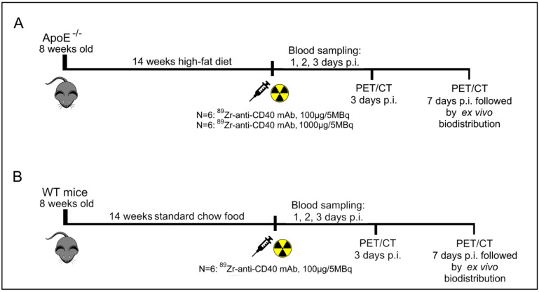
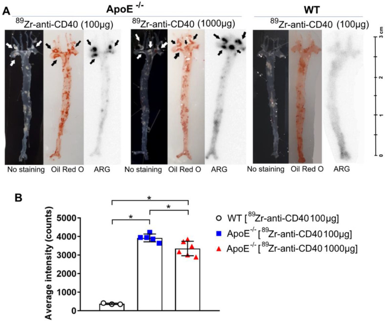
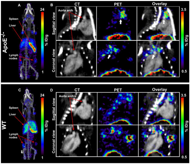
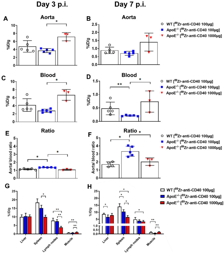
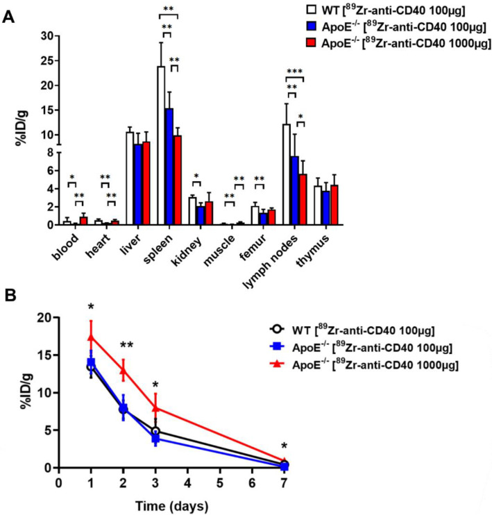
Non-invasive imaging of atherosclerosis can help in the identification of vulnerable plaque lesions. CD40 is a co-stimulatory molecule present on various immune and non-immune cells in the plaques and is linked to inflammation and plaque instability. We hypothesize that a Zr-labeled anti-CD40 monoclonal antibody (mAb) tracer has the potential to bind to cells present in atherosclerotic lesions and that CD40 Positron Emission Tomography (PET) can contribute to the detection of vulnerable atherosclerotic plaque lesions. To study this, wild-type (WT) and ApoE mice were fed a high cholesterol diet for 14 weeks to develop atherosclerosis. Mice were injected with [Zr]Zr-anti-CD40 mAb and the aortic uptake was evaluated and quantified using PET/Computed Tomography (CT) imaging. Ex vivo biodistribution was performed post-PET imaging and the uptake in the aorta was assessed with autoradiography and compared with Oil red O staining to determine the tracer potential to detect atherosclerotic plaques. On day 3 and 7 post injection, analysis of [Zr]Zr-anti-CD40 mAb PET/CT scans showed a more pronounced aortic signal in ApoE compared to WT mice with an increased aorta-to-blood uptake ratio. Autoradiography revealed [Zr]Zr-anti-CD40 mAb uptake in atherosclerotic plaque areas in ApoE mice, while no signal was found in WT mice. Clear overlap was observed between plaque areas as identified by Oil red O staining and autoradiography signal of [Zr]Zr-anti-CD40 mAb in ApoE mice. In this proof of concept study, we showed that PET/CT with [Zr]Zr-anti-CD40 mAb can detect atherosclerotic plaques. As CD40 is associated with plaque vulnerability, [Zr]Zr-anti-CD40 mAb has the potential to become a tracer to detect vulnerable atherosclerotic plaques.
动脉粥样硬化的非侵入性成像有助于识别易损斑块病变。CD40是一种共刺激分子,存在于斑块中的各种免疫和非免疫细胞上,与炎症和斑块不稳定性有关。我们假设,一种锆标记的抗CD40单克隆抗体(mAb)示踪剂有可能与动脉粥样硬化病变中的细胞结合,并且CD40正电子发射断层扫描(PET)有助于检测易损动脉粥样硬化斑块病变。为了研究这一点,将野生型(WT)小鼠和载脂蛋白E(ApoE)小鼠喂食高胆固醇饮食14周以诱发动脉粥样硬化。给小鼠注射[Zr]Zr-抗CD40 mAb,并使用PET/计算机断层扫描(CT)成像评估和量化主动脉摄取情况。PET成像后进行体外生物分布研究,用放射自显影评估主动脉摄取情况,并与油红O染色进行比较,以确定示踪剂检测动脉粥样硬化斑块的潜力。在注射后第3天和第7天,[Zr]Zr-抗CD40 mAb PET/CT扫描分析显示,与WT小鼠相比,ApoE小鼠的主动脉信号更明显,主动脉与血液的摄取比值增加。放射自显影显示ApoE小鼠的动脉粥样硬化斑块区域有[Zr]Zr-抗CD40 mAb摄取,而WT小鼠未发现信号。在ApoE小鼠中,油红O染色确定的斑块区域与[Zr]Zr-抗CD40 mAb的放射自显影信号之间观察到明显重叠。在这项概念验证研究中,我们表明,使用[Zr]Zr-抗CD40 mAb的PET/CT可以检测动脉粥样硬化斑块。由于CD40与斑块易损性相关,[Zr]Zr-抗CD40 mAb有可能成为检测易损动脉粥样硬化斑块的示踪剂。