Institute for Diagnostic and Interventional Neuroradiology, Support Center for Advanced Neuroimaging (SCAN), University of Bern, Bern, Switzerland.
Department of Radiology, University of Miami School of Medicine, Miami, Florida, USA.
Magn Reson Med. 2022 Jul;88(1):53-70. doi: 10.1002/mrm.29220. Epub 2022 Mar 28.
At ultra-high field (UHF), B -inhomogeneities and high specific absorption rate (SAR) of adiabatic slice-selective RF-pulses make spatial resolved spectral-editing extremely challenging with the conventional MEGA-approach. The purpose of the study was to develop a whole-brain resolved spectral-editing MRSI at UHF (UHF, B ≥ 7T) within clinical acceptable measurement-time and minimal chemical-shift-displacement-artifacts (CSDA) allowing for simultaneous GABA/Glx-, 2HG-, and PE-editing on a clinical approved 7T-scanner.
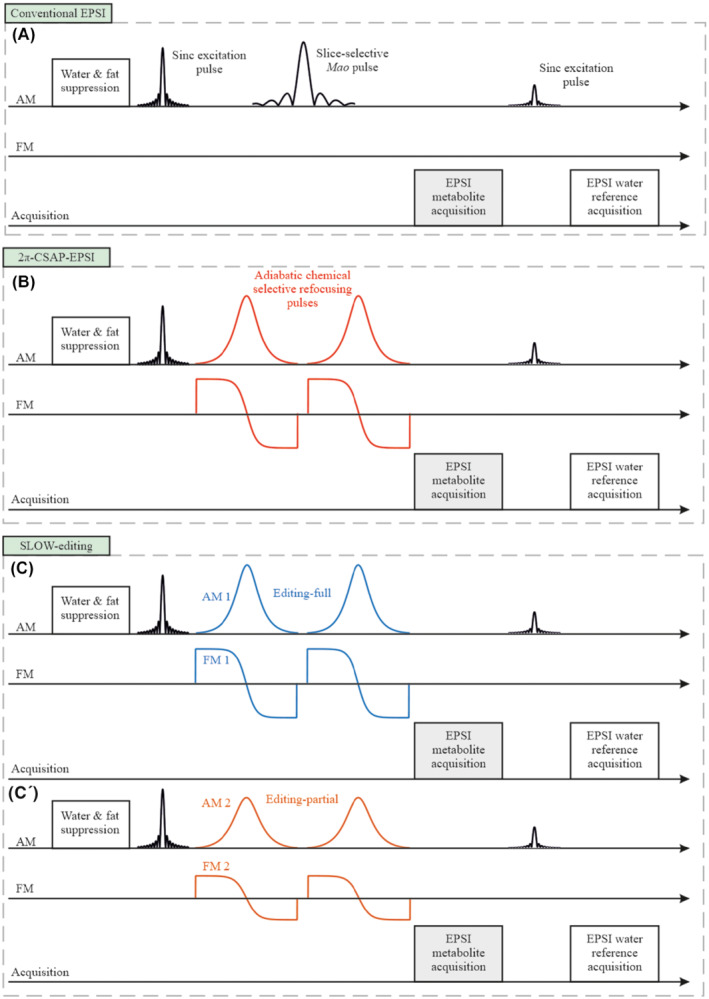
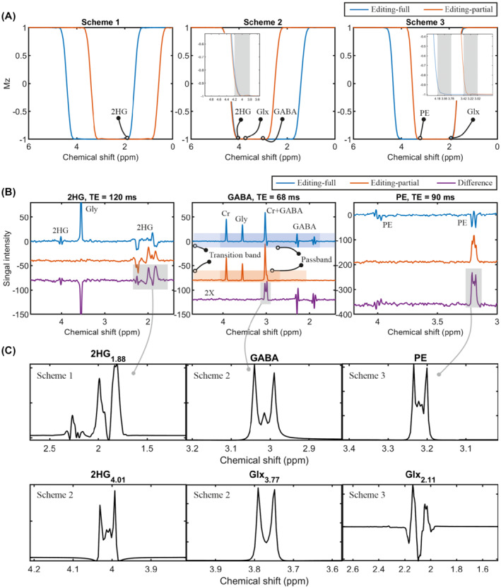
Slice-selective adiabatic refocusing RF-pulses (2π-SSAP) dominate the SAR to the patient in (semi)LASER based MEGA-editing sequences, causing large CSDA and long measurement times to fulfill SAR requirements, even using SAR-minimized GOIA-pulses. Therefore, a novel type of spectral-editing, called SLOW-editing, using two different pairs of phase-compensated chemical-shift selective adiabatic refocusing-pulses (2π-CSAP) with different refocusing bandwidths were investigated to overcome these problems.
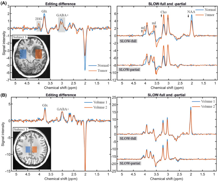
Compared to conventional echo-planar spectroscopic imaging (EPSI) and MEGA-editing, SLOW-editing shows robust refocusing and editing performance despite to B -inhomogeneity, and robustness to B -inhomogeneities (0.2 ppm ≥ ΔB ≥ -0.2 ppm). The narrow bandwidth (∼0.6-0.8 kHz) CSAP reduces the SAR by 92%, RF peak power by 84%, in-excitation slab CSDA by 77%, and has no in-plane CSDA. Furthermore, the CSAP implicitly dephases water, lipid and all the other signals outside of range (≥ 4.6 ppm and ≤1.4 ppm), resulting in additional water and lipid suppression (factors ≥ 1000s) at zero SAR-cost, and no spectral aliasing artifacts.
A new spectral-editing has been developed that is especially suitable for UHF, and was successfully applied for 2HG, GABA+, PE, and Glx-editing within 10 min clinical acceptable measurement time.
在超高场(UHF)下,B 不均匀性和高比吸收率(SAR)使得传统的 MEGA 方法在空间分辨率光谱编辑方面极具挑战性。本研究的目的是开发一种全脑分辨率的光谱编辑磁共振波谱成像(MRSI),在临床可接受的测量时间内,在最小的化学位移偏差伪影(CSDA)下,在临床批准的 7T 扫描仪上实现 GABA/Glx-、2HG-和 PE-编辑。
在基于(半)激光的 MEGA 编辑序列中,切片选择的绝热重聚焦 RF 脉冲(2π-SSAP)主导着患者的 SAR,导致大的 CSDA 和长的测量时间来满足 SAR 要求,即使使用 SAR 最小化的 GOIA 脉冲也是如此。因此,研究了一种新型的光谱编辑方法,称为 SLOW 编辑,使用两对具有不同重聚焦带宽的不同相位补偿化学位移选择绝热重聚焦脉冲(2π-CSAP)来克服这些问题。
与传统的回波平面波谱成像(EPSI)和 MEGA 编辑相比,SLOW 编辑在存在 B 不均匀性的情况下表现出稳健的重聚焦和编辑性能,并且对 B 不均匀性具有鲁棒性(0.2ppm≤ΔB≤-0.2ppm)。窄带宽(∼0.6-0.8kHz)的 CSAP 将 SAR 降低了 92%,RF 峰值功率降低了 84%,激发层 CSDA 降低了 77%,并且没有平面内 CSDA。此外,CSAP 隐含地使水、脂质和所有其他信号在范围外(≥4.6ppm 和≤1.4ppm)去相位,从而在零 SAR 成本下实现额外的水和脂质抑制(因子≥1000),并且没有光谱混叠伪影。
开发了一种新的光谱编辑方法,特别适用于 UHF,并成功应用于 2HG、GABA+、PE 和 Glx 编辑,测量时间在 10 分钟内可接受。