Chen Qiaofeng, Xiao Han, Gu Yunquan, Weng Zongpeng, Wei Lihong, Li Bin, Liao Bing, Li Jiali, Lin Jie, Hei Mengying, Peng Sui, Wang Wei, Kuang Ming, Chen Shuling
Department of Gastroenterology, the First Affiliated Hospital of Sun Yat-Sen University, Guangzhou, Guangdong, China.
Department of Medical Ultrasonics, Institute of Diagnostic and Interventional Ultrasound, the First Affiliated Hospital of Sun Yat-Sen University, No. 58, Zhongshan 2nd Road, Guangzhou, 510080, Guangdong, China.
Hepatol Int. 2022 Jun;16(3):590-602. doi: 10.1007/s12072-022-10323-w. Epub 2022 Mar 28.
Microvascular invasion (MVI) is essential for the management of hepatocellular carcinoma (HCC). However, MVI is hard to evaluate in patients without sufficient peri-tumoral tissue samples, which account for over a half of HCC patients.
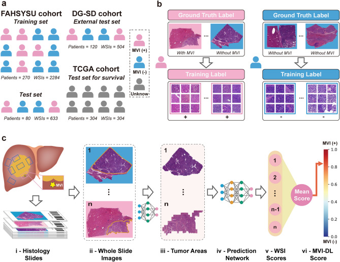
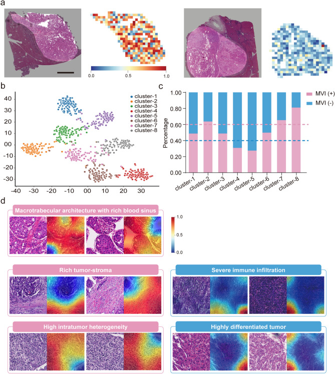
We established an MVI deep-learning (MVI-DL) model with a weakly supervised multiple-instance learning framework, to evaluate MVI status using only tumor tissues from the histological whole slide images (WSIs). A total of 350 HCC patients (2917 WSIs) from the First Affiliated Hospital of Sun Yat-sen University (FAHSYSU cohort) were divided into a training and test set. One hundred and twenty patients (504 WSIs) from Dongguan People's Hospital and Shunde Hospital of Southern Medical University (DG-SD cohort) formed an external test set. Unsupervised clustering and class activation mapping were applied to visualize the key histological features.
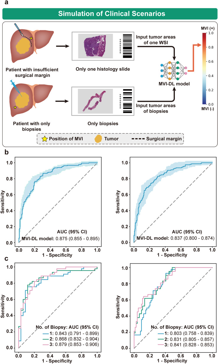
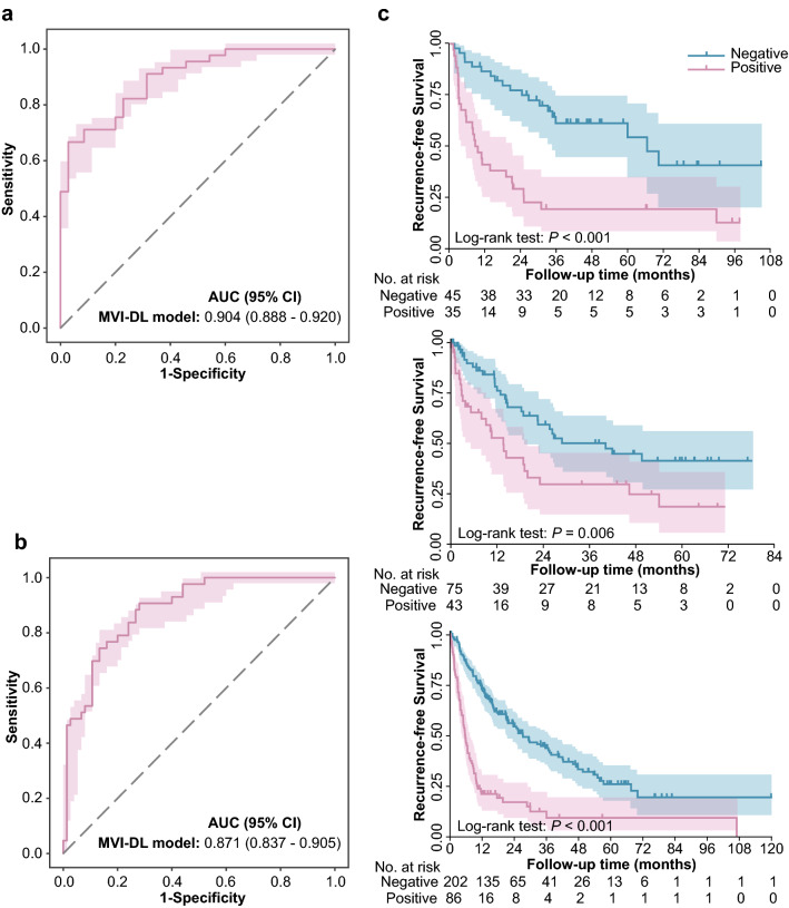
In the FAHSYSU and DG-SD test set, the MVI-DL model achieved an AUC of 0.904 (95% CI 0.888-0.920) and 0.871 (95% CI 0.837-0.905), respectively. Visualization results showed that macrotrabecular architecture with rich blood sinus, rich tumor stroma and high intratumor heterogeneity were identified as the key features associated with MVI ( +), whereas severe immune infiltration and highly differentiated tumor cells were associated with MVI (-). In the simulation of patients with only one WSI or biopsies only, the AUC of the MVI-DL model reached 0.875 (95% CI 0.855-0.895) and 0.879 (95% CI 0.853-0.906), respectively.
The effective, interpretable MVI-DL model has potential as an important tool with practical clinical applicability in evaluating MVI status from the tumor areas on the histological slides.
微血管侵犯(MVI)对于肝细胞癌(HCC)的治疗至关重要。然而,在没有足够肿瘤周围组织样本的患者中,MVI很难评估,而这类患者占HCC患者的一半以上。
我们建立了一个具有弱监督多实例学习框架的MVI深度学习(MVI-DL)模型,仅使用组织学全切片图像(WSIs)中的肿瘤组织来评估MVI状态。来自中山大学附属第一医院的350例HCC患者(2917张WSIs)(FAHSYSU队列)被分为训练集和测试集。来自东莞市人民医院和南方医科大学顺德医院的120例患者(504张WSIs)(DG-SD队列)组成外部测试集。应用无监督聚类和类激活映射来可视化关键组织学特征。
在FAHSYSU和DG-SD测试集中,MVI-DL模型的AUC分别为0.904(95%CI 0.888-0.920)和0.871(95%CI 0.837-0.905)。可视化结果显示,具有丰富血窦的大小梁结构、丰富的肿瘤间质和高肿瘤内异质性被确定为与MVI(+)相关的关键特征,而严重的免疫浸润和高分化肿瘤细胞与MVI(-)相关。在仅使用一张WSIs或仅进行活检的患者模拟中,MVI-DL模型的AUC分别达到0.875(95%CI 0.855-0.895)和0.879(95%CI 0.853-0.906)。
有效、可解释的MVI-DL模型有潜力成为一种重要工具,在从组织学切片上的肿瘤区域评估MVI状态方面具有实际临床适用性。