Perinatal Institute, Cincinnati Children's Hospital Medical Center, Cincinnati, OH, USA.
Division of Pulmonary Biology, Cincinnati Children's Hospital Medical Center, Cincinnati, OH, USA.
Sci Transl Med. 2022 Mar 30;14(638):eabl8574. doi: 10.1126/scitranslmed.abl8574.
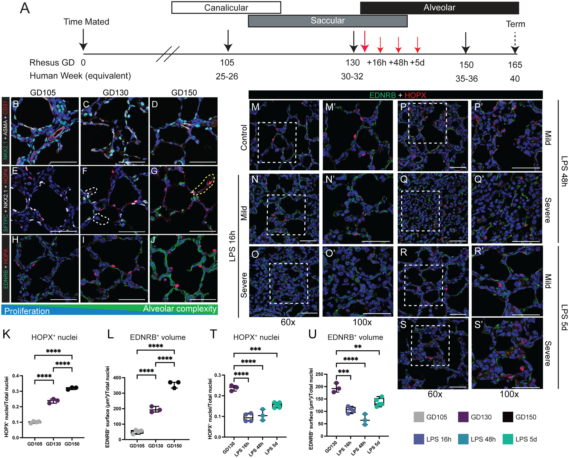
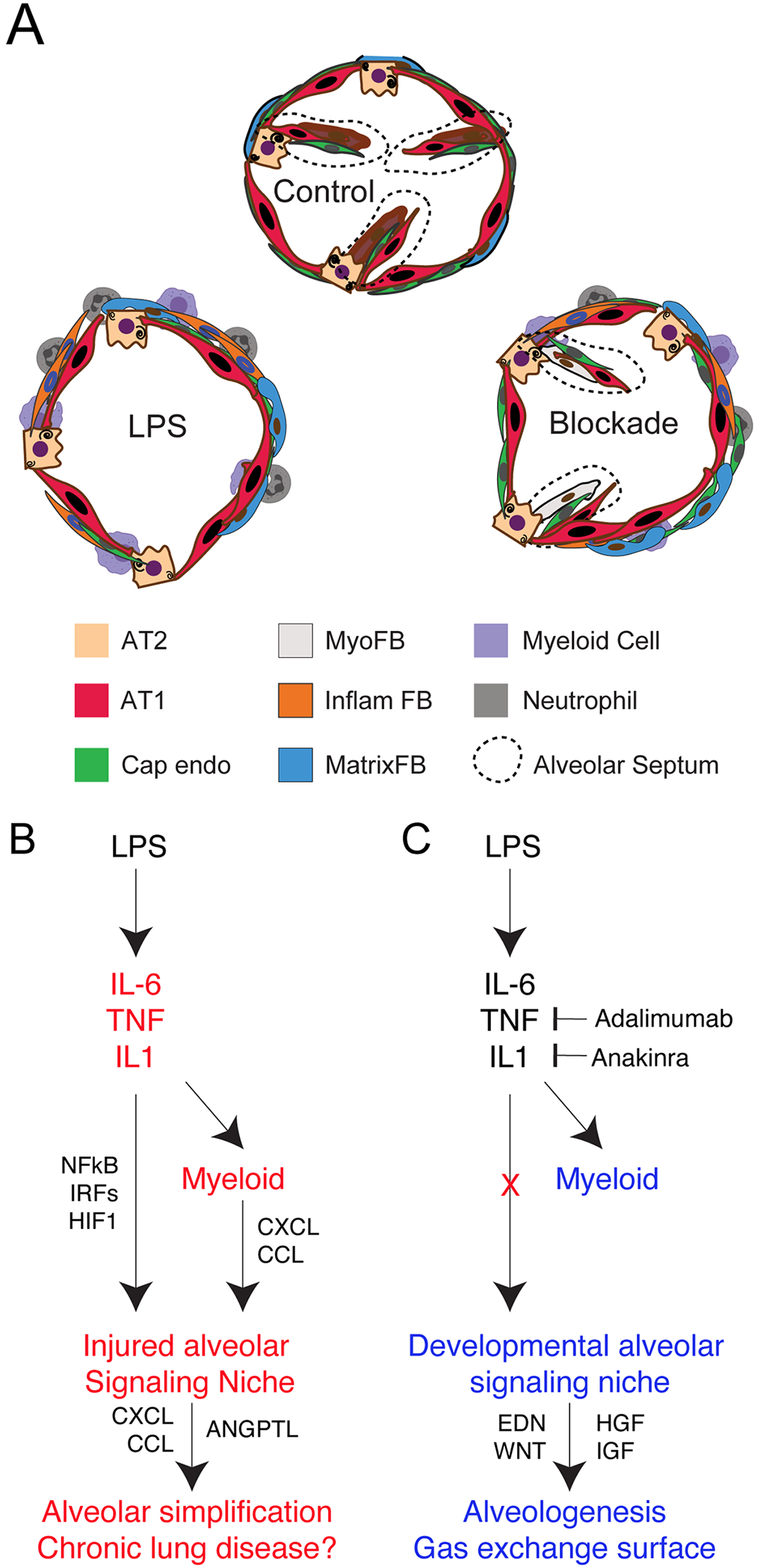
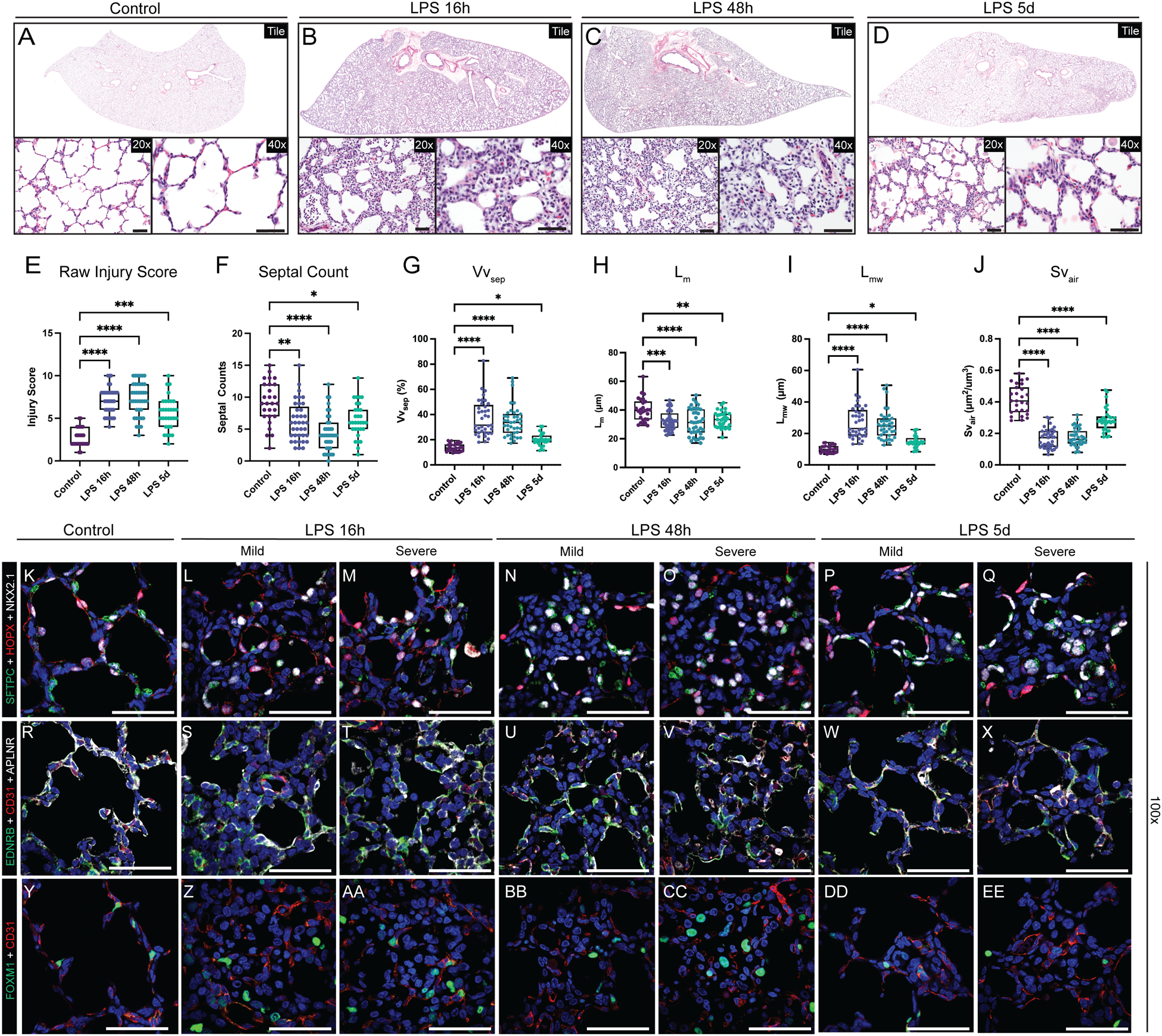
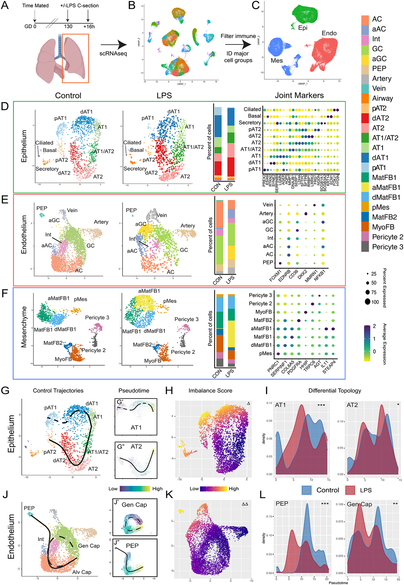
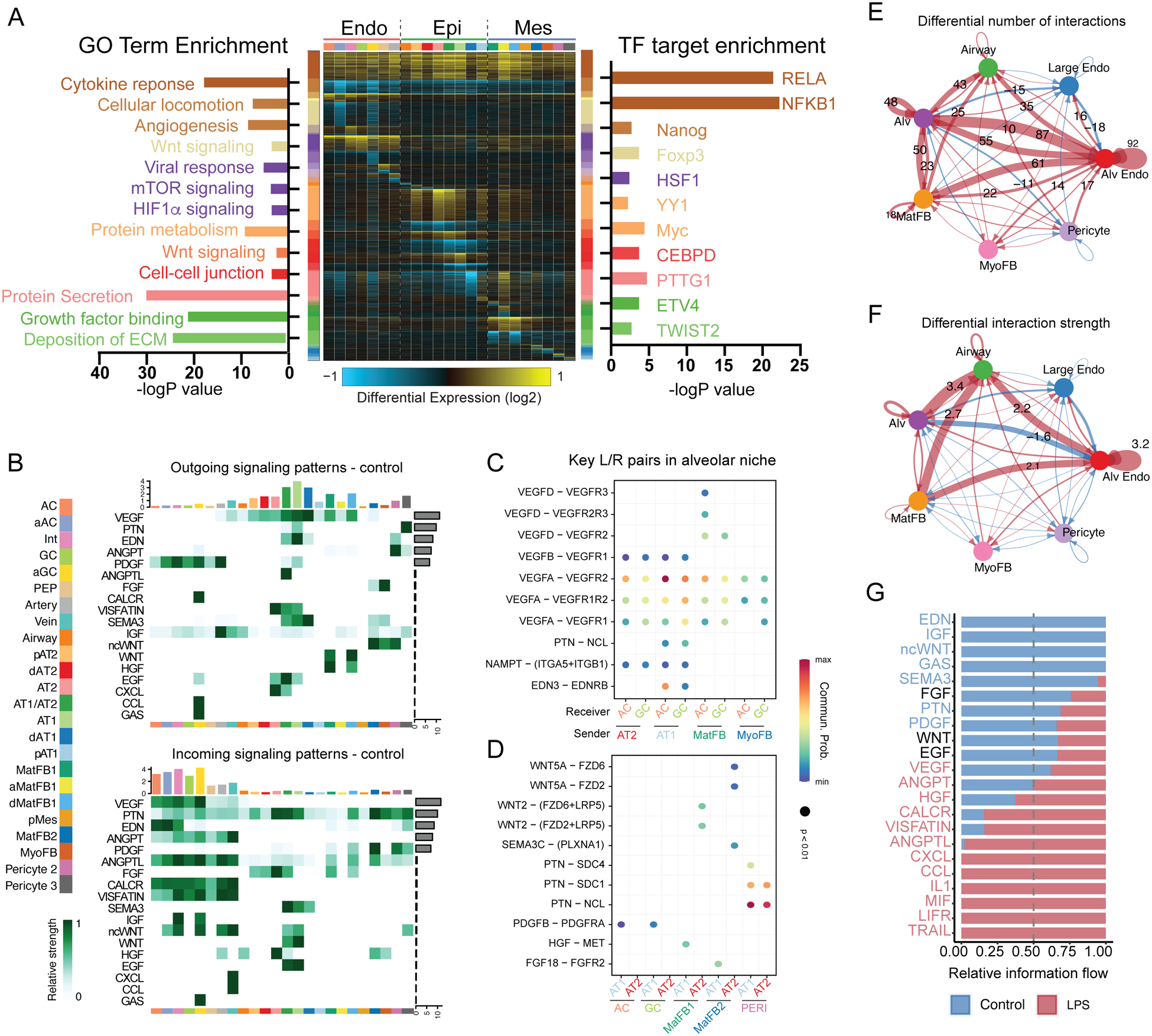
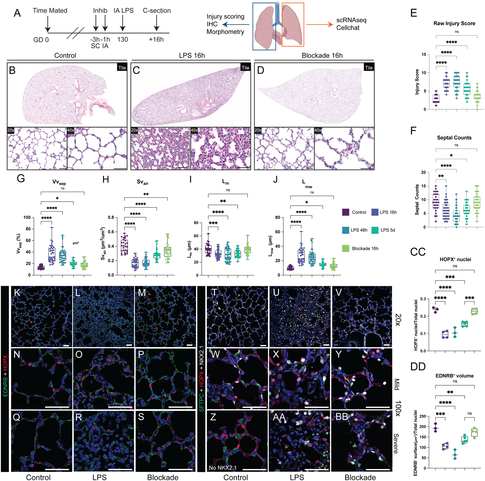
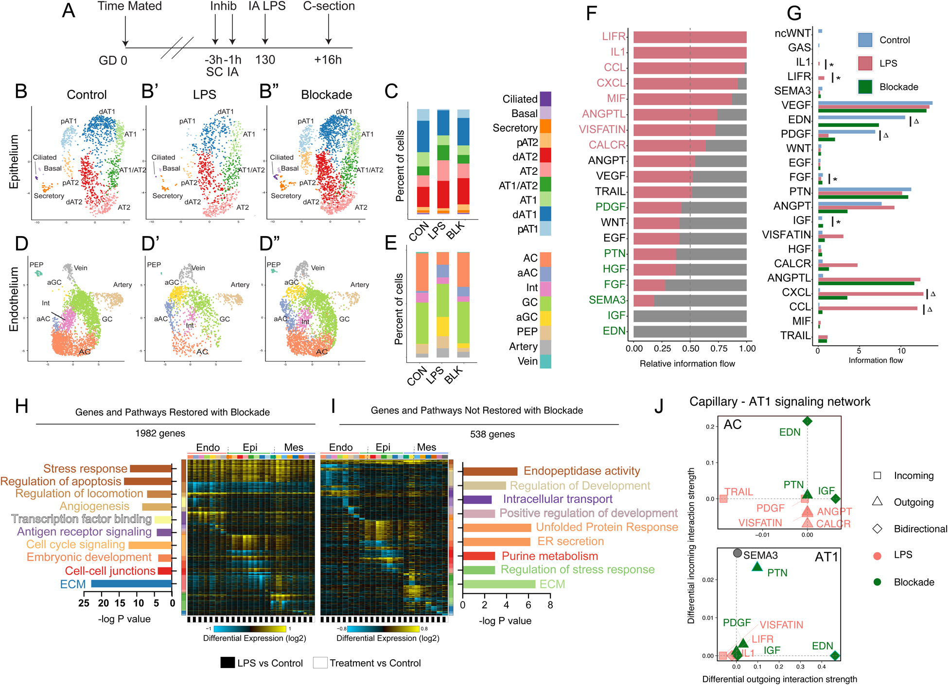
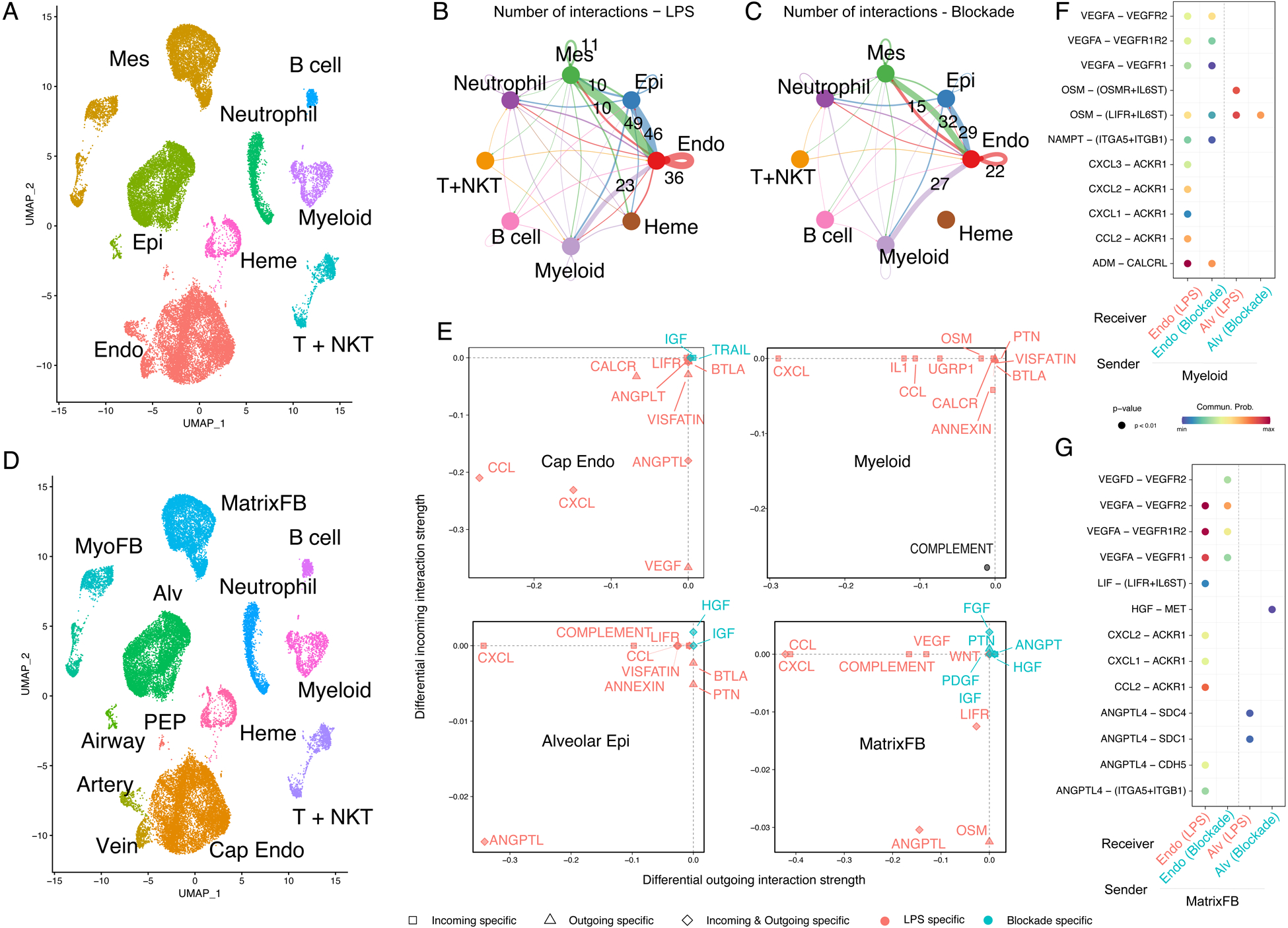
Perinatal inflammatory stress is associated with early life morbidity and lifelong consequences for pulmonary health. Chorioamnionitis, an inflammatory condition affecting the placenta and fluid surrounding the developing fetus, affects 25 to 40% of preterm births. Severe chorioamnionitis with preterm birth is associated with significantly increased risk of pulmonary disease and secondary infections in childhood, suggesting that fetal inflammation may markedly alter the development of the lung. Here, we used intra-amniotic lipopolysaccharide (LPS) challenge to induce experimental chorioamnionitis in a prenatal rhesus macaque () model that mirrors structural and temporal aspects of human lung development. Inflammatory injury directly disrupted the developing gas exchange surface of the primate lung, with extensive damage to alveolar structure, particularly the close association and coordinated differentiation of alveolar type 1 pneumocytes and specialized alveolar capillary endothelium. Single-cell RNA sequencing analysis defined a multicellular alveolar signaling niche driving alveologenesis that was extensively disrupted by perinatal inflammation, leading to a loss of gas exchange surface and alveolar simplification, with notable resemblance to chronic lung disease in newborns. Blockade of the inflammatory cytokines interleukin-1β and tumor necrosis factor-α ameliorated LPS-induced inflammatory lung injury by blunting stromal responses to inflammation and modulating innate immune activation in myeloid cells, restoring structural integrity and key signaling networks in the developing alveolus. These data provide new insight into the pathophysiology of developmental lung injury and suggest that modulating inflammation is a promising therapeutic approach to prevent fetal consequences of chorioamnionitis.
围产期炎症应激与生命早期发病率和肺部健康的终身后果有关。绒毛膜羊膜炎,一种影响胎盘和发育中胎儿周围液体的炎症状态,影响 25%至 40%的早产。伴有早产的严重绒毛膜羊膜炎与儿童期肺部疾病和继发感染的风险显著增加相关,这表明胎儿炎症可能显著改变肺部的发育。在这里,我们使用羊膜内脂多糖 (LPS) 挑战在模拟人类肺部发育的结构和时间方面的产前恒河猴 () 模型中诱导实验性绒毛膜羊膜炎。炎症损伤直接破坏了灵长类动物肺部的正在发育的气体交换表面,肺泡结构广泛受损,特别是肺泡 1 型细胞和特化的肺泡毛细血管内皮之间的紧密关联和协调分化。单细胞 RNA 测序分析定义了一个多细胞肺泡信号生态位,驱动肺泡发生,围产期炎症广泛破坏了这个生态位,导致气体交换表面丧失和肺泡简化,与新生儿慢性肺部疾病非常相似。阻断炎症细胞因子白细胞介素-1β和肿瘤坏死因子-α 通过抑制炎症对基质的反应和调节髓样细胞中的固有免疫激活,减轻 LPS 诱导的肺部炎症损伤,恢复发育中肺泡的结构完整性和关键信号网络。这些数据为发育性肺部损伤的病理生理学提供了新的见解,并表明调节炎症是预防绒毛膜羊膜炎胎儿后果的有前途的治疗方法。