Yusoff Farina Mohamad, Nakashima Ayumu, Kawano Ki-Ichiro, Kajikawa Masato, Kishimoto Shinji, Maruhashi Tatsuya, Ishiuchi Naoki, Abdul Wahid S Fadilah S, Higashi Yukihito
Department of Cardiovascular Regeneration and Medicine, Research Institute for Radiation Biology and Medicine, Hiroshima University, Hiroshima, Japan.
Department of Stem Cell Biology and Medicine, Hiroshima University Graduate School of Biomedical Sciences, Hiroshima, Japan.
Stem Cells Int. 2022 Mar 20;2022:6795274. doi: 10.1155/2022/6795274. eCollection 2022.
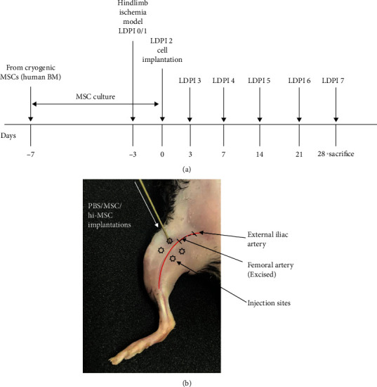
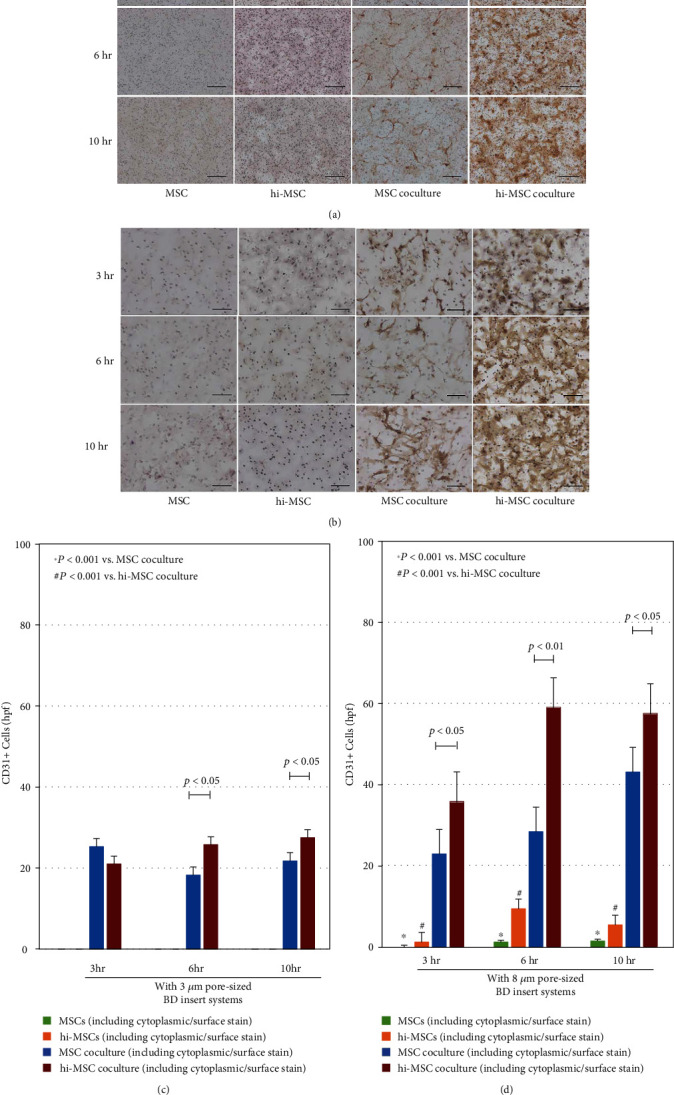
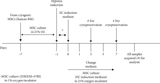
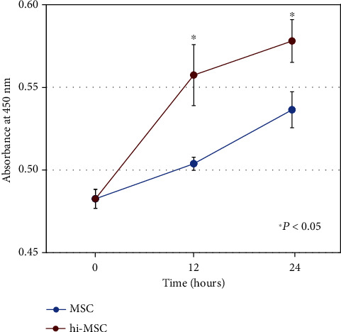
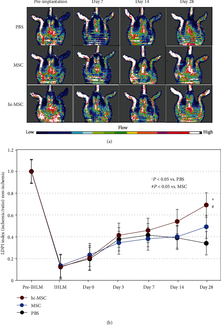
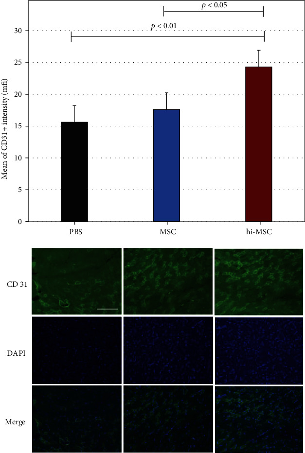
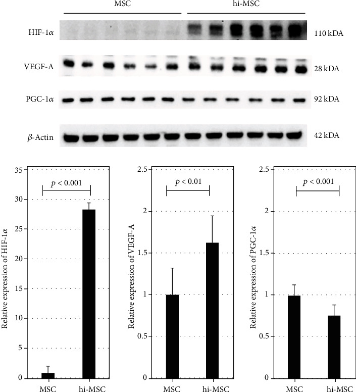
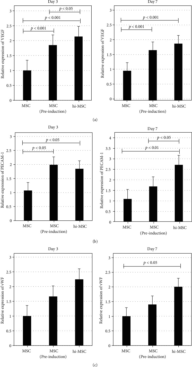
Hypoxia preconditioning enhances the paracrine abilities of mesenchymal stem cells (MSCs) for vascular regeneration and tissue healing. Implantation of hypoxia-induced mesenchymal stem cells (hi-MSCs) may further improve limb perfusion in a murine model of hindlimb ischemia. This study is aimed at determining whether implantation of hi-MSCs is an effective modality for improving outcomes of treatment of ischemic artery diseases. We evaluated the effects of human bone marrow-derived MSC implantation on limb blood flow in an ischemic hindlimb model. hi-MSCs were prepared by cell culture under 1% oxygen for 24 hours prior to implantation. A total of 1 × 10 MSCs and hi-MSCs and phosphate-buffered saline (PBS) were intramuscularly implanted into ischemic muscles at 36 hours after surgery. Restoration of blood flow and muscle perfusion was evaluated by laser Doppler perfusion imaging. Blood perfusion recovery, enhanced vessel densities, and improvement of function of the ischemia limb were significantly greater in the hi-MSC group than in the MSC or PBS group. Immunochemistry revealed that hi-MSCs had higher expression levels of hypoxia-inducible factor-1 alpha and vascular endothelial growth factor A than those in MSCs. In addition, an endothelial cell-inducing medium showed high expression levels of vascular endothelial growth factor, platelet endothelial cell adhesion molecule-1, and von Willebrand factor in hi-MSCs compared to those in MSCs. These findings suggest that pretreatment of MSCs with a hypoxia condition and implantation of hi-MSCs advances neovascularization capability with enhanced therapeutic angiogenic effects in a murine hindlimb ischemia model.
缺氧预处理可增强间充质干细胞(MSCs)的旁分泌能力,促进血管再生和组织愈合。植入缺氧诱导的间充质干细胞(hi-MSCs)可能会进一步改善后肢缺血小鼠模型的肢体灌注。本研究旨在确定植入hi-MSCs是否是改善缺血性动脉疾病治疗效果的有效方式。我们评估了人骨髓源MSCs植入对缺血后肢模型肢体血流的影响。hi-MSCs在植入前通过在1%氧气条件下细胞培养24小时制备。术后36小时,将总共1×10的MSCs、hi-MSCs和磷酸盐缓冲盐水(PBS)肌肉内植入缺血肌肉中。通过激光多普勒灌注成像评估血流和肌肉灌注的恢复情况。hi-MSC组的血流灌注恢复、血管密度增加以及缺血肢体功能改善均显著优于MSC组或PBS组。免疫化学分析显示,hi-MSCs中缺氧诱导因子-1α和血管内皮生长因子A的表达水平高于MSCs。此外,与MSCs相比,内皮细胞诱导培养基显示hi-MSCs中血管内皮生长因子、血小板内皮细胞黏附分子-1和血管性血友病因子的表达水平较高。这些发现表明,在小鼠后肢缺血模型中,对MSCs进行缺氧预处理并植入hi-MSCs可提高新生血管形成能力,并增强治疗性血管生成作用。