Jung Sung Yoon, Kim Hyeon Jun, Oh Kyu Taek
Department of Orthopedic Surgery, College of Medicine, Dong-A University, Busan, Korea.
Hip Pelvis. 2022 Mar;34(1):10-17. doi: 10.5371/hp.2022.34.1.10. Epub 2022 Mar 7.
This study was conducted in order to assess changes in hip muscles by comparing results of preoperative and postoperative computed tomography (CT) in older patients who underwent surgery for treatment of hip fracture.
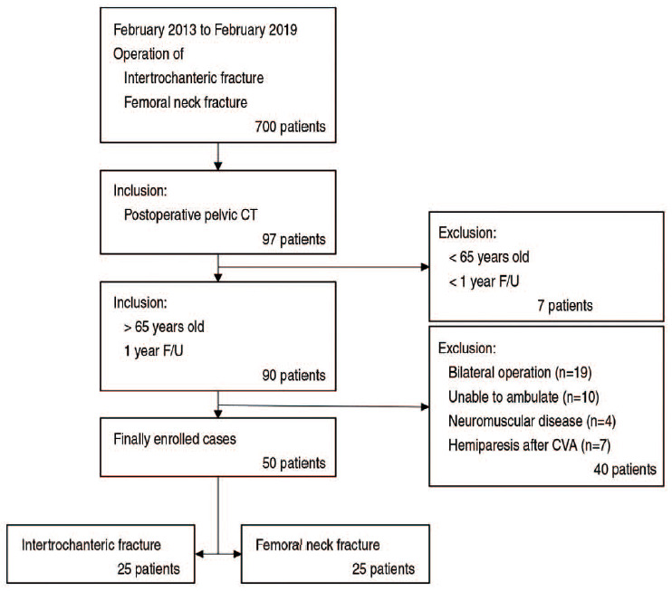
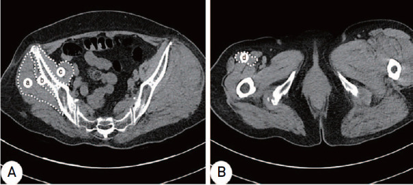
A total of 50 patients (aged ≥65 years) who underwent surgery for treatment of intertrochanteric fractures (25 patients) and femoral neck fractures (25 patients) between February 2013 and February 2019 and underwent preoperative and postoperative pelvic CT were enrolled in the study. The cross-sectional area, attenuation and estimates of muscle mass of the gluteus medius, gluteus minimus, iliopsoas, and rectus femoris on the uninjured side were measured. Basic patient data (sex, age, height, weight, body mass index [BMI], bone mineral density [BMD], Harris hip score [HHS], and length of follow-up) were collected from medical records.
No significant differences in sex, age, height, weight, BMI, BMD, HHS, and length of follow-up were observed between the two groups. No significant difference in the cross-sectional areas and attenuations of gluteus medius and gluteus minimus was observed after surgery; however, a statistically significant decrease was observed in those of iliopsoas and rectus femoris after surgery. Lower estimates with statistical significance of muscle mass of the iliopsoas and rectus femoris were observed on postoperative CT.
Muscle mass of the hip flexor (iliopsoas, rectus femoris) showed significant decreases on postoperative CT compared with preoperative CT. Based on these findings, selective strengthening exercise for hip flexor should be beneficial in rehabilitation of hip fractures.
本研究旨在通过比较接受髋部骨折手术治疗的老年患者术前和术后计算机断层扫描(CT)的结果,评估髋部肌肉的变化。
本研究纳入了2013年2月至2019年2月期间接受手术治疗转子间骨折(25例)和股骨颈骨折(25例)且术前行骨盆CT检查及术后行骨盆CT检查的50例患者(年龄≥65岁)。测量未受伤侧臀中肌、臀小肌、髂腰肌和股直肌的横截面积、衰减值和肌肉质量估计值。从病历中收集患者的基本数据(性别、年龄、身高、体重、体重指数[BMI]、骨密度[BMD]、Harris髋关节评分[HHS]和随访时间)。
两组患者在性别、年龄、身高、体重、BMI、BMD、HHS和随访时间方面均未观察到显著差异。术后臀中肌和臀小肌的横截面积和衰减值未见显著差异;然而,术后髂腰肌和股直肌的横截面积和衰减值出现了统计学上的显著下降。术后CT显示髂腰肌和股直肌的肌肉质量估计值较低且具有统计学意义。
与术前CT相比,术后CT显示髋部屈肌(髂腰肌、股直肌)的肌肉质量显著下降。基于这些发现,对髋部屈肌进行选择性强化锻炼可能有助于髋部骨折的康复。