• 文献检索
  • 文档翻译
  • 深度研究
  • 学术资讯
  • Suppr Zotero 插件Zotero 插件
  • 邀请有礼
  • 套餐&价格
  • 历史记录
应用&插件
Suppr Zotero 插件Zotero 插件浏览器插件Mac 客户端Windows 客户端微信小程序
定价
高级版会员购买积分包购买API积分包
服务
文献检索文档翻译深度研究API 文档MCP 服务
关于我们
关于 Suppr公司介绍联系我们用户协议隐私条款
关注我们

Suppr 超能文献

核心技术专利:CN118964589B侵权必究
粤ICP备2023148730 号-1Suppr @ 2026

文献检索

告别复杂PubMed语法,用中文像聊天一样搜索,搜遍4000万医学文献。AI智能推荐,让科研检索更轻松。

立即免费搜索

文件翻译

保留排版,准确专业,支持PDF/Word/PPT等文件格式,支持 12+语言互译。

免费翻译文档

深度研究

AI帮你快速写综述,25分钟生成高质量综述,智能提取关键信息,辅助科研写作。

立即免费体验

新生仔猪心肌梗死后心外膜传导动力学及自然心脏再生的电生理保留

Electrophysiologic Conservation of Epicardial Conduction Dynamics After Myocardial Infarction and Natural Heart Regeneration in Newborn Piglets.

作者信息

Wang Hanjay, Pong Terrence, Obafemi Oluwatomisin O, Lucian Haley J, Aparicio-Valenzuela Joy, Tran Nicholas A, Mullis Danielle M, Elde Stefan, Tada Yuko, Baker Sam W, Wang Caroline Y, Cyr Kevin J, Paulsen Michael J, Zhu Yuanjia, Lee Anson M, Woo Y Joseph

机构信息

Department of Cardiothoracic Surgery, Stanford University, Stanford, CA, United States.

Stanford Cardiovascular Institute, Stanford University, Stanford, CA, United States.

出版信息

Front Cardiovasc Med. 2022 Mar 9;9:829546. doi: 10.3389/fcvm.2022.829546. eCollection 2022.

DOI:10.3389/fcvm.2022.829546
PMID:35355973
原文链接:https://pmc.ncbi.nlm.nih.gov/articles/PMC8959497/
Abstract

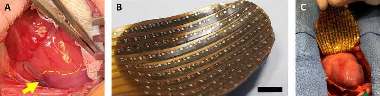
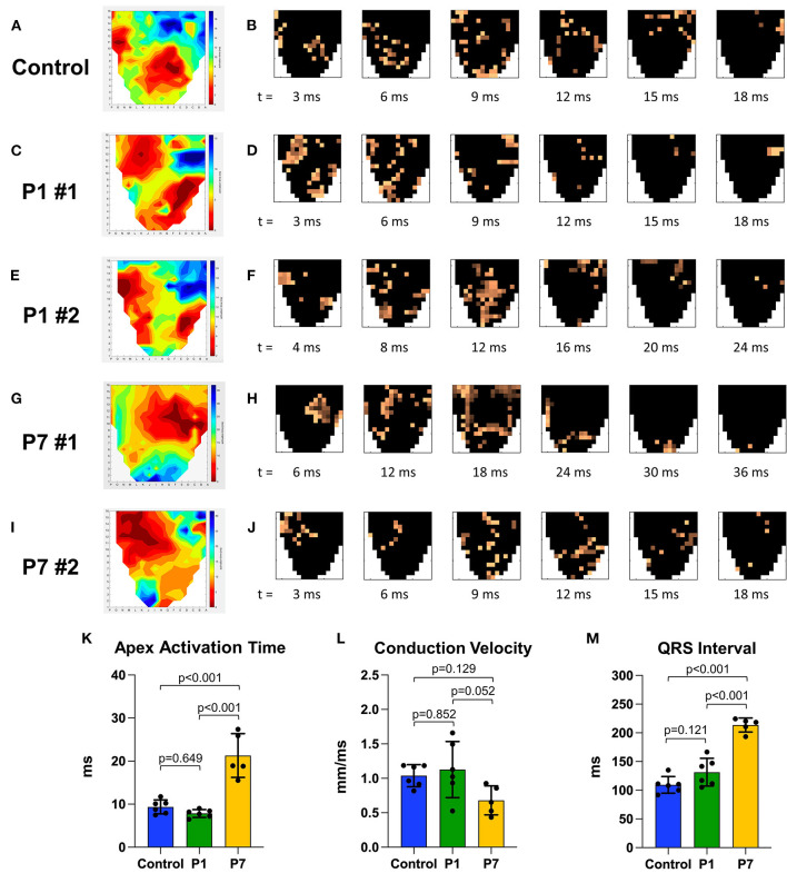
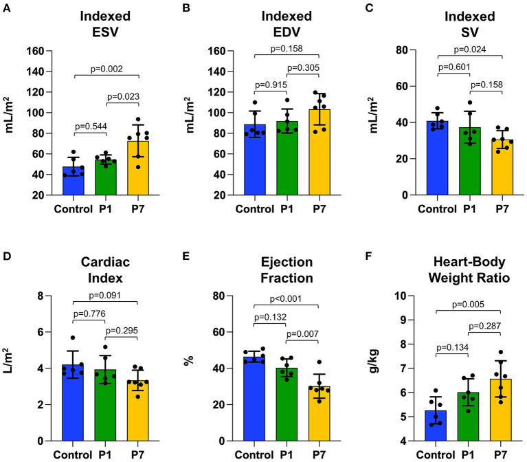
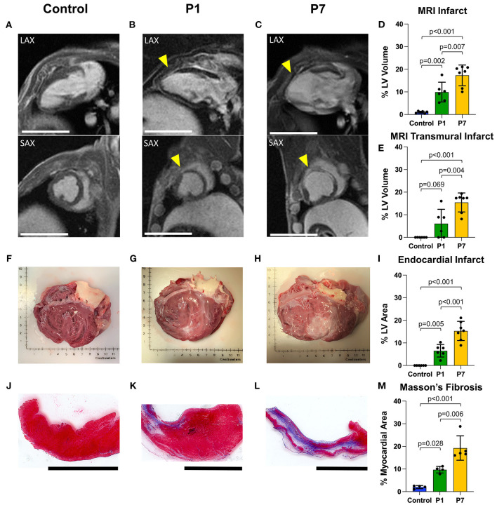
Newborn mammals, including piglets, exhibit natural heart regeneration after myocardial infarction (MI) on postnatal day 1 (P1), but this ability is lost by postnatal day 7 (P7). The electrophysiologic properties of this naturally regenerated myocardium have not been examined. We hypothesized that epicardial conduction is preserved after P1 MI in piglets. Yorkshire-Landrace piglets underwent left anterior descending coronary artery ligation at age P1 ( = 6) or P7 ( = 7), After 7 weeks, cardiac magnetic resonance imaging was performed with late gadolinium enhancement for analysis of fibrosis. Epicardial conduction mapping was performed using custom 3D-printed high-resolution mapping arrays. Age- and weight-matched healthy pigs served as controls ( = 6). At the study endpoint, left ventricular (LV) ejection fraction was similar for controls and P1 pigs (46.4 ± 3.0% vs. 40.3 ± 4.9%, = 0.132), but significantly depressed for P7 pigs (30.2 ± 6.6%, < 0.001 vs. control). The percentage of LV myocardial volume consisting of fibrotic scar was 1.0 ± 0.4% in controls, 9.9 ± 4.4% in P1 pigs ( = 0.002 vs. control), and 17.3 ± 4.6% in P7 pigs ( < 0.001 vs. control, = 0.007 vs. P1). Isochrone activation maps and apex activation time were similar between controls and P1 pigs (9.4 ± 1.6 vs. 7.8 ± 0.9 ms, = 0.649), but significantly prolonged in P7 pigs (21.3 ± 5.1 ms, < 0.001 vs. control, < 0.001 vs. P1). Conduction velocity was similar between controls and P1 pigs (1.0 ± 0.2 vs. 1.1 ± 0.4 mm/ms, = 0.852), but slower in P7 pigs (0.7 ± 0.2 mm/ms, = 0.129 vs. control, = 0.052 vs. P1). Overall, our data suggest that epicardial conduction dynamics are conserved in the setting of natural heart regeneration in piglets after P1 MI.

摘要

新生哺乳动物,包括仔猪,在出生后第1天(P1)发生心肌梗死后(MI)可表现出自然的心脏再生能力,但这种能力在出生后第7天(P7)会丧失。尚未对这种自然再生心肌的电生理特性进行研究。我们假设仔猪在P1期心肌梗死后心外膜传导得以保留。约克夏-长白仔猪在P1期(n = 6)或P7期(n = 7)接受左前降支冠状动脉结扎,7周后,进行心脏磁共振成像及钆延迟增强扫描以分析纤维化情况。使用定制的3D打印高分辨率标测阵列进行心外膜传导标测。年龄和体重匹配的健康猪作为对照(n = 6)。在研究终点,对照组和P1期仔猪的左心室(LV)射血分数相似(46.4±3.0%对40.3±4.9%,P = 0.132),但P7期仔猪的射血分数显著降低(30.2±6.6%,与对照组相比P<0.001)。对照组左心室心肌纤维化瘢痕体积占比为1.0±0.4%,P1期仔猪为9.9±4.4%(与对照组相比P = 0.002),P7期仔猪为17.3±4.6%(与对照组相比P<0.001,与P1期相比P = 0.007)。等时激活图和心尖激活时间在对照组和P1期仔猪之间相似(9.4±1.6对7.8±0.9毫秒,P = 0.649),但在P7期仔猪中显著延长(21.3±5.1毫秒,与对照组相比P<0.001,与P1期相比P<0.001)。传导速度在对照组和P1期仔猪之间相似(1.0±0.2对1.1±0.4毫米/毫秒,P = 0.852),但在P7期仔猪中较慢(0.7±0.2毫米/毫秒,与对照组相比P = 0.129,与P1期相比P = 0.052)。总体而言,我们的数据表明,仔猪在P1期心肌梗死后自然心脏再生过程中心外膜传导动力学得以保留。

https://cdn.ncbi.nlm.nih.gov/pmc/blobs/3abc/8959497/f6a5e6094701/fcvm-09-829546-g0004.jpg
https://cdn.ncbi.nlm.nih.gov/pmc/blobs/3abc/8959497/be334918dd88/fcvm-09-829546-g0001.jpg
https://cdn.ncbi.nlm.nih.gov/pmc/blobs/3abc/8959497/a82d892c9b33/fcvm-09-829546-g0002.jpg
https://cdn.ncbi.nlm.nih.gov/pmc/blobs/3abc/8959497/920d5f9ed362/fcvm-09-829546-g0003.jpg
https://cdn.ncbi.nlm.nih.gov/pmc/blobs/3abc/8959497/f6a5e6094701/fcvm-09-829546-g0004.jpg

相似文献

1
Electrophysiologic Conservation of Epicardial Conduction Dynamics After Myocardial Infarction and Natural Heart Regeneration in Newborn Piglets.新生仔猪心肌梗死后心外膜传导动力学及自然心脏再生的电生理保留
Front Cardiovasc Med. 2022 Mar 9;9:829546. doi: 10.3389/fcvm.2022.829546. eCollection 2022.
2
Multiaxial Lenticular Stress-Strain Relationship of Native Myocardium is Preserved by Infarct-Induced Natural Heart Regeneration in Neonatal Mice.新生小鼠心肌梗死诱导的自然心脏再生可保留天然心肌的多轴透镜状应力应变关系。
Sci Rep. 2020 Apr 30;10(1):7319. doi: 10.1038/s41598-020-63324-w.
3
A neonatal leporine model of age-dependent natural heart regeneration after myocardial infarction.一种用于研究心肌梗死后年龄依赖性自然心脏再生的新生兔模型。
J Thorac Cardiovasc Surg. 2022 Dec;164(6):e389-e405. doi: 10.1016/j.jtcvs.2021.08.013. Epub 2021 Aug 17.
4
Natural Heart Regeneration in a Neonatal Rat Myocardial Infarction Model.新生大鼠心肌梗死模型中的自然心脏再生。
Cells. 2020 Jan 16;9(1):229. doi: 10.3390/cells9010229.
5
Anisotropic conduction characteristics in ischemia-reperfusion induced chronic myocardial infarction.缺血再灌注诱导的慢性心肌梗死中的各向异性传导特征
Basic Res Cardiol. 1994 Mar-Apr;89(2):177-91. doi: 10.1007/BF00788736.
6
P2Y12 antagonists and cardiac repair post-myocardial infarction: global and regional heart function analysis and molecular assessments in pigs.P2Y12 拮抗剂与心肌梗死后的心脏修复:猪的整体和局部心脏功能分析及分子评估。
Cardiovasc Res. 2018 Dec 1;114(14):1860-1870. doi: 10.1093/cvr/cvy201.
7
Cardiovascular magnetic resonance imaging of functional and microstructural changes of the heart in a longitudinal pig model of acute to chronic myocardial infarction.心脏在急性至慢性心肌梗死的纵向猪模型中心功能和微观结构变化的心血管磁共振成像。
J Cardiovasc Magn Reson. 2021 Sep 20;23(1):103. doi: 10.1186/s12968-021-00794-5.
8
Inflammation in Remote Myocardium and Left Ventricular Remodeling After Acute Myocardial Infarction: A Pilot Study Using T2 Mapping.急性心肌梗死后远隔心肌炎症与左心室重构:一项使用T2映射的初步研究
J Magn Reson Imaging. 2022 Feb;55(2):555-564. doi: 10.1002/jmri.27827. Epub 2021 Jul 9.
9
ST segment elevation at the surface of a healed transmural myocardial infarction in pigs. Conditions for passive transmission from the ischemic peri-infarction zone.猪愈合透壁性心肌梗死表面的ST段抬高。缺血性梗死周边区被动传导的条件。
Circulation. 1995 Mar 1;91(5):1552-9. doi: 10.1161/01.cir.91.5.1552.
10
A complete heart regeneration model with inflammation as a key component.具有炎症关键组成部分的完整心脏再生模型。
Exp Anim. 2021 Nov 10;70(4):479-487. doi: 10.1538/expanim.20-0191. Epub 2021 Jun 15.

引用本文的文献

1
Cardiac conduction system regeneration prevents arrhythmias after myocardial infarction.心脏传导系统再生可预防心肌梗死后的心律失常。
Nat Cardiovasc Res. 2025 Feb;4(2):163-179. doi: 10.1038/s44161-024-00586-x. Epub 2025 Jan 3.
2
Structural, angiogenic, and immune responses influencing myocardial regeneration: a glimpse into the crucible.影响心肌再生的结构、血管生成和免疫反应:审视关键因素
NPJ Regen Med. 2024 Apr 30;9(1):18. doi: 10.1038/s41536-024-00357-z.
3
Electrophysiological mapping of the epicardium via 3D-printed flexible arrays.

本文引用的文献

1
Natural cardiac regeneration conserves native biaxial left ventricular biomechanics after myocardial infarction in neonatal rats.自然心脏再生可在新生大鼠心肌梗死后保留原生双轴左心室生物力学特性。
J Mech Behav Biomed Mater. 2022 Feb;126:105074. doi: 10.1016/j.jmbbm.2022.105074. Epub 2022 Jan 4.
2
A neonatal leporine model of age-dependent natural heart regeneration after myocardial infarction.一种用于研究心肌梗死后年龄依赖性自然心脏再生的新生兔模型。
J Thorac Cardiovasc Surg. 2022 Dec;164(6):e389-e405. doi: 10.1016/j.jtcvs.2021.08.013. Epub 2021 Aug 17.
3
A novel transgenic Cre allele to label mouse cardiac conduction system.
通过3D打印柔性阵列对心外膜进行电生理标测。
Bioeng Transl Med. 2023 Jul 19;8(6):e10575. doi: 10.1002/btm2.10575. eCollection 2023 Nov.
一种新型转基因 Cre 等位基因,用于标记小鼠心脏传导系统。
Dev Biol. 2021 Oct;478:163-172. doi: 10.1016/j.ydbio.2021.07.005. Epub 2021 Jul 8.
4
The Expanding Armamentarium of Innovative Bioengineered Strategies to Augment Cardiovascular Repair and Regeneration.用于增强心血管修复和再生的创新生物工程策略的不断扩展的手段
Front Bioeng Biotechnol. 2021 Jun 1;9:674172. doi: 10.3389/fbioe.2021.674172. eCollection 2021.
5
Navigating the Crossroads of Cell Therapy and Natural Heart Regeneration.探索细胞疗法与心脏自然再生的交叉点
Front Cell Dev Biol. 2021 May 11;9:674180. doi: 10.3389/fcell.2021.674180. eCollection 2021.
6
A Roadmap to Heart Regeneration Through Conserved Mechanisms in Zebrafish and Mammals.通过斑马鱼和哺乳动物中的保守机制实现心脏再生的路线图。
Curr Cardiol Rep. 2021 Mar 2;23(4):29. doi: 10.1007/s11886-021-01459-6.
7
Global Burden of Cardiovascular Diseases and Risk Factors, 1990-2019: Update From the GBD 2019 Study.全球心血管疾病负担及危险因素, 1990-2019:来自 GBD 2019 研究的更新。
J Am Coll Cardiol. 2020 Dec 22;76(25):2982-3021. doi: 10.1016/j.jacc.2020.11.010.
8
Cell-Type-Specific Gene Regulatory Networks Underlying Murine Neonatal Heart Regeneration at Single-Cell Resolution.单细胞分辨率下小鼠新生心脏再生的细胞类型特异性基因调控网络。
Cell Rep. 2020 Dec 8;33(10):108472. doi: 10.1016/j.celrep.2020.108472.
9
A Bioengineered Neuregulin-Hydrogel Therapy Reduces Scar Size and Enhances Post-Infarct Ventricular Contractility in an Ovine Large Animal Model.一种生物工程化的神经调节蛋白-水凝胶疗法可减小绵羊大型动物模型中的瘢痕大小并增强梗死后心室收缩力。
J Cardiovasc Dev Dis. 2020 Nov 17;7(4):53. doi: 10.3390/jcdd7040053.
10
Intrinsically stretchable electrode array enabled in vivo electrophysiological mapping of atrial fibrillation at cellular resolution.基于内在可拉伸电极阵列实现细胞分辨率的体内心房颤动电生理图谱绘制。
Proc Natl Acad Sci U S A. 2020 Jun 30;117(26):14769-14778. doi: 10.1073/pnas.2000207117. Epub 2020 Jun 15.