• 文献检索
  • 文档翻译
  • 深度研究
  • 学术资讯
  • Suppr Zotero 插件Zotero 插件
  • 邀请有礼
  • 套餐&价格
  • 历史记录
应用&插件
Suppr Zotero 插件Zotero 插件浏览器插件Mac 客户端Windows 客户端微信小程序
定价
高级版会员购买积分包购买API积分包
服务
文献检索文档翻译深度研究API 文档MCP 服务
关于我们
关于 Suppr公司介绍联系我们用户协议隐私条款
关注我们

Suppr 超能文献

核心技术专利:CN118964589B侵权必究
粤ICP备2023148730 号-1Suppr @ 2026

文献检索

告别复杂PubMed语法,用中文像聊天一样搜索,搜遍4000万医学文献。AI智能推荐,让科研检索更轻松。

立即免费搜索

文件翻译

保留排版,准确专业,支持PDF/Word/PPT等文件格式,支持 12+语言互译。

免费翻译文档

深度研究

AI帮你快速写综述,25分钟生成高质量综述,智能提取关键信息,辅助科研写作。

立即免费体验

单细胞分辨率下小鼠新生心脏再生的细胞类型特异性基因调控网络。

Cell-Type-Specific Gene Regulatory Networks Underlying Murine Neonatal Heart Regeneration at Single-Cell Resolution.

机构信息

Department of Molecular Biology, The Hamon Center for Regenerative Science and Medicine, and Sen. Paul D. Wellstone Muscular Dystrophy Cooperative Research Center, University of Texas Southwestern Medical Center, 5323 Harry Hines Boulevard, Dallas, TX 75390, USA.

Department of Molecular Biology, The Hamon Center for Regenerative Science and Medicine, and Sen. Paul D. Wellstone Muscular Dystrophy Cooperative Research Center, University of Texas Southwestern Medical Center, 5323 Harry Hines Boulevard, Dallas, TX 75390, USA.

出版信息

Cell Rep. 2020 Dec 8;33(10):108472. doi: 10.1016/j.celrep.2020.108472.

DOI:10.1016/j.celrep.2020.108472
PMID:33296652
原文链接:https://pmc.ncbi.nlm.nih.gov/articles/PMC7774872/
Abstract

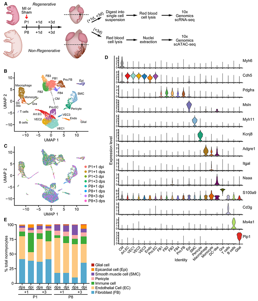
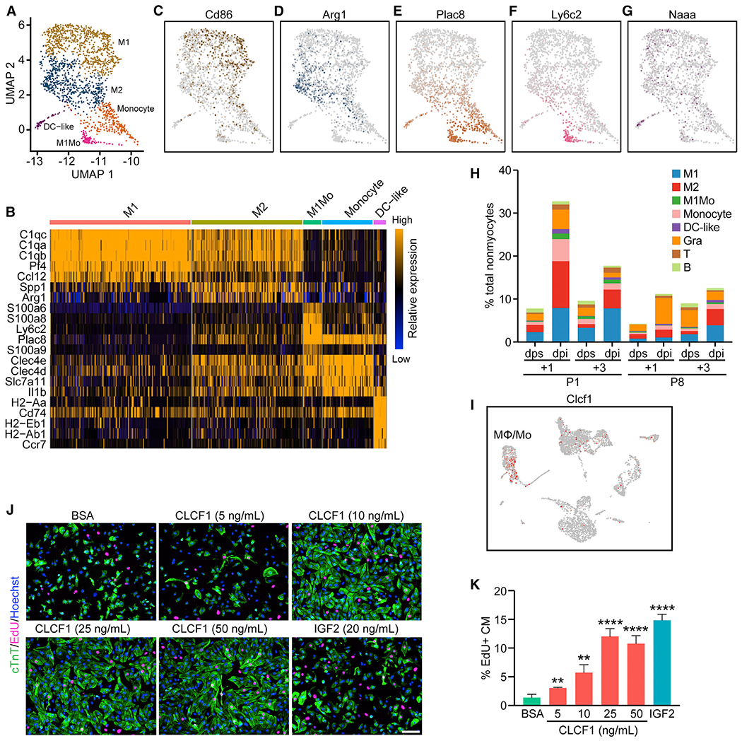
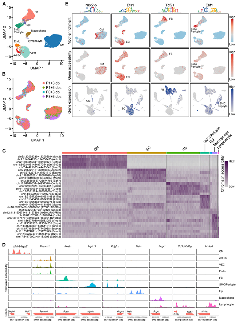
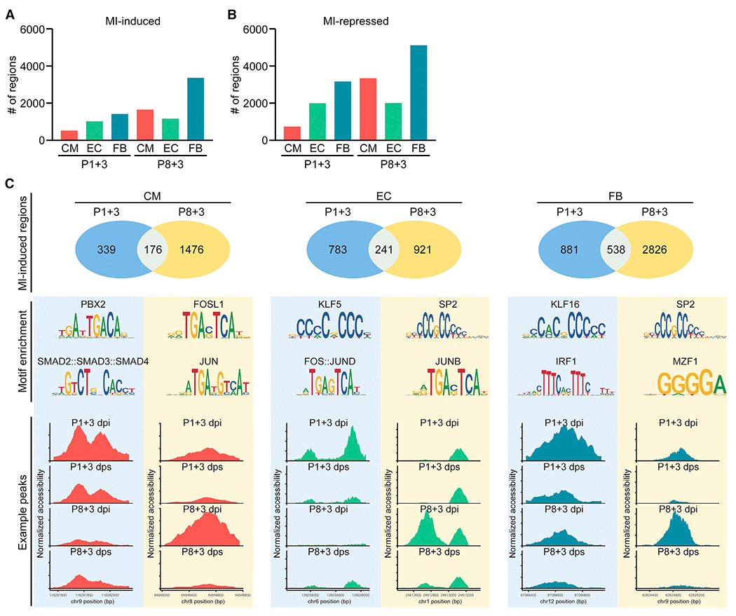
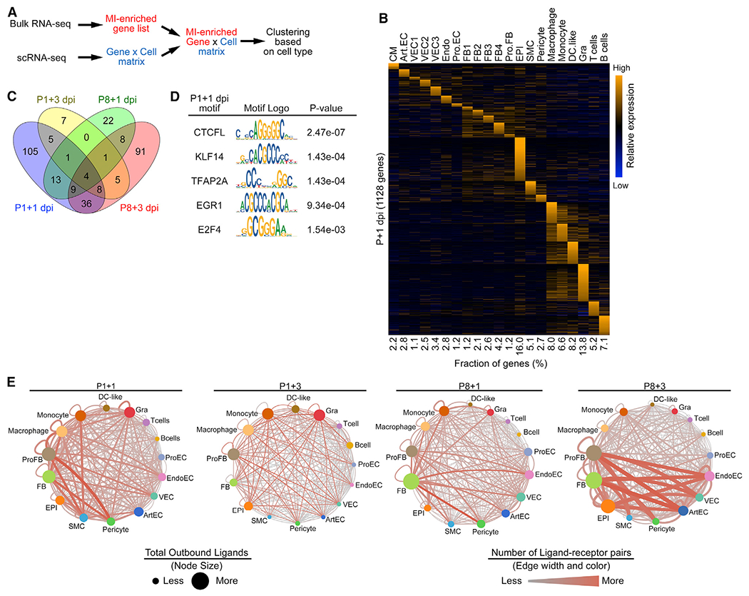
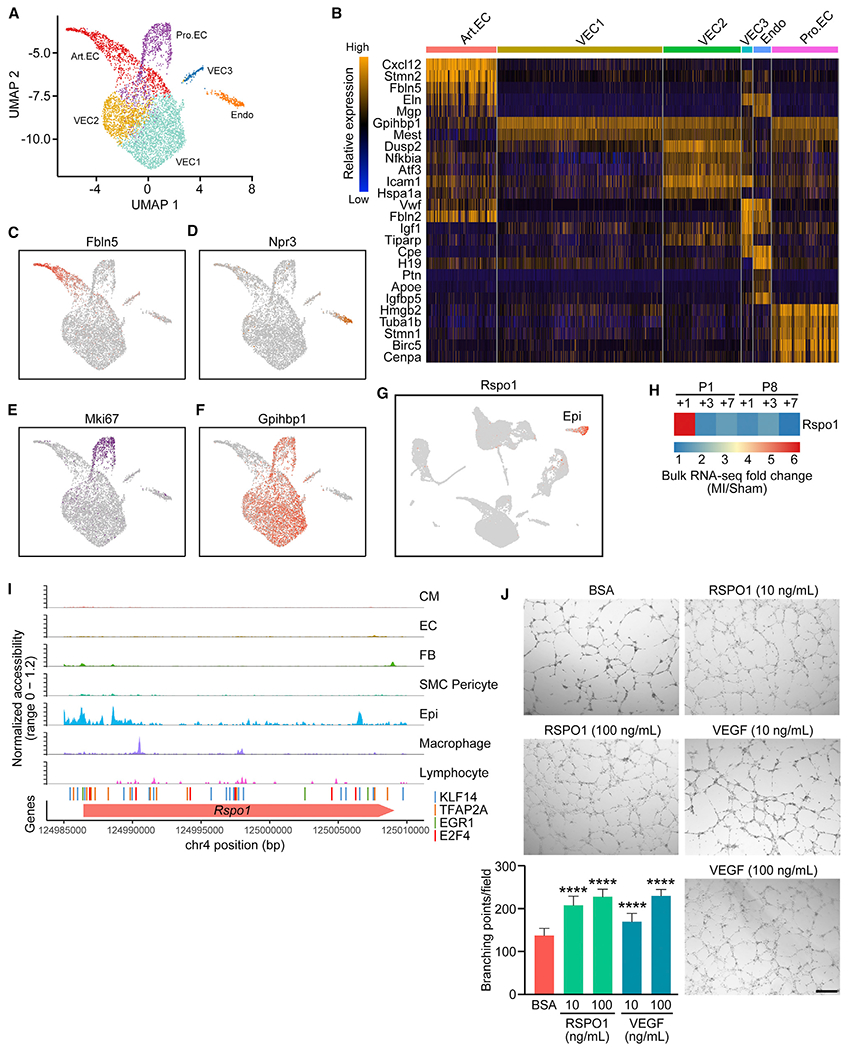
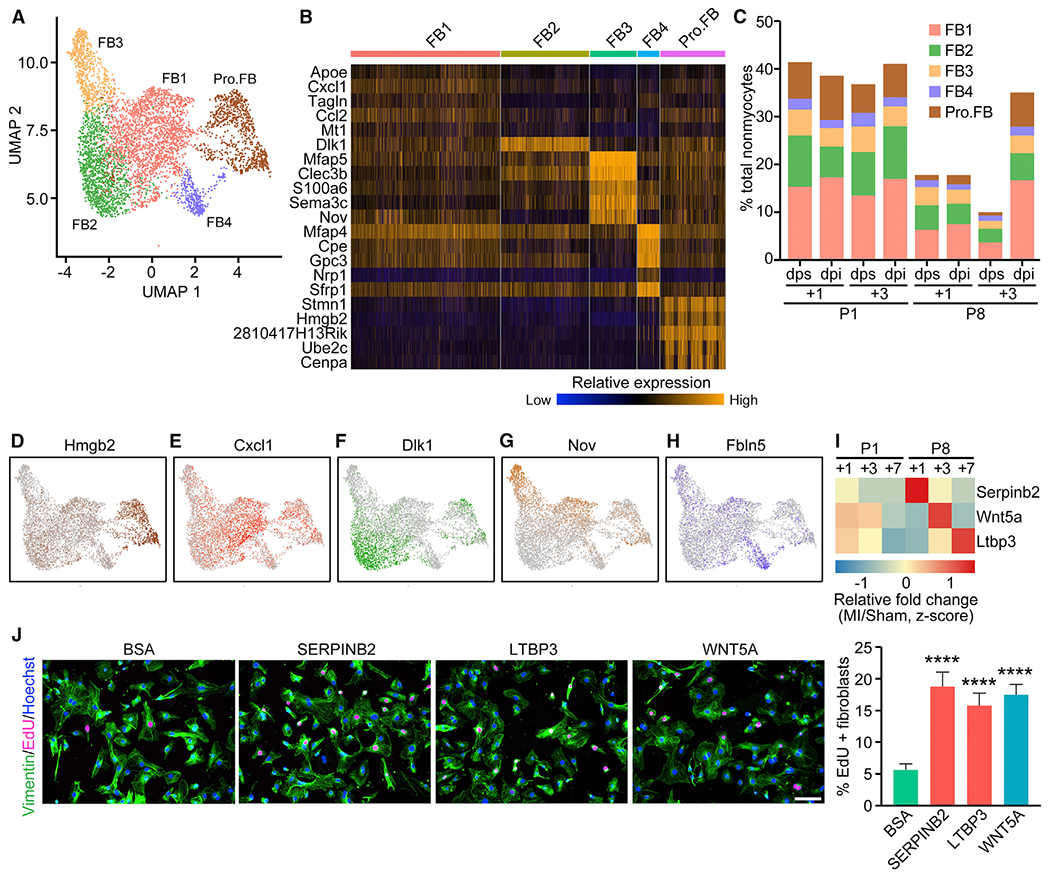
The adult mammalian heart has limited capacity for regeneration following injury, whereas the neonatal heart can readily regenerate within a short period after birth. Neonatal heart regeneration is orchestrated by multiple cell types intrinsic to the heart, as well as immune cells that infiltrate the heart after injury. To elucidate the transcriptional responses of the different cellular components of the mouse heart following injury, we perform single-cell RNA sequencing on neonatal hearts at various time points following myocardial infarction and couple the results with bulk tissue RNA-sequencing data collected at the same time points. Concomitant single-cell ATAC sequencing exposes underlying dynamics of open chromatin landscapes and regenerative gene regulatory networks of diverse cardiac cell types and reveals extracellular mediators of cardiomyocyte proliferation, angiogenesis, and fibroblast activation. Together, our data provide a transcriptional basis for neonatal heart regeneration at single-cell resolution and suggest strategies for enhancing cardiac function after injury.

摘要

成年哺乳动物的心脏在受伤后再生能力有限,而新生动物的心脏在出生后很短的时间内就能很容易地再生。新生儿心脏的再生是由心脏内的多种细胞类型以及受伤后浸润心脏的免疫细胞共同协调的。为了阐明损伤后小鼠心脏不同细胞成分的转录反应,我们对心肌梗死发生后不同时间点的新生鼠心脏进行单细胞 RNA 测序,并将结果与同时收集的大量组织 RNA 测序数据进行关联。伴随的单细胞 ATAC 测序揭示了不同心脏细胞类型的开放染色质景观和再生基因调控网络的潜在动态,并揭示了心肌细胞增殖、血管生成和成纤维细胞激活的细胞外介质。总之,我们的数据为单细胞分辨率下的新生儿心脏再生提供了转录基础,并为损伤后增强心脏功能提供了策略。

https://cdn.ncbi.nlm.nih.gov/pmc/blobs/3d6e/7774872/4319a38363b0/nihms-1653540-f0007.jpg
https://cdn.ncbi.nlm.nih.gov/pmc/blobs/3d6e/7774872/e95a07abd50b/nihms-1653540-f0001.jpg
https://cdn.ncbi.nlm.nih.gov/pmc/blobs/3d6e/7774872/ebe7ec36acd7/nihms-1653540-f0002.jpg
https://cdn.ncbi.nlm.nih.gov/pmc/blobs/3d6e/7774872/e83700b1ce94/nihms-1653540-f0003.jpg
https://cdn.ncbi.nlm.nih.gov/pmc/blobs/3d6e/7774872/ea701fe7425c/nihms-1653540-f0004.jpg
https://cdn.ncbi.nlm.nih.gov/pmc/blobs/3d6e/7774872/4257cf3d77e5/nihms-1653540-f0005.jpg
https://cdn.ncbi.nlm.nih.gov/pmc/blobs/3d6e/7774872/3990608296d0/nihms-1653540-f0006.jpg
https://cdn.ncbi.nlm.nih.gov/pmc/blobs/3d6e/7774872/4319a38363b0/nihms-1653540-f0007.jpg

相似文献

1
Cell-Type-Specific Gene Regulatory Networks Underlying Murine Neonatal Heart Regeneration at Single-Cell Resolution.单细胞分辨率下小鼠新生心脏再生的细胞类型特异性基因调控网络。
Cell Rep. 2020 Dec 8;33(10):108472. doi: 10.1016/j.celrep.2020.108472.
2
Multicellular Transcriptional Analysis of Mammalian Heart Regeneration.哺乳动物心脏再生的多细胞转录分析
Circulation. 2017 Sep 19;136(12):1123-1139. doi: 10.1161/CIRCULATIONAHA.117.028252. Epub 2017 Jul 21.
3
Mechanistic basis of neonatal heart regeneration revealed by transcriptome and histone modification profiling.转录组和组蛋白修饰谱分析揭示新生儿心脏再生的机制基础。
Proc Natl Acad Sci U S A. 2019 Sep 10;116(37):18455-18465. doi: 10.1073/pnas.1905824116. Epub 2019 Aug 26.
4
Regulatory T-cells regulate neonatal heart regeneration by potentiating cardiomyocyte proliferation in a paracrine manner.调节性 T 细胞通过旁分泌方式增强心肌细胞增殖来调节新生儿心脏再生。
Theranostics. 2019 Jun 9;9(15):4324-4341. doi: 10.7150/thno.32734. eCollection 2019.
5
Regenerative cross talk between cardiac cells and macrophages.心肌细胞与巨噬细胞间的再生对话。
Am J Physiol Heart Circ Physiol. 2021 Jun 1;320(6):H2211-H2221. doi: 10.1152/ajpheart.00056.2021. Epub 2021 Mar 26.
6
Dynamic Transcriptional Responses to Injury of Regenerative and Non-regenerative Cardiomyocytes Revealed by Single-Nucleus RNA Sequencing.单细胞 RNA 测序揭示再生和非再生心肌细胞损伤的动态转录反应。
Dev Cell. 2020 Apr 6;53(1):102-116.e8. doi: 10.1016/j.devcel.2020.02.019. Epub 2020 Mar 26.
7
Mydgf promotes Cardiomyocyte proliferation and Neonatal Heart regeneration.Mydgf 促进心肌细胞增殖和新生儿心脏再生。
Theranostics. 2020 Jul 11;10(20):9100-9112. doi: 10.7150/thno.44281. eCollection 2020.
8
RNA-Binding Protein LIN28a Regulates New Myocyte Formation in the Heart Through Long Noncoding RNA-H19.RNA 结合蛋白 LIN28a 通过长非编码 RNA-H19 调节心脏中的新心肌形成。
Circulation. 2023 Jan 24;147(4):324-337. doi: 10.1161/CIRCULATIONAHA.122.059346. Epub 2022 Oct 31.
9
Pressure Overload Greatly Promotes Neonatal Right Ventricular Cardiomyocyte Proliferation: A New Model for the Study of Heart Regeneration.压力超负荷极大地促进了新生儿右心室心肌细胞的增殖:心脏再生研究的新模型。
J Am Heart Assoc. 2020 Jun 2;9(11):e015574. doi: 10.1161/JAHA.119.015574. Epub 2020 May 30.
10
Regenerative potential of epicardium-derived extracellular vesicles mediated by conserved miRNA transfer.心肌外膜细胞外囊泡通过保守的 miRNA 转移介导的再生潜力。
Cardiovasc Res. 2022 Jan 29;118(2):597-611. doi: 10.1093/cvr/cvab054.

引用本文的文献

1
Transcriptional regulation in heart development, disease and regeneration: reassessing the fetal gene hypothesis.心脏发育、疾病与再生中的转录调控:重新审视胎儿基因假说。
Nat Rev Cardiol. 2025 Sep 5. doi: 10.1038/s41569-025-01205-3.
2
Generation of mature epicardium derived from human-induced pluripotent stem cells via inhibition of mTOR signaling.通过抑制mTOR信号通路从人诱导多能干细胞生成成熟心外膜。
Nat Commun. 2025 Jul 1;16(1):5902. doi: 10.1038/s41467-025-60934-8.
3
Sox9 in the epicardium: Implications for cell invasion, differentiation, and coronary vascular development.

本文引用的文献

1
An atlas of gene regulatory elements in adult mouse cerebrum.成年鼠大脑基因调控元件图谱
Nature. 2021 Oct;598(7879):129-136. doi: 10.1038/s41586-021-03604-1. Epub 2021 Oct 6.
2
Cells of the adult human heart.成人心脏细胞。
Nature. 2020 Dec;588(7838):466-472. doi: 10.1038/s41586-020-2797-4. Epub 2020 Sep 24.
3
Transcriptional and Cellular Diversity of the Human Heart.人类心脏的转录和细胞多样性。
心外膜中的Sox9:对细胞侵袭、分化及冠状动脉血管发育的影响
PLoS One. 2025 Jun 23;20(6):e0325852. doi: 10.1371/journal.pone.0325852. eCollection 2025.
4
An organ-wide spatiotemporal transcriptomic and cellular atlas of the regenerating zebrafish heart.再生斑马鱼心脏的全器官时空转录组学和细胞图谱
Nat Commun. 2025 Apr 19;16(1):3716. doi: 10.1038/s41467-025-59070-0.
5
Cardiac enhancers: Gateway to the regulatory mechanisms of heart regeneration.心脏增强子:通向心脏再生调控机制的大门。
Semin Cell Dev Biol. 2025 Jun;170:103610. doi: 10.1016/j.semcdb.2025.103610. Epub 2025 Apr 10.
6
Molecular gatekeepers of endogenous adult mammalian cardiomyocyte proliferation.成年哺乳动物内源性心肌细胞增殖的分子守门人。
Nat Rev Cardiol. 2025 Apr 7. doi: 10.1038/s41569-025-01145-y.
7
The paradigm shifts of periodontal regeneration strategy: From reparative manipulation to developmental engineering.牙周再生策略的范式转变:从修复性操作到发育工程。
Bioact Mater. 2025 Mar 18;49:418-436. doi: 10.1016/j.bioactmat.2025.03.009. eCollection 2025 Jul.
8
Uncovering Hippo pathway-related biomarkers in acute myocardial infarction via scRNA-seq binding transcriptomics.通过单细胞RNA测序结合转录组学揭示急性心肌梗死中与河马通路相关的生物标志物
Sci Rep. 2025 Mar 26;15(1):10368. doi: 10.1038/s41598-025-94820-6.
9
Dissecting endothelial cell heterogeneity with new tools.用新工具剖析内皮细胞异质性。
Cell Regen. 2025 Mar 23;14(1):10. doi: 10.1186/s13619-025-00223-3.
10
Early-age efferocytosis directs macrophage arachidonic acid metabolism for tissue regeneration.早期的胞葬作用引导巨噬细胞花生四烯酸代谢以促进组织再生。
Immunity. 2025 Feb 11;58(2):344-361.e7. doi: 10.1016/j.immuni.2024.11.018.
Circulation. 2020 Aug 4;142(5):466-482. doi: 10.1161/CIRCULATIONAHA.119.045401. Epub 2020 May 14.
4
Dynamic Transcriptional Responses to Injury of Regenerative and Non-regenerative Cardiomyocytes Revealed by Single-Nucleus RNA Sequencing.单细胞 RNA 测序揭示再生和非再生心肌细胞损伤的动态转录反应。
Dev Cell. 2020 Apr 6;53(1):102-116.e8. doi: 10.1016/j.devcel.2020.02.019. Epub 2020 Mar 26.
5
Dynamic Interstitial Cell Response during Myocardial Infarction Predicts Resilience to Rupture in Genetically Diverse Mice.心肌梗死后的动态间质细胞反应可预测遗传多样性小鼠对破裂的抵抗力。
Cell Rep. 2020 Mar 3;30(9):3149-3163.e6. doi: 10.1016/j.celrep.2020.02.008.
6
Phosphoproteomic Analysis of Neonatal Regenerative Myocardium Revealed Important Roles of Checkpoint Kinase 1 via Activating Mammalian Target of Rapamycin C1/Ribosomal Protein S6 Kinase b-1 Pathway.磷酸化蛋白质组学分析揭示了细胞周期检验点激酶 1 通过激活雷帕霉素靶蛋白 C1/核糖体蛋白 S6 激酶 b-1 通路在新生儿再生心肌中的重要作用。
Circulation. 2020 May 12;141(19):1554-1569. doi: 10.1161/CIRCULATIONAHA.119.040747. Epub 2020 Feb 26.
7
Single-Cell Transcriptome Atlas of Murine Endothelial Cells.单细胞转录组图谱:鼠类血管内皮细胞
Cell. 2020 Feb 20;180(4):764-779.e20. doi: 10.1016/j.cell.2020.01.015. Epub 2020 Feb 13.
8
The Role of the Epicardium During Heart Development and Repair.心外膜在心脏发育和修复中的作用。
Circ Res. 2020 Jan 31;126(3):377-394. doi: 10.1161/CIRCRESAHA.119.315857. Epub 2020 Jan 30.
9
Single-cell reconstruction of the adult human heart during heart failure and recovery reveals the cellular landscape underlying cardiac function.单细胞重构人类心力衰竭和恢复过程中的心脏,揭示了心脏功能的细胞基础。
Nat Cell Biol. 2020 Jan;22(1):108-119. doi: 10.1038/s41556-019-0446-7. Epub 2020 Jan 8.
10
A Spatiotemporal Organ-Wide Gene Expression and Cell Atlas of the Developing Human Heart.人类心脏发育的时空器官全基因表达和细胞图谱
Cell. 2019 Dec 12;179(7):1647-1660.e19. doi: 10.1016/j.cell.2019.11.025.