Department of Physiology, Anatomy and Genetics, University of Oxford, Sherrington Building, Parks Road, Oxford, OX1 3PT, UK.
Unit of Cardiac Physiology, Division of Cardiovascular Sciences, University of Manchester, Manchester, UK.
Basic Res Cardiol. 2022 Mar 31;117(1):17. doi: 10.1007/s00395-022-00924-9.
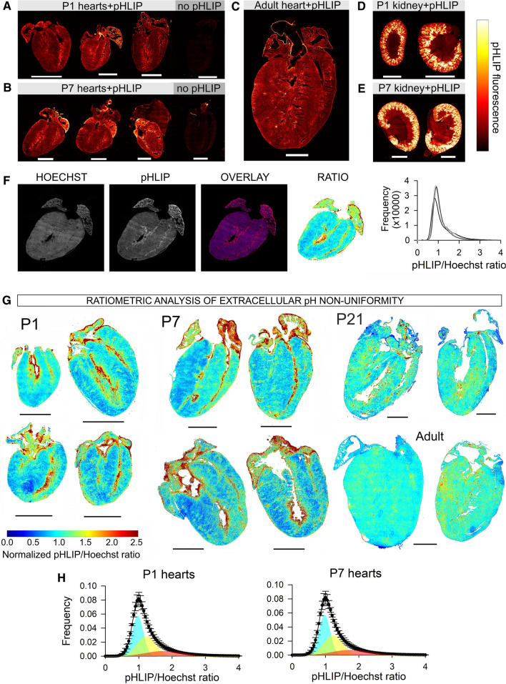
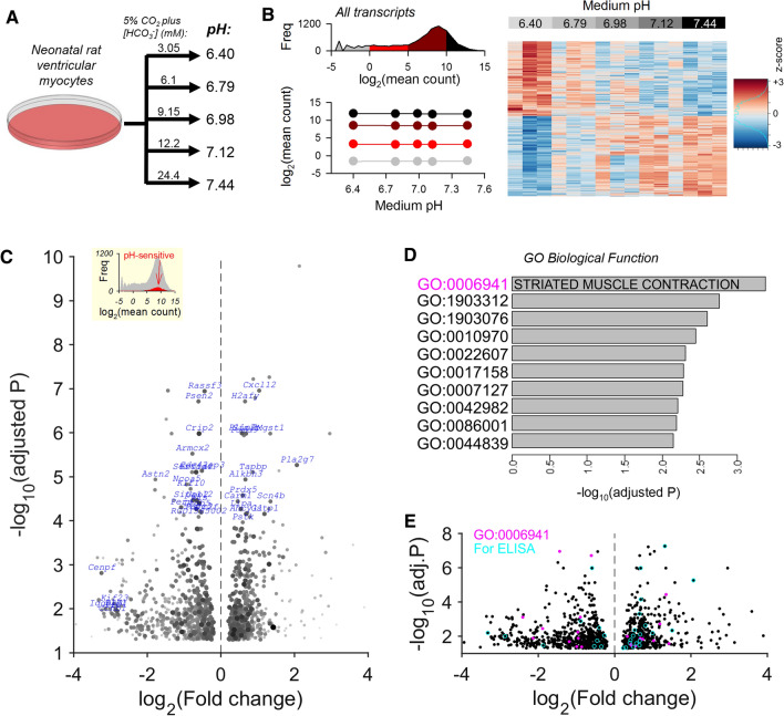
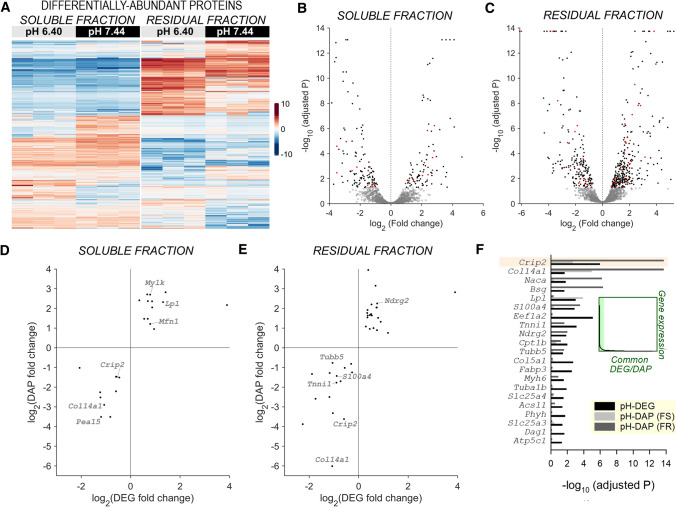
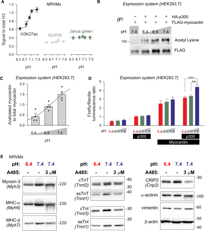
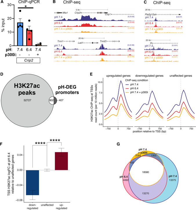
Cardiac contractile strength is recognised as being highly pH-sensitive, but less is known about the influence of pH on cardiac gene expression, which may become relevant in response to changes in myocardial metabolism or vascularization during development or disease. We sought evidence for pH-responsive cardiac genes, and a physiological context for this form of transcriptional regulation. pHLIP, a peptide-based reporter of acidity, revealed a non-uniform pH landscape in early-postnatal myocardium, dissipating in later life. pH-responsive differentially expressed genes (pH-DEGs) were identified by transcriptomics of neonatal cardiomyocytes cultured over a range of pH. Enrichment analysis indicated "striated muscle contraction" as a pH-responsive biological process. Label-free proteomics verified fifty-four pH-responsive gene-products, including contractile elements and the adaptor protein CRIP2. Using transcriptional assays, acidity was found to reduce p300/CBP acetylase activity and, its a functional readout, inhibit myocardin, a co-activator of cardiac gene expression. In cultured myocytes, acid-inhibition of p300/CBP reduced H3K27 acetylation, as demonstrated by chromatin immunoprecipitation. H3K27ac levels were more strongly reduced at promoters of acid-downregulated DEGs, implicating an epigenetic mechanism of pH-sensitive gene expression. By tandem cytoplasmic/nuclear pH imaging, the cardiac nucleus was found to exercise a degree of control over its pH through Na/H exchangers at the nuclear envelope. Thus, we describe how extracellular pH signals gain access to the nucleus and regulate the expression of a subset of cardiac genes, notably those coding for contractile proteins and CRIP2. Acting as a proxy of a well-perfused myocardium, alkaline conditions are permissive for expressing genes related to the contractile apparatus.
心肌收缩力被认为对 pH 值非常敏感,但对于 pH 值对心脏基因表达的影响知之甚少,而这种影响在发育或疾病过程中心肌代谢或血管生成发生变化时可能变得相关。我们试图寻找 pH 值反应性心脏基因的证据,并为这种转录调控形式提供生理背景。基于肽的酸度报告器 pHLIP 揭示了早期出生后心肌中的非均匀 pH 景观,在晚年消散。通过在一系列 pH 值下培养新生心肌细胞的转录组学鉴定 pH 反应性差异表达基因 (pH-DEGs)。富集分析表明“横纹肌收缩”是 pH 反应性的生物学过程。无标记蛋白质组学验证了五十四个 pH 反应性基因产物,包括收缩元件和衔接蛋白 CRIP2。使用转录测定法,发现酸度降低了 p300/CBP 乙酰转移酶的活性,其功能读数抑制了心肌基因表达的共激活因子 myocardin。在培养的心肌细胞中,如染色质免疫沉淀所示,酸抑制 p300/CBP 降低了 H3K27 乙酰化。在酸下调的 DEGs 的启动子处,H3K27ac 水平降低得更为强烈,表明 pH 值敏感基因表达存在表观遗传机制。通过串联细胞质/核 pH 成像,发现心脏核通过核膜上的 Na/H 交换器对其 pH 值行使一定程度的控制。因此,我们描述了细胞外 pH 值信号如何进入细胞核并调节一组心脏基因的表达,特别是那些编码收缩蛋白和 CRIP2 的基因。作为灌注良好的心肌的代理,碱性条件有利于表达与收缩装置相关的基因。