• 文献检索
  • 文档翻译
  • 深度研究
  • 学术资讯
  • Suppr Zotero 插件Zotero 插件
  • 邀请有礼
  • 套餐&价格
  • 历史记录
应用&插件
Suppr Zotero 插件Zotero 插件浏览器插件Mac 客户端Windows 客户端微信小程序
定价
高级版会员购买积分包购买API积分包
服务
文献检索文档翻译深度研究API 文档MCP 服务
关于我们
关于 Suppr公司介绍联系我们用户协议隐私条款
关注我们

Suppr 超能文献

核心技术专利:CN118964589B侵权必究
粤ICP备2023148730 号-1Suppr @ 2026

文献检索

告别复杂PubMed语法,用中文像聊天一样搜索,搜遍4000万医学文献。AI智能推荐,让科研检索更轻松。

立即免费搜索

文件翻译

保留排版,准确专业,支持PDF/Word/PPT等文件格式,支持 12+语言互译。

免费翻译文档

深度研究

AI帮你快速写综述,25分钟生成高质量综述,智能提取关键信息,辅助科研写作。

立即免费体验

自发荧光对基于荧光的研究小胶质细胞技术的影响。

Impacts of autofluorescence on fluorescence based techniques to study microglia.

机构信息

Department of Pharmacology, School of Basic Medical Sciences, Zhengzhou University, Zhengzhou, Henan, China.

Translational Medicine Research Center, People's Hospital Of Zhengzhou, Zhengzhou, Henan, China.

出版信息

BMC Neurosci. 2022 Mar 31;23(1):21. doi: 10.1186/s12868-022-00703-1.

DOI:10.1186/s12868-022-00703-1
PMID:35361108
原文链接:https://pmc.ncbi.nlm.nih.gov/articles/PMC8973892/
Abstract

BACKGROUND

Microglia, the resident immune cells in the central nervous system, accrue autofluorescent granules inside their cytoplasm throughout their lifespan. In this report, we studied the impacts of autofluorescence on widely used fluorescence-based techniques to study microglia, including flow cytometry, immunofluorescence staining, and live imaging.

RESULTS

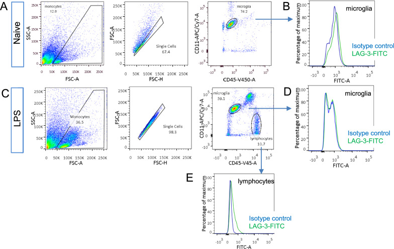
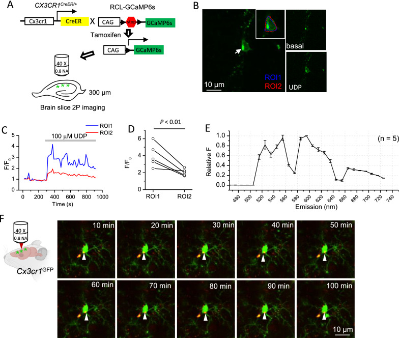
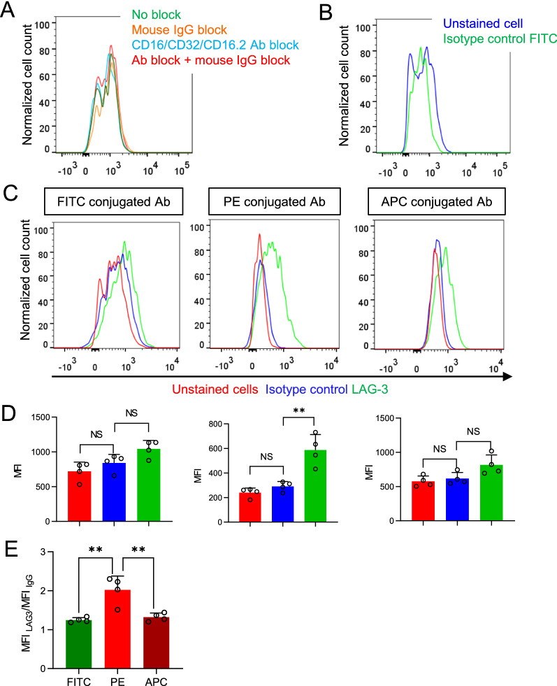
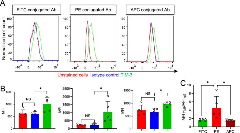
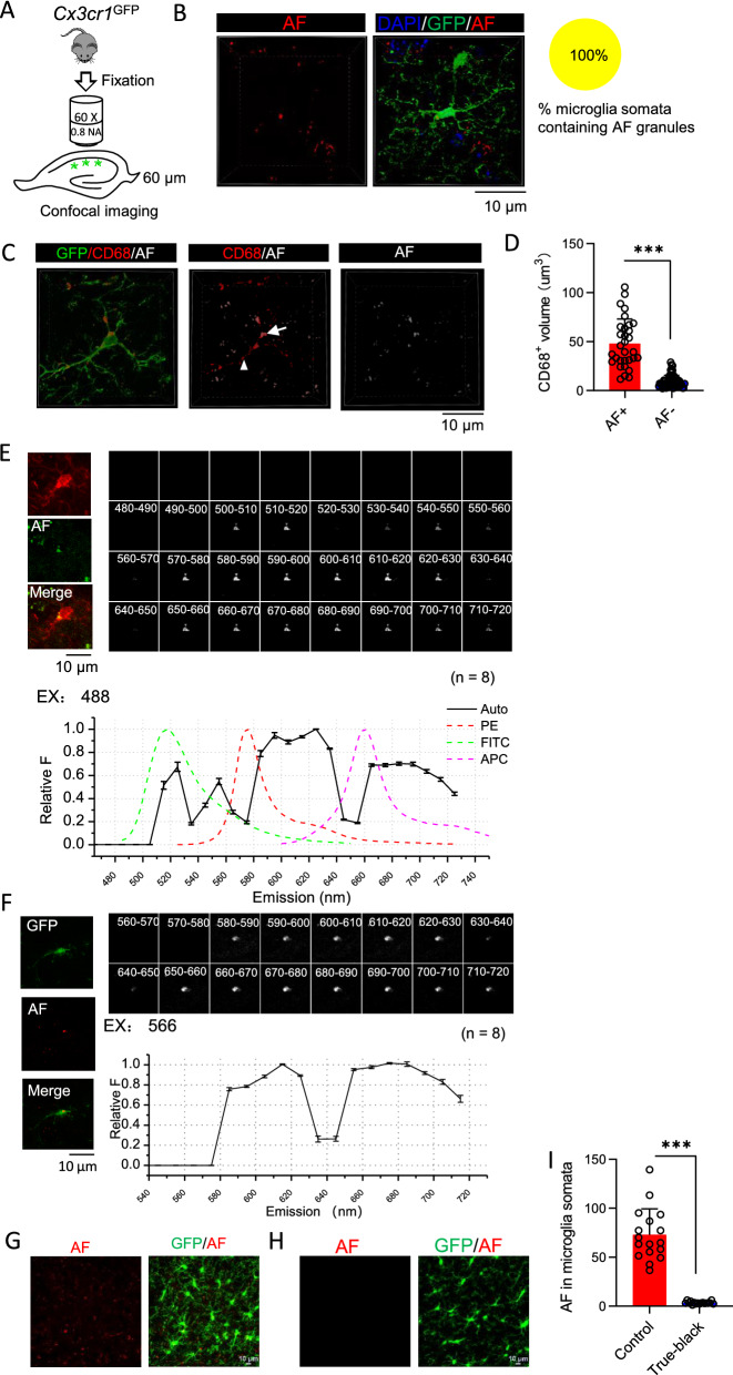
The failed attempt of using fluorescein isothiocyanate (FITC) conjugated antibody to detect lymphocyte-activation gene 3 protein in microglia prompted us to compare the sensitivity of FITC, phycoerythrin (PE) and allophycocyanin (APC) conjugated antibodies to detect surface protein expression in microglia. We found that PE outperformed FITC and APC as the fluorophore conjugated to antibody for flow cytometry by overcoming the interference from microglia autofluorescence. To identify the location and source of microglia autofluorescence, we did confocal imaging and spectral analysis of microglia autofluorescence on fixed brain tissues, revealing that microglia autofluorescence emitted from cytoplasmic granules and displayed a multi-peak emission spectrum. We recommended removing autofluorescence by lipofuscin removing agents when staining intracellular proteins in microglia with the immunofluorescence techniques. On live brain slices, autofluorescent granules reduced the amplitudes of calcium signals in microglial somata derived from GCaMP6s fluorescence and thus needed to be excluded when selecting regions of interest (ROI).

CONCLUSIONS

In conclusion, autofluorescence is a critical factor to consider when designing experiments and interpreting results based on fluorescence-based techniques to study microglia.

摘要

背景

小胶质细胞是中枢神经系统中的固有免疫细胞,在其整个生命周期中,其细胞质内会积累自发荧光颗粒。在本报告中,我们研究了自发荧光对广泛用于研究小胶质细胞的荧光基技术的影响,包括流式细胞术、免疫荧光染色和活体成像。

结果

使用异硫氰酸荧光素(FITC)缀合抗体检测小胶质细胞中的淋巴细胞激活基因 3 蛋白的尝试失败促使我们比较了 FITC、藻红蛋白(PE)和别藻青蛋白(APC)缀合抗体检测小胶质细胞表面蛋白表达的灵敏度。我们发现,PE 作为抗体缀合的荧光团优于 FITC 和 APC,可用于流式细胞术,克服了小胶质细胞自发荧光的干扰。为了确定小胶质细胞自发荧光的位置和来源,我们对固定脑组织中的小胶质细胞自发荧光进行了共聚焦成像和光谱分析,结果表明小胶质细胞自发荧光来自细胞质颗粒,并显示出多峰发射光谱。我们建议在用免疫荧光技术染色小胶质细胞内蛋白时,使用脂褐素去除剂去除自发荧光。在活体脑切片上,自发荧光颗粒降低了源自 GCaMP6s 荧光的小胶质细胞体钙信号的幅度,因此在选择感兴趣区域(ROI)时需要将其排除。

结论

总之,在设计实验和基于荧光基技术解释小胶质细胞研究结果时,自发荧光是一个需要考虑的关键因素。

https://cdn.ncbi.nlm.nih.gov/pmc/blobs/1edc/8973892/666e255f17d1/12868_2022_703_Fig5_HTML.jpg
https://cdn.ncbi.nlm.nih.gov/pmc/blobs/1edc/8973892/52ffe7f49b3d/12868_2022_703_Fig1_HTML.jpg
https://cdn.ncbi.nlm.nih.gov/pmc/blobs/1edc/8973892/d0f839f0f549/12868_2022_703_Fig2_HTML.jpg
https://cdn.ncbi.nlm.nih.gov/pmc/blobs/1edc/8973892/ac9b5fc46d61/12868_2022_703_Fig3_HTML.jpg
https://cdn.ncbi.nlm.nih.gov/pmc/blobs/1edc/8973892/9ee47acdcd64/12868_2022_703_Fig4_HTML.jpg
https://cdn.ncbi.nlm.nih.gov/pmc/blobs/1edc/8973892/666e255f17d1/12868_2022_703_Fig5_HTML.jpg

相似文献

1
Impacts of autofluorescence on fluorescence based techniques to study microglia.自发荧光对基于荧光的研究小胶质细胞技术的影响。
BMC Neurosci. 2022 Mar 31;23(1):21. doi: 10.1186/s12868-022-00703-1.
2
Enhanced immunofluorescence measurement resolution of surface antigens on highly autofluorescent, glutaraldehyde-fixed cells analyzed by phase-sensitive flow cytometry.通过相敏流式细胞术分析戊二醛固定的高自发荧光细胞表面抗原时增强免疫荧光测量分辨率。
Cytometry. 1999 Dec 1;37(4):275-83. doi: 10.1002/(sici)1097-0320(19991201)37:4<275::aid-cyto4>3.0.co;2-u.
3
Flow cytometry analysis using the Becton Dickinson FACS Calibur.使用贝克曼库尔特FACS Calibur进行流式细胞术分析。
Curr Protoc Immunol. 2002 Aug;Chapter 5:Unit 5.4. doi: 10.1002/0471142735.im0504s49.
4
Phycoerythrin-allophycocyanin: a resonance energy transfer fluorochrome for immunofluorescence.藻红蛋白-别藻蓝蛋白:一种用于免疫荧光的共振能量转移荧光染料。
Cytometry. 2001 May 1;44(1):24-9.
5
Experimental and model investigations of bleaching and saturation of fluorescence in flow cytometry.流式细胞术中荧光漂白与饱和的实验及模型研究。
Cytometry. 1997 Nov 1;29(3):204-14. doi: 10.1002/(sici)1097-0320(19971101)29:3<204::aid-cyto3>3.0.co;2-b.
6
Single laser three color immunofluorescence staining procedures based on energy transfer between phycoerythrin and cyanine 5.基于藻红蛋白与花青素5之间能量转移的单激光三色免疫荧光染色程序
Cytometry. 1991;12(8):723-30. doi: 10.1002/cyto.990120806.
7
Measurement of lymphocyte subset proliferation by three-color immunofluorescence and DNA flow cytometry.通过三色免疫荧光和DNA流式细胞术测量淋巴细胞亚群增殖。
J Immunol Methods. 2000 Feb 21;235(1-2):121-31. doi: 10.1016/s0022-1759(99)00225-2.
8
Cy7PE and Cy7APC: bright new probes for immunofluorescence.Cy7PE和Cy7APC:用于免疫荧光的新型明亮探针。
Cytometry. 1996 Jul 1;24(3):191-7. doi: 10.1002/(SICI)1097-0320(19960701)24:3<191::AID-CYTO1>3.0.CO;2-L.
9
Detection of triple antibody-binding lymphocytes in standard single laser flow cytometry using colloidal gold, fluorescein and phycoerythrin as labels.使用胶体金、荧光素和藻红蛋白作为标记物,在标准单激光流式细胞术中检测三抗体结合淋巴细胞。
J Immunol Methods. 1987 Jul 16;101(1):23-8. doi: 10.1016/0022-1759(87)90211-0.
10
Application of a flow cytometric method using autofluorescence and a tandem fluorescent dye to analyze human alveolar macrophage surface markers.应用一种利用自发荧光和串联荧光染料的流式细胞术方法来分析人肺泡巨噬细胞表面标志物。
J Immunol Methods. 1994 Jun 3;172(1):17-24. doi: 10.1016/0022-1759(94)90374-3.

引用本文的文献

1
Feasibility of Nanostructured Lipid Carrier Loaded with Alpha-Mangostin and Clove Oil for Canine Periodontal Therapy.负载α-山竹黄酮和丁香油的纳米结构脂质载体用于犬牙周治疗的可行性
Animals (Basel). 2024 Jul 17;14(14):2084. doi: 10.3390/ani14142084.
2
Multiparameter quantitative analyses of diagnostic cells in brain tissues from tuberous sclerosis complex.结节性硬化症脑组织中诊断性细胞的多参数定量分析。
Cytometry B Clin Cytom. 2025 Jan;108(1):35-54. doi: 10.1002/cyto.b.22194. Epub 2024 Jul 2.
3
Synthesis and Characterization of Click Chemical Probes for Single-Cell Resolution Detection of Epichaperomes in Neurodegenerative Disorders.

本文引用的文献

1
Microglial calcium signaling is attuned to neuronal activity in awake mice.小胶质细胞钙信号在清醒小鼠中与神经元活动相协调。
Elife. 2020 Jul 27;9:e56502. doi: 10.7554/eLife.56502.
2
Astrocytes and microglia play orchestrated roles and respect phagocytic territories during neuronal corpse removal in vivo.星形胶质细胞和小胶质细胞在体内神经元尸骸清除过程中发挥协调作用,并尊重吞噬作用领域。
Sci Adv. 2020 Jun 26;6(26):eaba3239. doi: 10.1126/sciadv.aba3239. eCollection 2020 Jun.
3
Differential accumulation of storage bodies with aging defines discrete subsets of microglia in the healthy brain.
用于神经退行性疾病中表观伴侣蛋白单细胞分辨率检测的点击化学探针的合成与表征
Biomedicines. 2024 Jun 4;12(6):1252. doi: 10.3390/biomedicines12061252.
4
Lipofuscin-like autofluorescence within microglia and its impact on studying microglial engulfment.内体样脂褐素样自发荧光在小胶质细胞内的存在及其对研究小胶质细胞吞噬作用的影响。
Nat Commun. 2023 Nov 3;14(1):7060. doi: 10.1038/s41467-023-42809-y.
5
Volumetric live-cell autofluorescence imaging using Fourier light-field microscopy.使用傅里叶光场显微镜进行体积活细胞自发荧光成像。
Biomed Opt Express. 2023 Jul 25;14(8):4237-4245. doi: 10.1364/BOE.495506. eCollection 2023 Aug 1.
6
Comparing Performance of Spectral Image Analysis Approaches for Detection of Cellular Signals in Time-Lapse Hyperspectral Imaging Fluorescence Excitation-Scanning Microscopy.比较光谱图像分析方法在延时高光谱成像荧光激发扫描显微镜中检测细胞信号的性能。
Bioengineering (Basel). 2023 May 25;10(6):642. doi: 10.3390/bioengineering10060642.
7
Label-free macrophage phenotype classification using machine learning methods.基于机器学习方法的无标记巨噬细胞表型分类。
Sci Rep. 2023 Mar 30;13(1):5202. doi: 10.1038/s41598-023-32158-7.
随着年龄的增长,储存体的差异积累定义了健康大脑中小胶质细胞的离散亚群。
Elife. 2020 Jun 24;9:e57495. doi: 10.7554/eLife.57495.
4
Microglia mediate forgetting via complement-dependent synaptic elimination.小胶质细胞通过补体依赖性突触消除来介导遗忘。
Science. 2020 Feb 7;367(6478):688-694. doi: 10.1126/science.aaz2288.
5
Targeted Complement Inhibition at Synapses Prevents Microglial Synaptic Engulfment and Synapse Loss in Demyelinating Disease.靶向突触补体抑制可预防脱髓鞘疾病中的小胶质细胞突触吞噬和突触丢失。
Immunity. 2020 Jan 14;52(1):167-182.e7. doi: 10.1016/j.immuni.2019.12.004. Epub 2019 Dec 26.
6
Complement C3aR Inactivation Attenuates Tau Pathology and Reverses an Immune Network Deregulated in Tauopathy Models and Alzheimer's Disease.补体 C3aR 失活可减轻 Tau 病理学并逆转 Tau 病模型和阿尔茨海默病中失调的免疫网络。
Neuron. 2018 Dec 19;100(6):1337-1353.e5. doi: 10.1016/j.neuron.2018.10.031. Epub 2018 Nov 8.
7
Changes in the Synaptic Proteome in Tauopathy and Rescue of Tau-Induced Synapse Loss by C1q Antibodies.tau 病中的突触蛋白组变化及 C1q 抗体对 tau 诱导的突触丢失的挽救作用。
Neuron. 2018 Dec 19;100(6):1322-1336.e7. doi: 10.1016/j.neuron.2018.10.014. Epub 2018 Nov 1.
8
Mitochondrial contribution to lipofuscin formation.线粒体在脂褐素形成中的作用。
Redox Biol. 2017 Apr;11:673-681. doi: 10.1016/j.redox.2017.01.017. Epub 2017 Jan 25.
9
Neurotoxic reactive astrocytes are induced by activated microglia.神经毒性反应性星形胶质细胞由活化的小胶质细胞诱导产生。
Nature. 2017 Jan 26;541(7638):481-487. doi: 10.1038/nature21029. Epub 2017 Jan 18.
10
P2X4 Receptor Reporter Mice: Sparse Brain Expression and Feeding-Related Presynaptic Facilitation in the Arcuate Nucleus.P2X4受体报告基因小鼠:大脑中的稀疏表达以及弓状核中与进食相关的突触前易化
J Neurosci. 2016 Aug 24;36(34):8902-20. doi: 10.1523/JNEUROSCI.1496-16.2016.