Lukashenko Mariia V, Gavrilova Natalia Y, Bregovskaya Anna V, Soprun Lidiia A, Churilov Leonid P, Petropoulos Ioannis N, Malik Rayaz A, Shoenfeld Yehuda
Laboratory Mosaic Autoimmunity, St. Petersburg State University, 199304 St. Petersburg, Russia.
Department of Phthisiopulmonology, St. Petersburg Scientific Research Institute of Phthisiopulmonology, 199304 St. Petersburg, Russia.
Pathophysiology. 2021 Dec 26;29(1):1-8. doi: 10.3390/pathophysiology29010001.
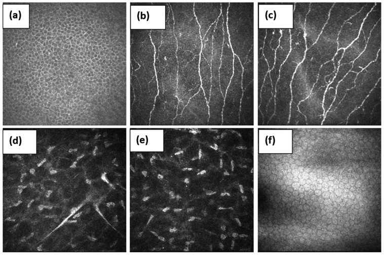
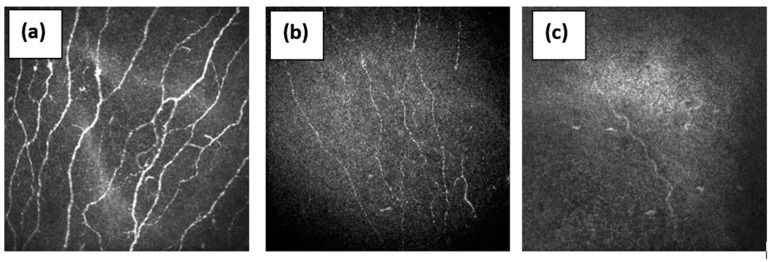
Chronic pain may affect 30-50% of the world's population and an important cause is small fiber neuropathy (SFN). Recent research suggests that autoimmune diseases may be one of the most common causes of small nerve fiber damage. There is low awareness of SFN among patients and clinicians and it is difficult to diagnose as routine electrophysiological methods only detect large fiber abnormalities, and specialized small fiber tests, like skin biopsy and quantitative sensory testing, are not routinely available. Corneal confocal microscopy (CCM) is a rapid, non-invasive, reproducible method for quantifying small nerve fiber degeneration and regeneration, and could be an important tool for diagnosing SFN. This review considers the advantages and disadvantages of CCM and highlights the evolution of this technique from a research tool to a diagnostic test for small fiber damage, which can be a valuable contribution to the study and management of autoimmune disease.
慢性疼痛可能影响全球30%至50%的人口,一个重要原因是小纤维神经病变(SFN)。最近的研究表明,自身免疫性疾病可能是小神经纤维损伤最常见的原因之一。患者和临床医生对SFN的认识较低,且难以诊断,因为常规电生理方法只能检测大纤维异常,而专门的小纤维检测,如皮肤活检和定量感觉测试,并非常规可用。角膜共焦显微镜检查(CCM)是一种快速、无创、可重复的定量小神经纤维变性和再生的方法,可能成为诊断SFN的重要工具。本文综述了CCM的优缺点,并强调了该技术从研究工具到小纤维损伤诊断测试的演变,这可能对自身免疫性疾病的研究和管理做出有价值的贡献。