Ferdousi Maryam, Kalteniece Alise, Azmi Shazli, Petropoulos Ioannis N, Ponirakis Georgios, Alam Uazman, Asghar Omar, Marshall Andrew, Fullwood Catherine, Jeziorska Maria, Abbott Caroline, Lauria Giuseppe, Faber Catharina G, Soran Handrean, Efron Nathan, Boulton Andrew J M, Malik Rayaz A
Institute of Cardiovascular Sciences, Cardiac Centre, Faculty of Medical and Human Sciences, The University of Manchester and NIHR/Wellcome Trust Clinical Research Facility, Manchester, U.K.
Research Division, Weill Cornell Medicine-Qatar, Qatar Foundation, Education City, Doha, Qatar.
Diabetes Care. 2021 Jan;44(1):150-156. doi: 10.2337/dc20-1482. Epub 2020 Nov 3.
To assess the diagnostic utility of corneal confocal microscopy (CCM) for diabetic peripheral neuropathy (DPN) and the risk factors for corneal nerve loss.
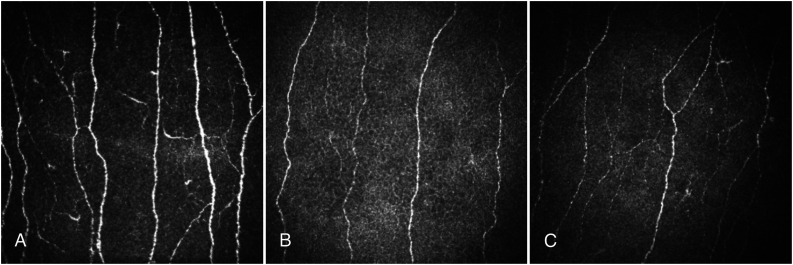
A total of 490 participants, including 72 healthy control subjects, 149 with type 1 diabetes, and 269 with type 2 diabetes, underwent detailed assessment of peripheral neuropathy and CCM in relation to risk factors.
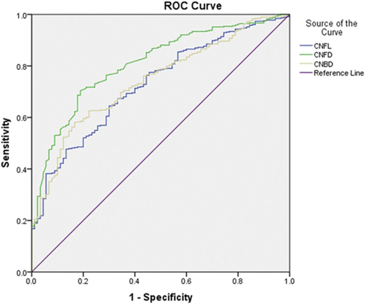
Corneal nerve fiber density (CNFD) ( < 0.0001 and < 0.0001), corneal nerve fiber branch density (CNBD) ( < 0.0001 and < 0.0001), and corneal nerve fiber length (CNFL) ( < 0.0001 and = 0.02) were significantly lower in patients with type 1 and type 2 diabetes compared with control subjects. CNFD ( < 0.0001), CNBD ( < 0.0001), and CNFL ( < 0.0001) were lower in type 1 diabetes compared with type 2 diabetes. Receiver operating characteristic curve analysis for the diagnosis of DPN demonstrated a good area under the curve for CNFD of 0.81, CNBD of 0.74, and CNFL of 0.73. Multivariable regression analysis showed a significant association among reduced CNFL with age (β = -0.27, = 0.007), HbA (β = -1.1; = 0.01), and weight (β = -0.14; = 0.03) in patients with type 2 diabetes and with duration of diabetes (β = -0.13; = 0.02), LDL cholesterol (β = 1.8, = 0.04), and triglycerides (β = -2.87; = 0.009) in patients with type 1 diabetes.
CCM identifies more severe corneal nerve loss in patients with type 1 diabetes compared with type 2 diabetes and shows good diagnostic accuracy for DPN. Furthermore, the risk factors for a reduction in corneal nerve fiber length differ between type 1 and type 2 diabetes.
评估角膜共焦显微镜(CCM)对糖尿病周围神经病变(DPN)的诊断效用以及角膜神经损伤的危险因素。
共有490名参与者,包括72名健康对照者、149名1型糖尿病患者和269名2型糖尿病患者,接受了与危险因素相关的周围神经病变和CCM的详细评估。
与对照者相比,1型和2型糖尿病患者的角膜神经纤维密度(CNFD)(<0.0001和<0.0001)、角膜神经纤维分支密度(CNBD)(<0.0001和<0.0001)以及角膜神经纤维长度(CNFL)(<0.0001和=0.02)显著降低。与2型糖尿病相比,1型糖尿病患者的CNFD(<0.0001)、CNBD(<0.0001)和CNFL(<0.0001)更低。用于诊断DPN的受试者工作特征曲线分析显示,CNFD的曲线下面积为0.81,CNBD为0.74,CNFL为0.73,具有良好的诊断准确性。多变量回归分析显示,在2型糖尿病患者中,CNFL降低与年龄(β=-0.27,=0.007)、糖化血红蛋白(β=-1.1;=0.01)和体重(β=-0.14;=0.03)显著相关;在1型糖尿病患者中,CNFL降低与糖尿病病程(β=-0.13;=0.02)、低密度脂蛋白胆固醇(β=1.8,=0.04)和甘油三酯(β=-2.87;=0.009)显著相关。
与2型糖尿病患者相比,CCM显示1型糖尿病患者的角膜神经损伤更严重,并且对DPN具有良好的诊断准确性。此外,1型和2型糖尿病患者角膜神经纤维长度降低的危险因素有所不同。