Replicor Inc.MontrealQuebecCanada.
Abbott DiagnosticsAbbott ParkIllinoisUSA.
Hepatol Commun. 2022 Aug;6(8):1870-1880. doi: 10.1002/hep4.1951. Epub 2022 Apr 2.
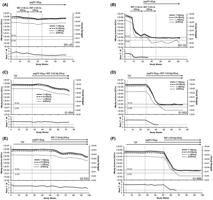
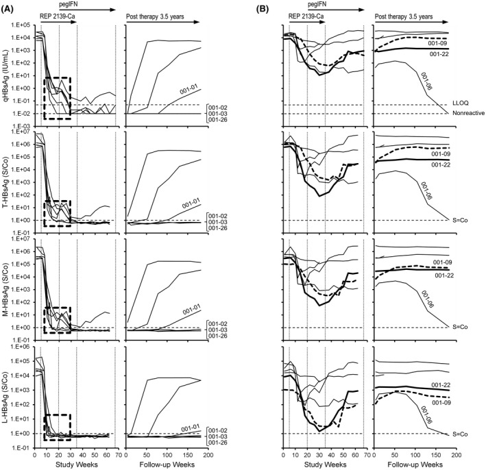
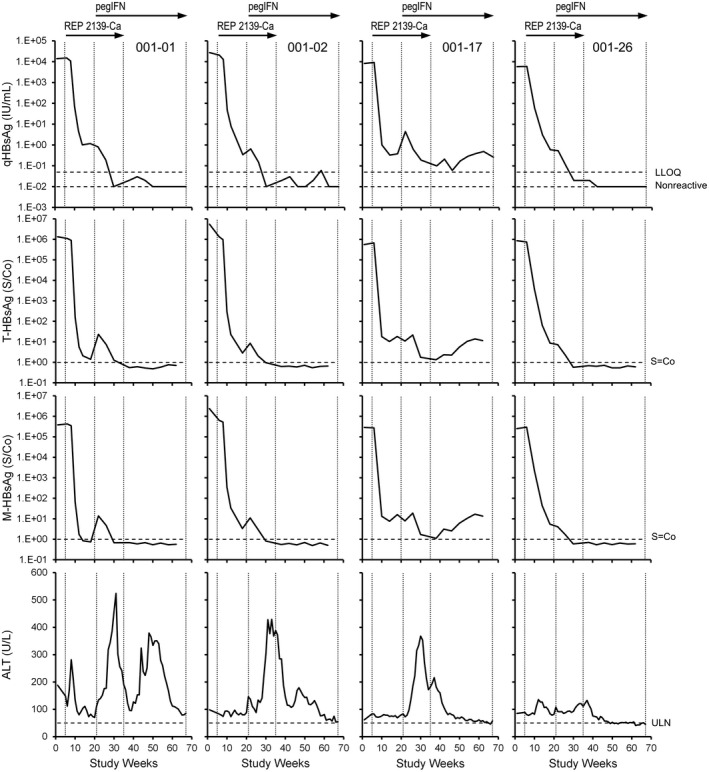
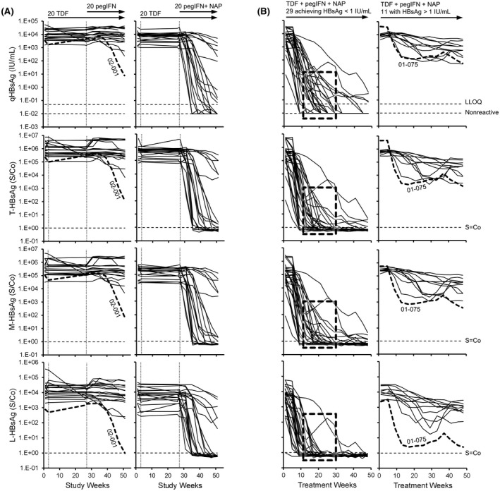
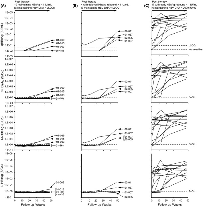
Nucleic acid polymers block the assembly of hepatitis B virus (HBV) subviral particles, effectively preventing hepatitis B surface antigen (HBsAg) replenishment in the circulation. Nucleic acid polymer (NAP)-based combination therapy of HBV infection or HBV/hepatitis D virus (HDV) co-infection is accompanied by HBsAg clearance and seroconversion, HDV-RNA clearance in co-infection, and persistent functional cure of HBV (HBsAg < 0.05 IU/ml, HBV-DNA target not dected, normal alanine aminotransferase) and persistent clearance of HDV RNA. An analysis of HBsAg isoform changes during quantitative HBsAg declines (qHBsAg), and subsequent treatment-free follow-up in the REP 301/REP 301-LTF (HBV/HDV) and REP 401 (HBV) studies was conducted. HBsAg isoforms were analyzed from frozen serum samples using Abbott Research Use Only assays for HBsAg isoforms (large [L], medium [M], and total [T]). The relative change over time in small HBsAg relative to the other isoforms was inferred by the change in the ratio over time of T-HBsAg to M-HBsAg. HBsAg isoform declines followed qHBsAg declines in all participants. No HBsAg isoforms were detectable in any participants with functional cure. HBsAg declines > 2 log IU/ml from baseline were correlated with selective clearance of S-HBsAg in 39 of 42 participants. Selective S-HBsAg decline was absent in 9 of 10 participants with HBsAg decline < 2 log IU/ml from baseline. Mild qHBsAg rebound during follow-up <10 IU/ml consisted mostly of S-HBsAg and M-HBsAg and not accompanied by significant covalently closed circular DNA activity. Conclusion: The faster observed declines in S-HBsAg indicate the selective clearance of subviral particles from the circulation, consistent with previous mechanistic studies on NAPs. Trace HBsAg rebound in the absence of HBV DNA may reflect HBsAg derived from integrated HBV DNA and not rebound of viral infection.
核酸聚合物阻止乙型肝炎病毒 (HBV) 亚病毒颗粒的组装,有效防止乙型肝炎表面抗原 (HBsAg) 在循环中补充。基于核酸聚合物 (NAP) 的 HBV 感染或 HBV/丁型肝炎病毒 (HDV) 合并感染的联合治疗伴有 HBsAg 清除和血清转换、合并感染中 HDV-RNA 清除以及 HBV 的持续功能性治愈 (HBsAg <0.05IU/ml,HBV-DNA 检测不到,丙氨酸氨基转移酶正常) 和 HDV RNA 的持续清除。对定量 HBsAg 下降 (qHBsAg) 期间 HBsAg 表型变化的分析,以及随后在 REP 301/REP 301-LTF (HBV/HDV) 和 REP 401 (HBV) 研究中的无治疗随访。使用 Abbott 研究专用 HBsAg 表型分析试剂盒 (用于 HBsAg 表型的大 [L]、中 [M] 和总 [T]) 分析冷冻血清样本中的 HBsAg 表型。通过 T-HBsAg 与 M-HBsAg 的比值随时间的变化推断小 HBsAg 与其他表型相比随时间的相对变化。所有参与者的 HBsAg 表型下降均跟随 qHBsAg 下降。所有功能性治愈的参与者均未检测到任何 HBsAg 表型。与基线相比,HBsAg 下降>2logIU/ml 与 42 名参与者中的 39 名 S-HBsAg 的选择性清除相关。与基线相比,HBsAg 下降<2logIU/ml 的 10 名参与者中,不存在 S-HBsAg 的选择性下降。随访期间轻度 qHBsAg 反弹<10IU/ml 主要由 S-HBsAg 和 M-HBsAg 组成,不伴有显著共价闭合环状 DNA 活性。结论:观察到 S-HBsAg 更快下降表明亚病毒颗粒从循环中选择性清除,与之前关于 NAP 的机制研究一致。在没有 HBV DNA 的情况下,HBsAg 的微量反弹可能反映了源自整合 HBV DNA 的 HBsAg,而不是病毒感染的反弹。