• 文献检索
  • 文档翻译
  • 深度研究
  • 学术资讯
  • Suppr Zotero 插件Zotero 插件
  • 邀请有礼
  • 套餐&价格
  • 历史记录
应用&插件
Suppr Zotero 插件Zotero 插件浏览器插件Mac 客户端Windows 客户端微信小程序
定价
高级版会员购买积分包购买API积分包
服务
文献检索文档翻译深度研究API 文档MCP 服务
关于我们
关于 Suppr公司介绍联系我们用户协议隐私条款
关注我们

Suppr 超能文献

核心技术专利:CN118964589B侵权必究
粤ICP备2023148730 号-1Suppr @ 2026

文献检索

告别复杂PubMed语法,用中文像聊天一样搜索,搜遍4000万医学文献。AI智能推荐,让科研检索更轻松。

立即免费搜索

文件翻译

保留排版,准确专业,支持PDF/Word/PPT等文件格式,支持 12+语言互译。

免费翻译文档

深度研究

AI帮你快速写综述,25分钟生成高质量综述,智能提取关键信息,辅助科研写作。

立即免费体验

基于深度学习的 X 射线预测肺纤维化。

Prediction of Pulmonary Fibrosis Based on X-Rays by Deep Neural Network.

机构信息

Dalian University of Technology, Dalian 116024, China.

The First Affiliated Hospital of Dalian Medical University, Dalian 116011, China.

出版信息

J Healthc Eng. 2022 Mar 26;2022:3845008. doi: 10.1155/2022/3845008. eCollection 2022.

DOI:10.1155/2022/3845008
PMID:35378944
原文链接:https://pmc.ncbi.nlm.nih.gov/articles/PMC8976624/
Abstract

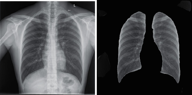
As a fatal lung disease, pulmonary fibrosis can cause irreversible damage to the lung, affect normal lung function, and eventually lead to death. At present, the pathogenesis of this kind of disease is not completely clear, and there is no radical cure. The main purpose of the treatment of this disease is to slow down the deterioration of pulmonary fibrosis. For this kind of disease, if it can be found early, it can be treated as soon as possible and the life of patients will be prolonged. Clinically, the diagnosis of pulmonary fibrosis depends on the relevant imaging examination, lung biopsy, lung function examination, and so on. Imaging data such as X-rays is a common examination means in clinical medicine and also plays an important role in the prediction of pulmonary fibrosis. Through X-ray, radiologists can clearly see the relevant lung lesions so as to make the relevant diagnosis. Based on the common medical image data, this paper designs related models to complete the prediction of pulmonary fibrosis. The model designed in this paper is mainly divided into two parts: first, this paper uses a neural network to complete the segmentation of lung organs; second, the neural network of image classification is designed to complete the process from lung image to disease prediction. In the design of these two parts, this paper improves on the basis of previous research methods. Through the design of a neural network with higher performance, more optimized results are achieved on the key indicators which can be applied to the real scene of pulmonary fibrosis prediction.

摘要

作为一种致命的肺部疾病,肺纤维化会对肺部造成不可逆转的损伤,影响正常的肺功能,最终导致死亡。目前,这种疾病的发病机制尚不完全清楚,也没有根治方法。治疗这种疾病的主要目的是减缓肺纤维化的恶化。对于这种疾病,如果能早期发现,就可以尽早进行治疗,延长患者的生命。临床上,肺纤维化的诊断依赖于相关的影像学检查、肺活检、肺功能检查等。X 射线等影像学数据是临床医学中的一种常见检查手段,对肺纤维化的预测也起着重要作用。通过 X 射线,放射科医生可以清楚地看到相关的肺部病变,从而做出相关诊断。本文基于常见的医学影像数据,设计相关模型来完成肺纤维化的预测。本文设计的模型主要分为两部分:第一部分,本文使用神经网络完成肺器官的分割;第二部分,设计图像分类的神经网络,完成从肺部图像到疾病预测的过程。在这两部分的设计中,本文在之前研究方法的基础上进行了改进。通过设计具有更高性能的神经网络,在关键指标上取得了更优化的结果,可应用于肺纤维化预测的真实场景。

https://cdn.ncbi.nlm.nih.gov/pmc/blobs/2ba2/8976624/7299004ece70/JHE2022-3845008.018.jpg
https://cdn.ncbi.nlm.nih.gov/pmc/blobs/2ba2/8976624/d39e848dd8a9/JHE2022-3845008.001.jpg
https://cdn.ncbi.nlm.nih.gov/pmc/blobs/2ba2/8976624/4399995cf51c/JHE2022-3845008.002.jpg
https://cdn.ncbi.nlm.nih.gov/pmc/blobs/2ba2/8976624/40b2c6952403/JHE2022-3845008.003.jpg
https://cdn.ncbi.nlm.nih.gov/pmc/blobs/2ba2/8976624/26dbc1bfde01/JHE2022-3845008.004.jpg
https://cdn.ncbi.nlm.nih.gov/pmc/blobs/2ba2/8976624/f54d7913709c/JHE2022-3845008.005.jpg
https://cdn.ncbi.nlm.nih.gov/pmc/blobs/2ba2/8976624/b8bbaac88819/JHE2022-3845008.006.jpg
https://cdn.ncbi.nlm.nih.gov/pmc/blobs/2ba2/8976624/ec85c35a0411/JHE2022-3845008.007.jpg
https://cdn.ncbi.nlm.nih.gov/pmc/blobs/2ba2/8976624/91c3a3995f8b/JHE2022-3845008.008.jpg
https://cdn.ncbi.nlm.nih.gov/pmc/blobs/2ba2/8976624/136a85dd64c4/JHE2022-3845008.009.jpg
https://cdn.ncbi.nlm.nih.gov/pmc/blobs/2ba2/8976624/799ceb96ad3e/JHE2022-3845008.010.jpg
https://cdn.ncbi.nlm.nih.gov/pmc/blobs/2ba2/8976624/9df60ec9b45b/JHE2022-3845008.011.jpg
https://cdn.ncbi.nlm.nih.gov/pmc/blobs/2ba2/8976624/3f3233621bd6/JHE2022-3845008.012.jpg
https://cdn.ncbi.nlm.nih.gov/pmc/blobs/2ba2/8976624/e6a6c7ed1b8a/JHE2022-3845008.013.jpg
https://cdn.ncbi.nlm.nih.gov/pmc/blobs/2ba2/8976624/c5f414351031/JHE2022-3845008.014.jpg
https://cdn.ncbi.nlm.nih.gov/pmc/blobs/2ba2/8976624/c5953cb23d69/JHE2022-3845008.015.jpg
https://cdn.ncbi.nlm.nih.gov/pmc/blobs/2ba2/8976624/e40e0f4a8b7b/JHE2022-3845008.016.jpg
https://cdn.ncbi.nlm.nih.gov/pmc/blobs/2ba2/8976624/26cd91b7317d/JHE2022-3845008.017.jpg
https://cdn.ncbi.nlm.nih.gov/pmc/blobs/2ba2/8976624/7299004ece70/JHE2022-3845008.018.jpg

相似文献

1
Prediction of Pulmonary Fibrosis Based on X-Rays by Deep Neural Network.基于深度学习的 X 射线预测肺纤维化。
J Healthc Eng. 2022 Mar 26;2022:3845008. doi: 10.1155/2022/3845008. eCollection 2022.
2
Computer-aided diagnosis of pulmonary diseases using x-ray darkfield radiography.使用X射线暗场摄影术对肺部疾病进行计算机辅助诊断。
Phys Med Biol. 2015 Dec 21;60(24):9253-68. doi: 10.1088/0031-9155/60/24/9253. Epub 2015 Nov 18.
3
Computer-Aided System for the Detection of Multicategory Pulmonary Tuberculosis in Radiographs.计算机辅助系统在 X 光片中检测多类肺结核。
J Healthc Eng. 2020 Aug 24;2020:9205082. doi: 10.1155/2020/9205082. eCollection 2020.
4
Lung contour detection in Chest X-ray images using Mask Region-based Convolutional Neural Network and Adaptive Closed Polyline Searching Method.基于掩模区域卷积神经网络和自适应封闭折线搜索方法的胸部 X 射线图像中的肺轮廓检测。
Annu Int Conf IEEE Eng Med Biol Soc. 2021 Nov;2021:2839-2842. doi: 10.1109/EMBC46164.2021.9630012.
5
Lung Region Segmentation in Chest X-Ray Images using Deep Convolutional Neural Networks.使用深度卷积神经网络对胸部X光图像中的肺部区域进行分割
Annu Int Conf IEEE Eng Med Biol Soc. 2020 Jul;2020:1246-1249. doi: 10.1109/EMBC44109.2020.9175478.
6
An Attention-Guided Deep Neural Network for Annotating Abnormalities in Chest X-ray Images: Visualization of Network Decision Basis.用于标注胸部X光图像异常的注意力引导深度神经网络:网络决策依据的可视化
Annu Int Conf IEEE Eng Med Biol Soc. 2020 Jul;2020:1258-1261. doi: 10.1109/EMBC44109.2020.9175378.
7
An automatic method for lung segmentation and reconstruction in chest X-ray using deep neural networks.使用深度神经网络的胸部 X 射线自动肺分割和重建方法。
Comput Methods Programs Biomed. 2019 Aug;177:285-296. doi: 10.1016/j.cmpb.2019.06.005. Epub 2019 Jun 6.
8
Fibrosis-Net: A Tailored Deep Convolutional Neural Network Design for Prediction of Pulmonary Fibrosis Progression From Chest CT Images.纤维化网络:一种用于从胸部CT图像预测肺纤维化进展的定制深度卷积神经网络设计。
Front Artif Intell. 2021 Nov 3;4:764047. doi: 10.3389/frai.2021.764047. eCollection 2021.
9
Automated vessel segmentation in lung CT and CTA images via deep neural networks.通过深度神经网络实现肺部CT和CTA图像中的血管自动分割。
J Xray Sci Technol. 2021;29(6):1123-1137. doi: 10.3233/XST-210955.
10
Small lung nodules detection based on local variance analysis and probabilistic neural network.基于局部方差分析和概率神经网络的肺部小结节检测。
Comput Methods Programs Biomed. 2018 Jul;161:173-180. doi: 10.1016/j.cmpb.2018.04.025. Epub 2018 Apr 28.

引用本文的文献

1
Artificial intelligence in clinical medicine: catalyzing a sustainable global healthcare paradigm.临床医学中的人工智能:催化可持续的全球医疗保健范式。
Front Artif Intell. 2023 Aug 29;6:1227091. doi: 10.3389/frai.2023.1227091. eCollection 2023.
2
Research Progress of Respiratory Disease and Idiopathic Pulmonary Fibrosis Based on Artificial Intelligence.基于人工智能的呼吸系统疾病与特发性肺纤维化的研究进展
Diagnostics (Basel). 2023 Jan 18;13(3):357. doi: 10.3390/diagnostics13030357.

本文引用的文献

1
Co-Learning Non-Negative Correlated and Uncorrelated Features for Multi-View Data.用于多视图数据的协同学习非负相关和不相关特征
IEEE Trans Neural Netw Learn Syst. 2021 Apr;32(4):1486-1496. doi: 10.1109/TNNLS.2020.2984810. Epub 2021 Apr 2.
2
Overview of idiopathic pulmonary fibrosis, evidence-based guidelines, and recent developments in the treatment landscape.特发性肺纤维化概述、循证指南以及治疗领域的最新进展。
Am J Manag Care. 2019 Jul;25(11 Suppl):S195-S203.
3
ICFS Clustering With Multiple Representatives for Large Data.用于大数据的具有多个代表的ICFS聚类
IEEE Trans Neural Netw Learn Syst. 2019 Mar;30(3):728-738. doi: 10.1109/TNNLS.2018.2851979. Epub 2018 Jul 26.
4
Lung segmentation in chest radiographs using anatomical atlases with nonrigid registration.基于解剖图谱的非刚性配准在胸部 X 光片中的肺分割。
IEEE Trans Med Imaging. 2014 Feb;33(2):577-90. doi: 10.1109/TMI.2013.2290491. Epub 2013 Nov 13.
5
Automatic tuberculosis screening using chest radiographs.利用胸部 X 光片进行自动结核病筛查。
IEEE Trans Med Imaging. 2014 Feb;33(2):233-45. doi: 10.1109/TMI.2013.2284099. Epub 2013 Oct 1.