Division of Cardiovascular Medicine, Department of Medicine, Hospital of the University of Pennsylvania, Philadelphia, PA, USA.
Department of Radiology, West China Hospital, Sichuan University, Chengdu, Sichuan, China.
J Cardiovasc Magn Reson. 2022 Apr 7;24(1):24. doi: 10.1186/s12968-022-00853-5.
Cardiac remodeling in rheumatic mitral stenosis (MS) is complex and incompletely understood. The objective of this study was to evaluate cardiac structural and functional changes in a cohort of patients with rheumatic MS using cardiovascular magnetic resonance (CMR).
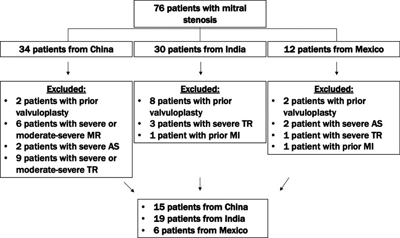
This retrospective study included 40 patients with rheumatic MS, consisting of 19 patients from India, 15 patients from China, and 6 patients from Mexico (median (interquartile range (IQR)) age: 45 years (34-55); 75% women). Twenty patients were included in the control group. CMR variables pertaining to morphology and function were collected. Late gadolinium enhancement (LGE) sequences were acquired for tissue characterization. Statistical analyses were performed using the Kruskal-Wallis test and the chi-square test.
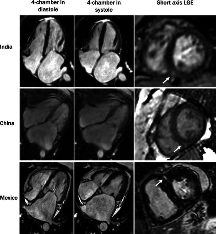
Compared to the control group, patients with MS had lower left ventricular (LV) ejection fraction (51% (42%-55%) vs 60% (57%-65%), p < 0.001), lower right ventricular (RV) ejection fraction (44% (40%-52%) vs 64% (59%-67%), p < 0.001), higher RV end-diastolic volume (72 (58-87) mL/m vs 59 (49-69) mL/m, p = 0.003), larger left atrial volume (87 (67-108) mL/m vs 29 (22-34) mL/m, p < 0.001), and right atrial areas (20 (16-23) cm vs 13 (12-16) cm, p < 0.001). LGE was prevalent in patients with rheumatic MS (82%), and was commonly located at the RV insertion sites. Furthermore, the patient cohorts from India, China, and Mexico were heterogeneous in terms of baseline characteristics and cardiac remodeling.
Our findings demonstrated that biventricular dysfunction, right and left atrial remodeling, and LGE at the RV insertion sites are underappreciated in contemporary rheumatic MS. Further studies are needed to elucidate the prognostic implications of these findings.
风湿性二尖瓣狭窄(MS)中的心脏重构是复杂且尚未完全被理解的。本研究的目的是使用心血管磁共振(CMR)评估一组风湿性 MS 患者的心脏结构和功能变化。
本回顾性研究纳入了 40 例风湿性 MS 患者,其中包括 19 例来自印度的患者、15 例来自中国的患者和 6 例来自墨西哥的患者(中位数(四分位距(IQR))年龄:45 岁(34-55);75%为女性)。20 例患者被纳入对照组。采集了与形态和功能相关的 CMR 变量。采集了钆延迟增强(LGE)序列以进行组织特征分析。使用 Kruskal-Wallis 检验和卡方检验进行统计分析。
与对照组相比,MS 患者的左心室(LV)射血分数更低(51%(42%-55%) vs 60%(57%-65%),p<0.001),右心室(RV)射血分数更低(44%(40%-52%) vs 64%(59%-67%),p<0.001),RV 舒张末期容积更大(72(58-87)mL/m 比 59(49-69)mL/m,p=0.003),左心房容积更大(87(67-108)mL/m 比 29(22-34)mL/m,p<0.001),右心房面积更大(20(16-23)cm 比 13(12-16)cm,p<0.001)。风湿性 MS 患者的 LGE 较为常见(82%),且常见于 RV 插入部位。此外,来自印度、中国和墨西哥的患者队列在基线特征和心脏重构方面存在异质性。
本研究结果表明,双心室功能障碍、左右心房重构以及 RV 插入部位的 LGE 在当前风湿性 MS 中被低估了。需要进一步的研究来阐明这些发现的预后意义。