• 文献检索
  • 文档翻译
  • 深度研究
  • 学术资讯
  • Suppr Zotero 插件Zotero 插件
  • 邀请有礼
  • 套餐&价格
  • 历史记录
应用&插件
Suppr Zotero 插件Zotero 插件浏览器插件Mac 客户端Windows 客户端微信小程序
定价
高级版会员购买积分包购买API积分包
服务
文献检索文档翻译深度研究API 文档MCP 服务
关于我们
关于 Suppr公司介绍联系我们用户协议隐私条款
关注我们

Suppr 超能文献

核心技术专利:CN118964589B侵权必究
粤ICP备2023148730 号-1Suppr @ 2026

文献检索

告别复杂PubMed语法,用中文像聊天一样搜索,搜遍4000万医学文献。AI智能推荐,让科研检索更轻松。

立即免费搜索

文件翻译

保留排版,准确专业,支持PDF/Word/PPT等文件格式,支持 12+语言互译。

免费翻译文档

深度研究

AI帮你快速写综述,25分钟生成高质量综述,智能提取关键信息,辅助科研写作。

立即免费体验

在小梁切除术后的第一周内,基于 Bruch 膜开口的形态计量学的短期变化。

Short-term changes in Bruch's membrane opening-based morphometrics during the first week after trabeculectomy.

机构信息

Department of Ophthalmology, Faculty of Medicine and University Hospital Cologne, University of Cologne, Kerpener Straße 62, 50924, Cologne, Germany.

Glaucoma Imaging Center University of Cologne (GICC), Faculty of Medicine and University Hospital Cologne, Kerpener Straße 62, 50924, Cologne, Germany.

出版信息

Graefes Arch Clin Exp Ophthalmol. 2022 Oct;260(10):3321-3329. doi: 10.1007/s00417-022-05644-3. Epub 2022 Apr 8.

DOI:10.1007/s00417-022-05644-3
PMID:35394209
原文链接:https://pmc.ncbi.nlm.nih.gov/articles/PMC8990274/
Abstract

PURPOSE

To evaluate the dynamics of Bruch's membrane opening-based morphometrics of the optic nerve head (ONH) using spectral-domain optical coherence tomography (SD-OCT) during the first week after glaucoma surgery by trabeculectomy with mitomycin C.

METHODS

Prospective, longitudinal analysis of 25 eyes of 25 patients treated by trabeculectomy. Twenty-four eyes had evaluable postoperative SD-OCT examinations. Bruch's membrane opening minimum rim width (BMO-MRW) and peripapillary retinal nerve fiber layer (RNFL) thickness were analyzed at baseline before surgery, 1 day, 2 to 3 days, and 1 week after surgery. Changes compared to baseline were correlated to intraocular pressure (IOP).

RESULTS

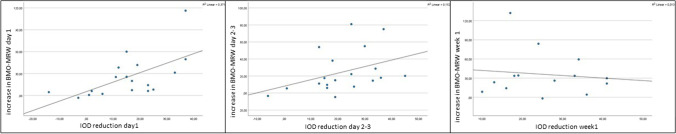
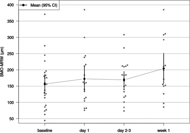
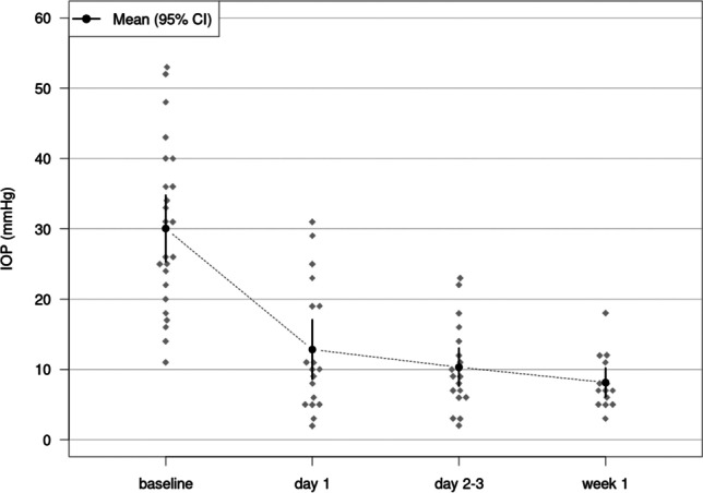
One day after surgery, the mean BMO-MRW changed by + 26.17 µm, p = 0.001 (mean IOP reduction by 17.01 mmHg). This increase persisted on day 2-3 with a mean increase of BMO-MRW of + 25.33 µm, p = 0.001 (mean IOP reduction by 20.46 mmHg) and by week 1 with a mean BMO-MRW increase of + 33.17 µm, p < 0.001 (mean IOP reduction by 22.55 mmHg). The increase in BMO-MRW correlated significantly with the reduction of IOP on day 1 (Spearman's rho ρ = 0.656, p = 0.003) and d2-3 (Spearman's rho ρ = 0.479, p = 0.038). There was no statistically significant correlation found between the IOP and the increase in BMO-MRW in week 1. RNFL thickness showed no significant changes at day 1 as well as days 2-3 (p ≥ 0.078, respectively). It showed a small but significant increase in week 1 by 3.94 µm, p = 0.015.

CONCLUSIONS

Structural reversal of disc cupping in BMO-MRW occurs as early as 1 day after trabeculectomy and correlates to the extent of the IOP reduction. During the whole first week after surgery, a strong increase in BMO-MRW can be noted. The changes in BMO-based parameters need to be considered when evaluating patients' longitudinal follow-up.

摘要

目的

利用频域光学相干断层扫描(SD-OCT)评估青光眼手术后第 1 周内,应用丝裂霉素 C 的小梁切除术对神经眼(ONH)的 Bruch 膜开口形态测量的动力学。

方法

对 25 例 25 眼接受小梁切除术的患者进行前瞻性、纵向分析。24 只眼可进行术后 SD-OCT 检查。在手术前、手术后第 1 天、第 2-3 天和第 1 周,分析 Bruch 膜开口最小边缘宽度(BMO-MRW)和视盘周围视网膜神经纤维层(RNFL)厚度。与基线相比,将变化与眼内压(IOP)相关联。

结果

术后第 1 天,BMO-MRW 平均增加 26.17µm,p=0.001(平均 IOP 降低 17.01mmHg)。第 2-3 天,这种增加持续存在,BMO-MRW 平均增加 25.33µm,p=0.001(平均 IOP 降低 20.46mmHg),第 1 周,BMO-MRW 平均增加 33.17µm,p<0.001(平均 IOP 降低 22.55mmHg)。BMO-MRW 的增加与第 1 天(Spearman 的 rho ρ=0.656,p=0.003)和第 2-3 天(Spearman 的 rho ρ=0.479,p=0.038)IOP 的降低显著相关。在第 1 周,未发现 IOP 与 BMO-MRW 增加之间存在统计学显著相关性。RNFL 厚度在第 1 天以及第 2-3 天均无明显变化(分别为 p≥0.078)。第 1 周,它的厚度增加了 3.94µm,p=0.015,有统计学意义。

结论

小梁切除术后第 1 天即可出现 Bruch 膜开口最小边缘宽度的盘凹陷结构反转,与 IOP 降低程度相关。术后第 1 周,BMO-MRW 明显增加。在评估患者的纵向随访时,需要考虑基于 Bruch 膜的参数变化。

https://cdn.ncbi.nlm.nih.gov/pmc/blobs/9168/9477950/6453dae0ee0e/417_2022_5644_Fig4_HTML.jpg
https://cdn.ncbi.nlm.nih.gov/pmc/blobs/9168/9477950/d5b61dc5db88/417_2022_5644_Fig1_HTML.jpg
https://cdn.ncbi.nlm.nih.gov/pmc/blobs/9168/9477950/dbaf05746d89/417_2022_5644_Fig2_HTML.jpg
https://cdn.ncbi.nlm.nih.gov/pmc/blobs/9168/9477950/4ad56f9144dc/417_2022_5644_Fig3_HTML.jpg
https://cdn.ncbi.nlm.nih.gov/pmc/blobs/9168/9477950/6453dae0ee0e/417_2022_5644_Fig4_HTML.jpg

相似文献

1
Short-term changes in Bruch's membrane opening-based morphometrics during the first week after trabeculectomy.在小梁切除术后的第一周内,基于 Bruch 膜开口的形态计量学的短期变化。
Graefes Arch Clin Exp Ophthalmol. 2022 Oct;260(10):3321-3329. doi: 10.1007/s00417-022-05644-3. Epub 2022 Apr 8.
2
Impact of ab-interno trabeculectomy on Bruch's membrane opening-based morphometry of the optic nerve head for glaucoma progression analysis.内路小梁切除术对基于布鲁赫膜开口的视神经乳头形态测量在青光眼病情进展分析中的影响。
Graefes Arch Clin Exp Ophthalmol. 2019 Feb;257(2):339-347. doi: 10.1007/s00417-018-4187-2. Epub 2018 Nov 28.
3
Dynamics of structural reversal in Bruch's membrane opening-based morphometrics after glaucoma drainage device surgery.青光眼引流装置手术后基于布鲁赫膜开口的形态测量学中结构逆转的动态变化
Graefes Arch Clin Exp Ophthalmol. 2020 Jun;258(6):1227-1236. doi: 10.1007/s00417-020-04621-y. Epub 2020 Mar 5.
4
Structural Reversal of Disc Cupping After Trabeculectomy Alters Bruch Membrane Opening-Based Parameters to Assess Neuroretinal Rim.经小梁切除术的盘状杯反转改变了基于脉络膜上腔开口的参数来评估神经视网膜边缘。
Am J Ophthalmol. 2018 Oct;194:143-152. doi: 10.1016/j.ajo.2018.07.016. Epub 2018 Jul 25.
5
Novel Bruch's Membrane Opening Minimum Rim Area Equalizes Disc Size Dependency and Offers High Diagnostic Power for Glaucoma.新型布鲁赫膜开口最小边缘面积可平衡视盘大小依赖性并为青光眼提供高诊断效能。
Invest Ophthalmol Vis Sci. 2016 Dec 1;57(15):6596-6603. doi: 10.1167/iovs.16-20561.
6
Neuroretinal rim response to transient changes in intraocular pressure in healthy non-human primate eyes.健康非人类灵长类动物眼内压瞬态变化的神经视网膜边缘反应。
Exp Eye Res. 2020 Apr;193:107978. doi: 10.1016/j.exer.2020.107978. Epub 2020 Feb 17.
7
Evaluation of two-dimensional Bruch's membrane opening minimum rim area for glaucoma diagnostics in a large patient cohort.大样本患者群体中二维 Bruch 膜开口最小边缘区域在青光眼诊断中的评估。
Acta Ophthalmol. 2019 Feb;97(1):60-67. doi: 10.1111/aos.13698. Epub 2018 Mar 24.
8
Analysis of peripapillary vessel density and Bruch's membrane opening-based neuroretinal rim parameters in glaucoma using OCT and OCT-angiography.利用 OCT 和 OCT 血管成像分析青光眼的视盘周围血管密度和基于 Bruch 膜开口的神经视网膜边缘参数。
Eye (Lond). 2020 Jun;34(6):1086-1093. doi: 10.1038/s41433-019-0631-8. Epub 2019 Oct 24.
9
The use of Bruch's membrane opening-based optical coherence tomography of the optic nerve head for glaucoma detection in microdiscs.基于布鲁赫膜开口的视神经乳头光学相干断层扫描技术在微小视盘中检测青光眼的应用。
Br J Ophthalmol. 2017 Apr;101(4):530-535. doi: 10.1136/bjophthalmol-2016-308957. Epub 2016 Jul 19.
10
Factors Associated with Increased Neuroretinal Rim Thickness Measured Based on Bruch's Membrane Opening-Minimum Rim Width after Trabeculectomy.小梁切除术后基于布鲁赫膜开口-最小视盘边缘宽度测量的神经视网膜边缘厚度增加的相关因素。
J Clin Med. 2021 Aug 18;10(16):3646. doi: 10.3390/jcm10163646.

引用本文的文献

1
Reversal of cup-to-disc ratio following trabeculectomy using optical coherence tomography imaging: A case series.使用光学相干断层扫描成像观察小梁切除术后杯盘比的逆转:病例系列
Saudi J Ophthalmol. 2024 Dec 2;38(4):391-395. doi: 10.4103/sjopt.sjopt_165_24. eCollection 2024 Oct-Dec.
2
Structural reversal of disc cupping measured in Bruch's membrane opening-based OCT morphometry after PRESERFLO microshunt implantation for open-angle glaucoma.在采用 PRESERFLO 微型分流器植入术治疗开角型青光眼后,基于布鲁赫膜开口的光学相干断层扫描形态测量法所测得的视盘杯状凹陷结构逆转。
BMC Ophthalmol. 2025 Jan 17;25(1):26. doi: 10.1186/s12886-024-03838-3.
3

本文引用的文献

1
Dynamics of structural reversal in Bruch's membrane opening-based morphometrics after glaucoma drainage device surgery.青光眼引流装置手术后基于布鲁赫膜开口的形态测量学中结构逆转的动态变化
Graefes Arch Clin Exp Ophthalmol. 2020 Jun;258(6):1227-1236. doi: 10.1007/s00417-020-04621-y. Epub 2020 Mar 5.
2
Impact of ab-interno trabeculectomy on Bruch's membrane opening-based morphometry of the optic nerve head for glaucoma progression analysis.内路小梁切除术对基于布鲁赫膜开口的视神经乳头形态测量在青光眼病情进展分析中的影响。
Graefes Arch Clin Exp Ophthalmol. 2019 Feb;257(2):339-347. doi: 10.1007/s00417-018-4187-2. Epub 2018 Nov 28.
3
Variation of Bruch's membrane opening in response to intraocular pressure change during scleral lens wear, in a population with keratoconus.
圆锥角膜患者佩戴巩膜镜期间,布鲁赫膜开口随眼压变化的情况
Ophthalmic Physiol Opt. 2025 Mar;45(2):405-415. doi: 10.1111/opo.13431. Epub 2024 Dec 6.
4
Susceptibility of optic nerve head in children with posture-related elevation of intraocular pressure.与姿势相关的眼压升高儿童的视神经头易感性。
Int Ophthalmol. 2024 Apr 16;44(1):182. doi: 10.1007/s10792-024-03109-6.
5
Neuroretinal Rim Response to Transient Intraocular Pressure Challenge Predicts the Extent of Retinal Ganglion Cell Loss in Experimental Glaucoma.神经视网膜边缘对瞬态眼压挑战的反应可预测实验性青光眼中视网膜神经节细胞损失的程度。
Invest Ophthalmol Vis Sci. 2023 May 1;64(5):30. doi: 10.1167/iovs.64.5.30.
6
Changes of Optical Coherence Tomography Parameters after Cataract Surgery in Primary Open-Angle Glaucoma Eyes.白内障术后原发性开角型青光眼眼的光相干断层扫描参数变化。
Korean J Ophthalmol. 2023 Feb;37(1):1-11. doi: 10.3341/kjo.2022.0102. Epub 2022 Oct 25.
Structural Reversal of Disc Cupping After Trabeculectomy Alters Bruch Membrane Opening-Based Parameters to Assess Neuroretinal Rim.
经小梁切除术的盘状杯反转改变了基于脉络膜上腔开口的参数来评估神经视网膜边缘。
Am J Ophthalmol. 2018 Oct;194:143-152. doi: 10.1016/j.ajo.2018.07.016. Epub 2018 Jul 25.
4
Evaluation of two-dimensional Bruch's membrane opening minimum rim area for glaucoma diagnostics in a large patient cohort.大样本患者群体中二维 Bruch 膜开口最小边缘区域在青光眼诊断中的评估。
Acta Ophthalmol. 2019 Feb;97(1):60-67. doi: 10.1111/aos.13698. Epub 2018 Mar 24.
5
Bruch's membrane opening-based optical coherence tomography of the optic nerve head: a useful diagnostic tool to detect glaucoma in macrodiscs.基于神经盘大视杯 Bruch 膜裂孔的视神经头光学相干断层扫描:一种探测青光眼的有用诊断工具。
Eye (Lond). 2018 Feb;32(2):314-323. doi: 10.1038/eye.2017.306. Epub 2018 Jan 26.
6
Detecting Structural Progression in Glaucoma with Optical Coherence Tomography.利用光学相干断层扫描技术检测青光眼的结构进展
Ophthalmology. 2017 Dec;124(12S):S57-S65. doi: 10.1016/j.ophtha.2017.07.015.
7
Intraday Repeatability of Bruch's Membrane Opening-Based Neuroretinal Rim Measurements.基于布鲁赫膜开口的神经视网膜边缘测量的日内重复性
Invest Ophthalmol Vis Sci. 2017 Oct 1;58(12):5195-5200. doi: 10.1167/iovs.17-22812.
8
Estimating Optical Coherence Tomography Structural Measurement Floors to Improve Detection of Progression in Advanced Glaucoma.估计光学相干断层扫描结构测量阈值以改善晚期青光眼进展的检测
Am J Ophthalmol. 2017 Mar;175:37-44. doi: 10.1016/j.ajo.2016.11.010. Epub 2016 Nov 30.
9
Automated Segmentation Errors When Using Optical Coherence Tomography to Measure Retinal Nerve Fiber Layer Thickness in Glaucoma.使用光学相干断层扫描测量青光眼视网膜神经纤维层厚度时的自动分割误差
Am J Ophthalmol. 2017 Feb;174:1-8. doi: 10.1016/j.ajo.2016.10.020. Epub 2016 Nov 4.
10
The use of Bruch's membrane opening-based optical coherence tomography of the optic nerve head for glaucoma detection in microdiscs.基于布鲁赫膜开口的视神经乳头光学相干断层扫描技术在微小视盘中检测青光眼的应用。
Br J Ophthalmol. 2017 Apr;101(4):530-535. doi: 10.1136/bjophthalmol-2016-308957. Epub 2016 Jul 19.