Tipping William J, Wilson Liam T, An Connie, Leventi Aristea A, Wark Alastair W, Wetherill Corinna, Tomkinson Nicholas C O, Faulds Karen, Graham Duncan
Centre for Molecular Nanometrology, WestCHEM, Department of Pure and Applied Chemistry, Technology and Innovation Centre, University of Strathclyde Glasgow G1 1RD UK
Department of Pure and Applied Chemistry, University of Strathclyde Glasgow G1 1XL UK
Chem Sci. 2022 Feb 25;13(12):3468-3476. doi: 10.1039/d1sc06976d. eCollection 2022 Mar 24.
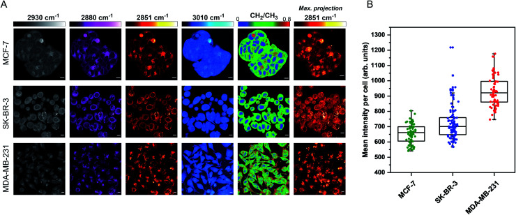
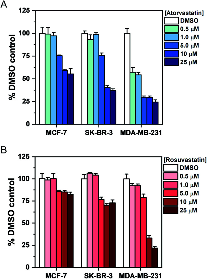
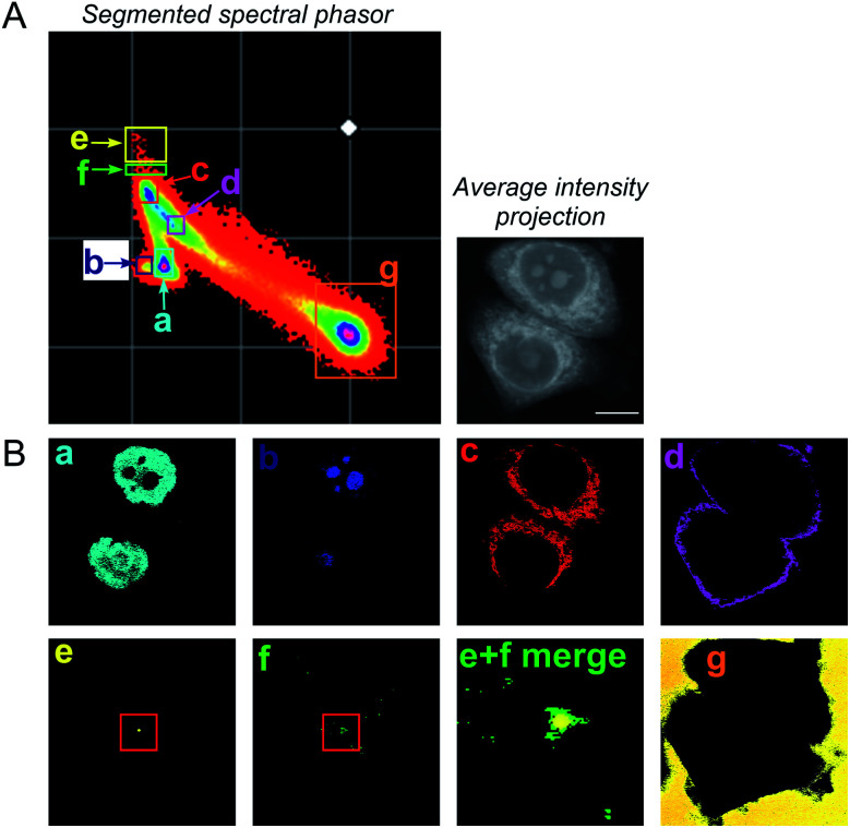
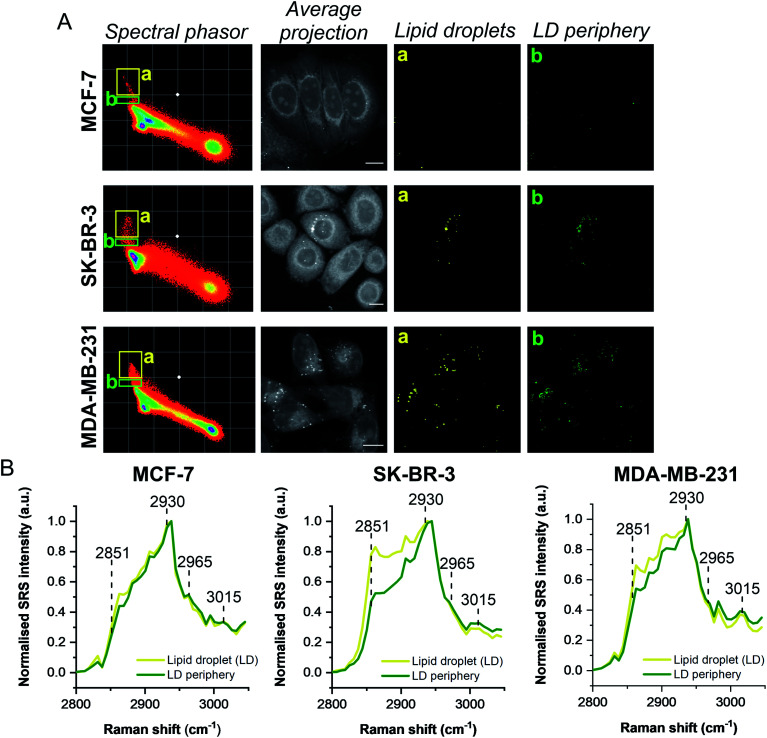
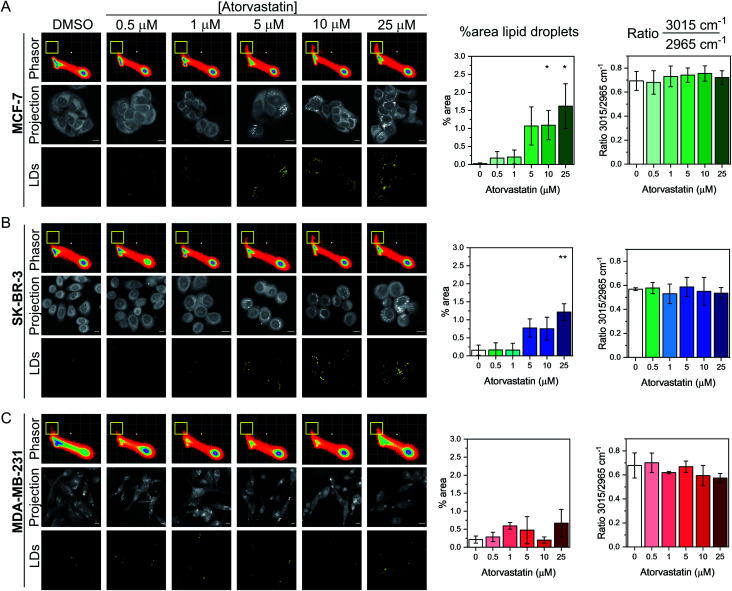
Statins have displayed significant, although heterogeneous, anti-tumour activity in breast cancer disease progression and recurrence. They offer promise as a class of drugs, normally used for cardiovascular disease control, that could have a significant impact on the treatment of cancer. Understanding their mode of action and accurately assessing their efficacy on live cancer cells is an important and significant challenge. Stimulated Raman scattering (SRS) microscopy is a powerful, label-free imaging technique that can rapidly characterise the biochemical responses of live cell populations following drug treatment. Here, we demonstrate multi-wavelength SRS imaging together with spectral phasor analysis to characterise a panel of breast cancer cell lines (MCF-7, SK-BR-3 and MDA-MB-231 cells) treated with two clinically relevant statins, atorvastatin and rosuvastatin. Label-free SRS imaging within the high wavenumber region of the Raman spectrum (2800-3050 cm) revealed the lipid droplet distribution throughout populations of live breast cancer cells using biocompatible imaging conditions. A spectral phasor analysis of the hyperspectral dataset enables rapid differentiation of discrete cellular compartments based on their intrinsic SRS characteristics. Applying the spectral phasor method to studying statin treated cells identified a lipid accumulating phenotype in cell populations which displayed the lowest sensitivity to statin treatment, whilst a weaker lipid accumulating phenotype was associated with a potent reduction in cell viability. This study provides an insight into potential resistance mechanisms of specific cancer cells towards treatment with statins. Label-free SRS imaging provides a novel and innovative technique for phenotypic assessment of drug-induced effects across different cellular populations and enables effective analysis of drug-cell interactions at the subcellular scale.
他汀类药物在乳腺癌疾病进展和复发中已显示出显著的抗肿瘤活性,尽管这种活性存在异质性。作为一类通常用于控制心血管疾病的药物,它们有望对癌症治疗产生重大影响。了解其作用方式并准确评估其对活癌细胞的疗效是一项重要且具有挑战性的任务。受激拉曼散射(SRS)显微镜是一种强大的无标记成像技术,可快速表征药物处理后活细胞群体的生化反应。在此,我们展示了多波长SRS成像以及光谱相量分析,以表征用两种临床相关他汀类药物阿托伐他汀和瑞舒伐他汀处理的一组乳腺癌细胞系(MCF-7、SK-BR-3和MDA-MB-231细胞)。在拉曼光谱的高波数区域(2800 - 3050 cm)内进行的无标记SRS成像,使用生物相容性成像条件揭示了活乳腺癌细胞群体中脂滴的分布。对高光谱数据集进行光谱相量分析能够基于离散细胞区室的内在SRS特征快速区分它们。将光谱相量方法应用于研究他汀类药物处理的细胞,在对他汀类药物治疗敏感性最低的细胞群体中鉴定出脂质积累表型,而较弱的脂质积累表型与细胞活力的显著降低相关。这项研究深入了解了特定癌细胞对他汀类药物治疗的潜在耐药机制。无标记SRS成像为跨不同细胞群体的药物诱导效应的表型评估提供了一种新颖的创新技术,并能够在亚细胞尺度上有效分析药物与细胞的相互作用。