• 文献检索
  • 文档翻译
  • 深度研究
  • 学术资讯
  • Suppr Zotero 插件Zotero 插件
  • 邀请有礼
  • 套餐&价格
  • 历史记录
应用&插件
Suppr Zotero 插件Zotero 插件浏览器插件Mac 客户端Windows 客户端微信小程序
定价
高级版会员购买积分包购买API积分包
服务
文献检索文档翻译深度研究API 文档MCP 服务
关于我们
关于 Suppr公司介绍联系我们用户协议隐私条款
关注我们

Suppr 超能文献

核心技术专利:CN118964589B侵权必究
粤ICP备2023148730 号-1Suppr @ 2026

文献检索

告别复杂PubMed语法,用中文像聊天一样搜索,搜遍4000万医学文献。AI智能推荐,让科研检索更轻松。

立即免费搜索

文件翻译

保留排版,准确专业,支持PDF/Word/PPT等文件格式,支持 12+语言互译。

免费翻译文档

深度研究

AI帮你快速写综述,25分钟生成高质量综述,智能提取关键信息,辅助科研写作。

立即免费体验

通过生物正交受激拉曼散射显微镜成像药物摄取。

Imaging drug uptake by bioorthogonal stimulated Raman scattering microscopy.

作者信息

Tipping William J, Lee Martin, Serrels Alan, Brunton Valerie G, Hulme Alison N

机构信息

EaStCHEM School of Chemistry , The University of Edinburgh , Joseph Black Building, David Brewster Road , Edinburgh , EH9 3FJ , UK . Email:

Edinburgh Cancer Research Centre , Institute of Genetics and Molecular Medicine , The University of Edinburgh , Crewe Road South , Edinburgh , EH4 2XR , UK . Email:

出版信息

Chem Sci. 2017 Aug 1;8(8):5606-5615. doi: 10.1039/c7sc01837a. Epub 2017 May 24.

DOI:10.1039/c7sc01837a
PMID:30155229
原文链接:https://pmc.ncbi.nlm.nih.gov/articles/PMC6103005/
Abstract

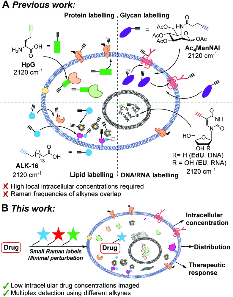
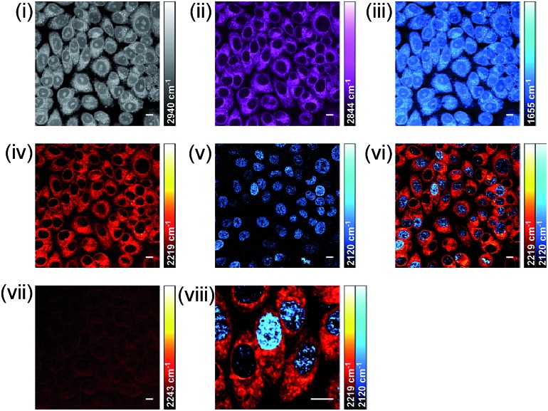
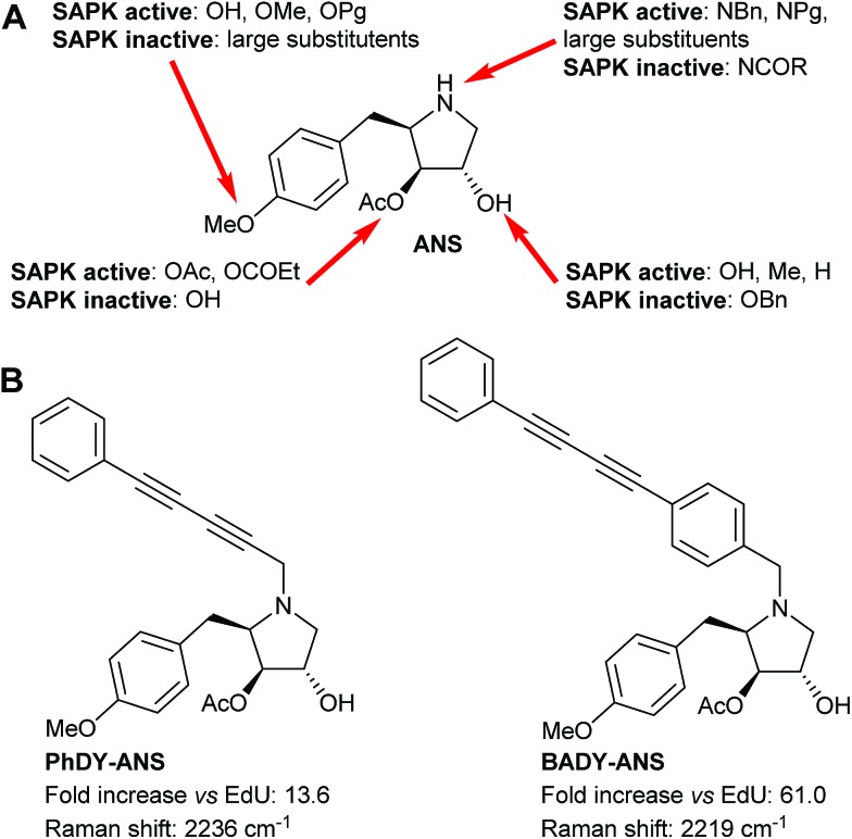
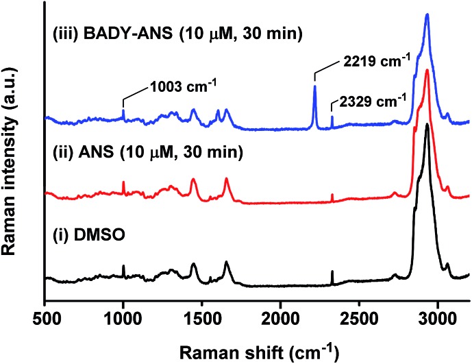
Stimulated Raman scattering (SRS) microscopy in tandem with bioorthogonal Raman labelling strategies is set to revolutionise the direct visualisation of intracellular drug uptake. Rational evaluation of a series of Raman-active labels has allowed the identification of highly active labels which have minimal perturbation on the biological efficacy of the parent drug. Drug uptake has been correlated with markers of cellular composition and cell cycle status, and mapped across intracellular structures using dual-colour and multi-modal imaging. The minimal phototoxicity and low photobleaching associated with SRS microscopy has enabled real-time imaging in live cells. These studies demonstrate the potential for SRS microscopy in the drug development process.

摘要

受激拉曼散射(SRS)显微镜与生物正交拉曼标记策略相结合,有望彻底改变细胞内药物摄取的直接可视化。对一系列拉曼活性标记物的合理评估,已确定了高活性标记物,这些标记物对母体药物的生物学功效影响最小。药物摄取已与细胞组成和细胞周期状态的标志物相关联,并通过双色和多模态成像在细胞内结构上进行了映射。SRS显微镜相关的最小光毒性和低光漂白特性,使得在活细胞中进行实时成像成为可能。这些研究证明了SRS显微镜在药物开发过程中的潜力。

https://cdn.ncbi.nlm.nih.gov/pmc/blobs/46d0/6103005/9919256ddbaf/c7sc01837a-f8.jpg
https://cdn.ncbi.nlm.nih.gov/pmc/blobs/46d0/6103005/09c23755ed37/c7sc01837a-f1.jpg
https://cdn.ncbi.nlm.nih.gov/pmc/blobs/46d0/6103005/78978213669b/c7sc01837a-f2.jpg
https://cdn.ncbi.nlm.nih.gov/pmc/blobs/46d0/6103005/a0c0ad3f3bf2/c7sc01837a-f3.jpg
https://cdn.ncbi.nlm.nih.gov/pmc/blobs/46d0/6103005/a95921360fc9/c7sc01837a-f4.jpg
https://cdn.ncbi.nlm.nih.gov/pmc/blobs/46d0/6103005/14a2f89dfdff/c7sc01837a-f5.jpg
https://cdn.ncbi.nlm.nih.gov/pmc/blobs/46d0/6103005/2e5113faeb47/c7sc01837a-f6.jpg
https://cdn.ncbi.nlm.nih.gov/pmc/blobs/46d0/6103005/930b7b63c148/c7sc01837a-f7.jpg
https://cdn.ncbi.nlm.nih.gov/pmc/blobs/46d0/6103005/9919256ddbaf/c7sc01837a-f8.jpg

相似文献

1
Imaging drug uptake by bioorthogonal stimulated Raman scattering microscopy.通过生物正交受激拉曼散射显微镜成像药物摄取。
Chem Sci. 2017 Aug 1;8(8):5606-5615. doi: 10.1039/c7sc01837a. Epub 2017 May 24.
2
Live-Cell Bioorthogonal Chemical Imaging: Stimulated Raman Scattering Microscopy of Vibrational Probes.活细胞生物正交化学成像:振动探针的受激拉曼散射显微镜。
Acc Chem Res. 2016 Aug 16;49(8):1494-502. doi: 10.1021/acs.accounts.6b00210. Epub 2016 Aug 3.
3
Fast vibrational imaging of single cells and tissues by stimulated Raman scattering microscopy.通过受激拉曼散射显微镜对单细胞和组织进行快速振动成像。
Acc Chem Res. 2014 Aug 19;47(8):2282-90. doi: 10.1021/ar400331q. Epub 2014 May 28.
4
Quantitative Drug Dynamics Visualized by Alkyne-Tagged Plasmonic-Enhanced Raman Microscopy.炔基标记的等离子体增强拉曼显微镜可视化定量药物动力学。
ACS Nano. 2020 Nov 24;14(11):15032-15041. doi: 10.1021/acsnano.0c05010. Epub 2020 Oct 20.
5
Real-Time Monitoring of Pharmacokinetics of Mitochondria-Targeting Molecules in Live Cells with Bioorthogonal Hyperspectral Stimulated Raman Scattering Microscopy.利用生物正交超分辨受激拉曼散射显微镜实时监测活细胞中线粒体靶向分子的药代动力学。
Anal Chem. 2020 Jan 7;92(1):740-748. doi: 10.1021/acs.analchem.9b02838. Epub 2019 Dec 9.
6
Temporal imaging of drug dynamics in live cells using stimulated Raman scattering microscopy and a perfusion cell culture system.使用受激拉曼散射显微镜和灌注细胞培养系统对活细胞中药物动力学进行时间成像。
RSC Chem Biol. 2022 Aug 9;3(9):1154-1164. doi: 10.1039/d2cb00160h. eCollection 2022 Aug 31.
7
Label-Free, Quantitative Imaging of MoS -Nanosheets in Live Cells with Simultaneous Stimulated Raman Scattering and Transient Absorption Microscopy.利用受激拉曼散射和瞬态吸收显微镜对活细胞中的二硫化钼纳米片进行无标记定量成像
Adv Biosyst. 2017 Apr;1(4):e1700013. doi: 10.1002/adbi.201700013. Epub 2017 Mar 31.
8
Advances in stimulated Raman scattering imaging for tissues and animals.用于组织和动物的受激拉曼散射成像进展
Quant Imaging Med Surg. 2021 Mar;11(3):1078-1101. doi: 10.21037/qims-20-712.
9
Live-cell stimulated Raman scattering imaging of alkyne-tagged biomolecules.炔基标记生物分子的活细胞受激拉曼散射成像。
Angew Chem Int Ed Engl. 2014 Jun 2;53(23):5827-31. doi: 10.1002/anie.201400328. Epub 2014 Apr 17.
10
Mitochondrial Imaging with Combined Fluorescence and Stimulated Raman Scattering Microscopy Using a Probe of the Aggregation-Induced Emission Characteristic.利用具有聚集诱导发光特性的探针的荧光和受激发射拉曼散射显微镜联合进行线粒体成像。
J Am Chem Soc. 2017 Nov 29;139(47):17022-17030. doi: 10.1021/jacs.7b06273. Epub 2017 Nov 16.

引用本文的文献

1
Illuminating life processes by vibrational probes.用振动探针揭示生命过程。
Nat Methods. 2025 May;22(5):928-944. doi: 10.1038/s41592-025-02689-0. Epub 2025 May 13.
2
Recent advances in the design of SERS substrates and sensing systems for (bio)sensing applications: Systems from single cell to single molecule detection.用于(生物)传感应用的表面增强拉曼散射(SERS)基底和传感系统设计的最新进展:从单细胞检测到单分子检测的系统
F1000Res. 2025 Mar 18;13:670. doi: 10.12688/f1000research.149263.2. eCollection 2024.
3
Raman active diyne-girder conformationally constrained p53 stapled peptides bind to MDM2 for visualisation without fluorophores.

本文引用的文献

1
Quantification of Lipid Metabolism in Living Cells through the Dynamics of Lipid Droplets Measured by Stimulated Raman Scattering Imaging.通过受激拉曼散射成像测量的脂滴动力学定量研究活细胞中的脂代谢。
Anal Chem. 2017 Apr 18;89(8):4502-4507. doi: 10.1021/acs.analchem.6b04699. Epub 2017 Apr 6.
2
In vivo imaging of the tumor and its associated microenvironment using combined CARS / 2-photon microscopy.使用相干反斯托克斯拉曼散射(CARS)/双光子显微镜联合技术对肿瘤及其相关微环境进行体内成像。
Intravital. 2015 Jun 8;4(1):e1055430. doi: 10.1080/21659087.2015.1055430. eCollection 2015 Jan-Apr.
3
Live-Cell Bioorthogonal Chemical Imaging: Stimulated Raman Scattering Microscopy of Vibrational Probes.
具有拉曼活性的二炔大梁构象受限的p53钉肽在无荧光团的情况下与MDM2结合用于可视化。
RSC Chem Biol. 2025 Jan 15;6(3):394-403. doi: 10.1039/d4cb00288a. eCollection 2025 Mar 5.
4
Recent Advances in Enhancement of Raman Scattering Intensity for Biological Applications.用于生物应用的拉曼散射强度增强的最新进展
Chem Biomed Imaging. 2023 Apr 22;1(7):575-589. doi: 10.1021/cbmi.3c00017. eCollection 2023 Oct 23.
5
Seeing is Believing: Developing Multimodal Metabolic Insights at the Molecular Level.眼见为实:在分子水平上开发多模态代谢见解。
ACS Cent Sci. 2024 Mar 21;10(4):758-774. doi: 10.1021/acscentsci.3c01438. eCollection 2024 Apr 24.
6
Biomedical applications, perspectives and tag design concepts in the cell - silent Raman window.细胞沉默拉曼窗口中的生物医学应用、前景及标签设计概念
RSC Chem Biol. 2024 Feb 12;5(4):273-292. doi: 10.1039/d3cb00217a. eCollection 2024 Apr 3.
7
A Palette of Minimally Tagged Sucrose Analogues for Real-Time Raman Imaging of Intracellular Plant Metabolism.用于植物细胞内代谢实时拉曼成像的一组最少标记的蔗糖类似物
Angew Chem Weinheim Bergstr Ger. 2021 Mar 29;133(14):7715-7720. doi: 10.1002/ange.202016802. Epub 2021 Feb 26.
8
VIBRANT: spectral profiling for single-cell drug responses.VIBRANT:单细胞药物反应的光谱分析
Nat Methods. 2024 Mar;21(3):501-511. doi: 10.1038/s41592-024-02185-x. Epub 2024 Feb 19.
9
Strategies and Progress of Raman Technologies for Cellular Uptake Analysis of the Drug Delivery Systems.药物传递系统细胞摄取分析的喇曼技术策略和进展。
Int J Nanomedicine. 2023 Nov 20;18:6883-6900. doi: 10.2147/IJN.S435087. eCollection 2023.
10
Expanding the Range of Bioorthogonal Tags for Multiplex Stimulated Raman Scattering Microscopy.扩展用于多重刺激拉曼散射显微镜的生物正交标签范围。
Angew Chem Int Ed Engl. 2023 Nov 27;62(48):e202311530. doi: 10.1002/anie.202311530. Epub 2023 Oct 27.
活细胞生物正交化学成像:振动探针的受激拉曼散射显微镜。
Acc Chem Res. 2016 Aug 16;49(8):1494-502. doi: 10.1021/acs.accounts.6b00210. Epub 2016 Aug 3.
4
Label-Free Neurosurgical Pathology with Stimulated Raman Imaging.基于受激拉曼成像的无标记神经外科病理学
Cancer Res. 2016 Jun 15;76(12):3451-62. doi: 10.1158/0008-5472.CAN-16-0270. Epub 2016 Apr 12.
5
Stimulated Raman scattering microscopy: an emerging tool for drug discovery.受激拉曼散射显微镜:药物发现的新兴工具。
Chem Soc Rev. 2016 Apr 21;45(8):2075-89. doi: 10.1039/c5cs00693g. Epub 2016 Feb 3.
6
Vibrational spectroscopic imaging of living systems: An emerging platform for biology and medicine.振动光谱成像在活体系统中的应用:生物学和医学的新兴平台。
Science. 2015 Nov 27;350(6264):aaa8870. doi: 10.1126/science.aaa8870.
7
Label-free DNA imaging in vivo with stimulated Raman scattering microscopy.利用受激拉曼散射显微镜进行体内无标记DNA成像。
Proc Natl Acad Sci U S A. 2015 Sep 15;112(37):11624-9. doi: 10.1073/pnas.1515121112. Epub 2015 Aug 31.
8
Molecular diffusion in the human nail measured by stimulated Raman scattering microscopy.通过受激拉曼散射显微镜测量人体指甲中的分子扩散。
Proc Natl Acad Sci U S A. 2015 Jun 23;112(25):7725-30. doi: 10.1073/pnas.1503791112. Epub 2015 Jun 8.
9
Assessing cholesterol storage in live cells and C. elegans by stimulated Raman scattering imaging of phenyl-Diyne cholesterol.通过苯基二炔胆固醇的受激拉曼散射成像评估活细胞和秀丽隐杆线虫中的胆固醇储存情况。
Sci Rep. 2015 Jan 22;5:7930. doi: 10.1038/srep07930.
10
Imaging complex protein metabolism in live organisms by stimulated Raman scattering microscopy with isotope labeling.通过同位素标记的受激拉曼散射显微镜对活生物体中的复杂蛋白质代谢进行成像。
ACS Chem Biol. 2015 Mar 20;10(3):901-8. doi: 10.1021/cb500787b. Epub 2015 Jan 15.