Department of Radiology, Wuhan Third Hospital (Tongren Hospital of Wuhan University), Wuhan, 430000, China.
BMC Med Imaging. 2022 Apr 22;22(1):74. doi: 10.1186/s12880-022-00802-9.
Multidetector CT is currently the best imaging method for detecting tracheal diverticulum (TD). Compared with CT, MRI is radiation-free and has higher resolution. However, the MRI characteristics of this disease have not been previously reported. The present retrospective study compared the MR and CT imaging features of TD, aiming to examine the role of MRI in TD diagnosis and management.
Imaging data were collected in 26 TD patients divided into two groups, including the uninfected and infected groups. The MR and CT imaging features (size/wall/channel) of uninfected patients were compared. The performances of MRI and CT in diagnosing and monitoring therapeutic efficacy in infected TD patients were comparatively assessed.
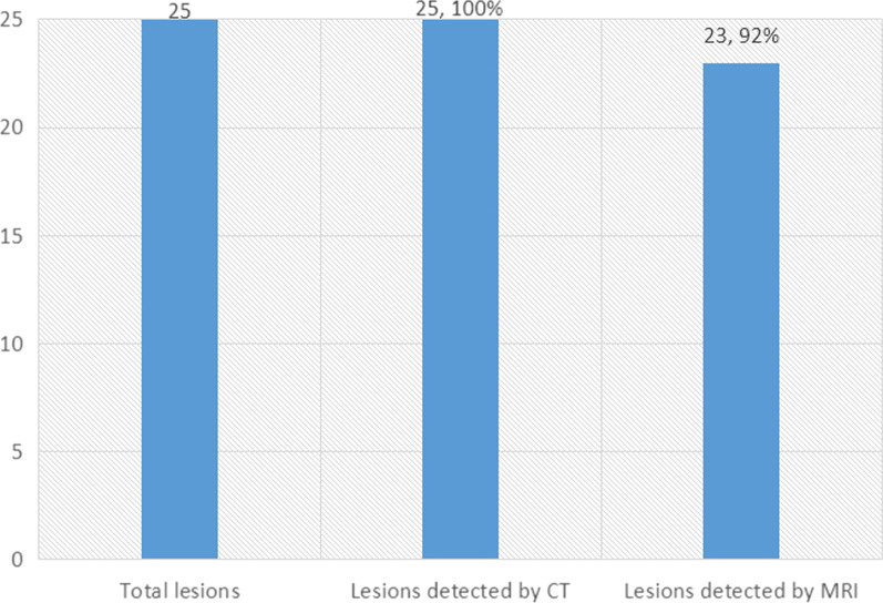
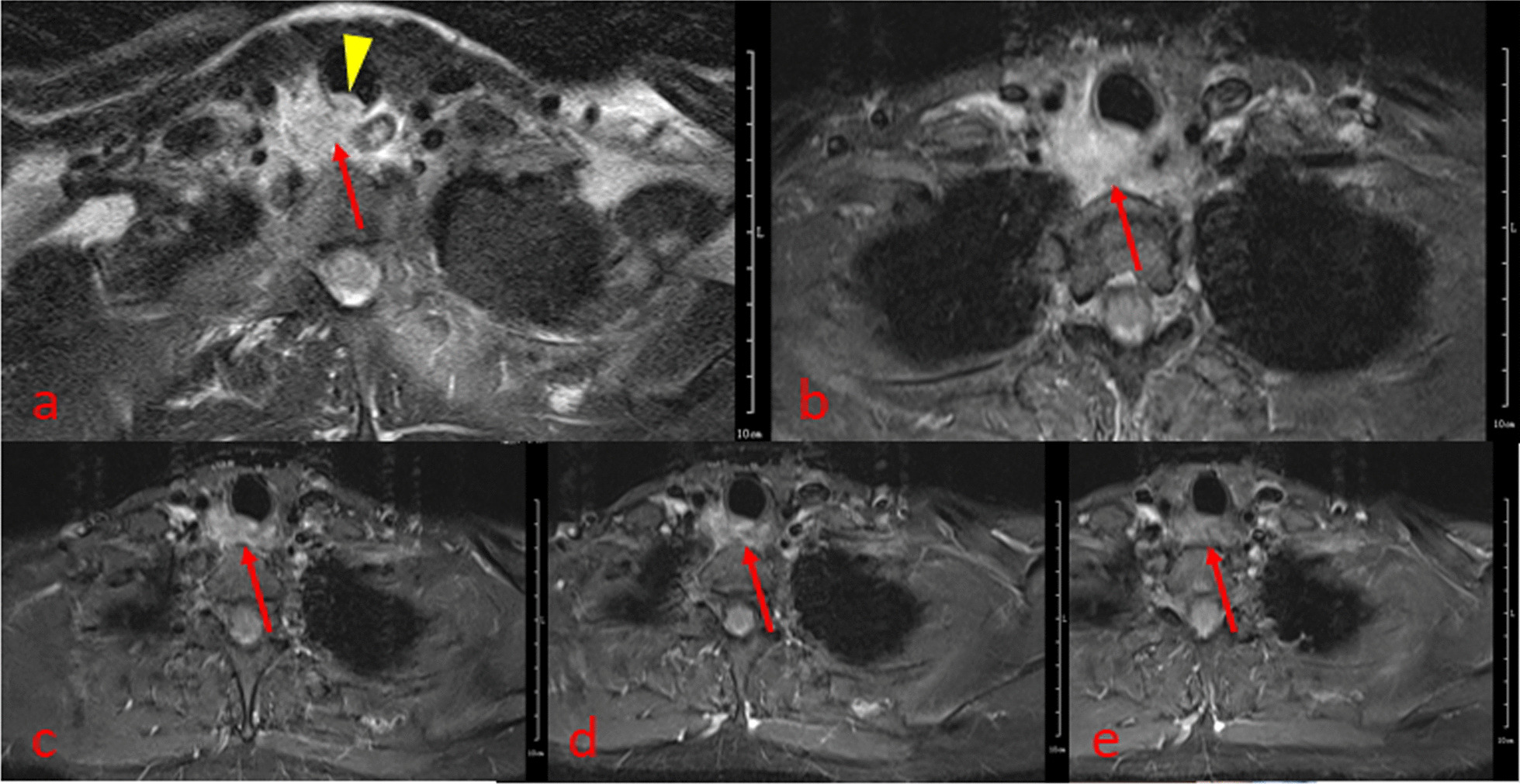
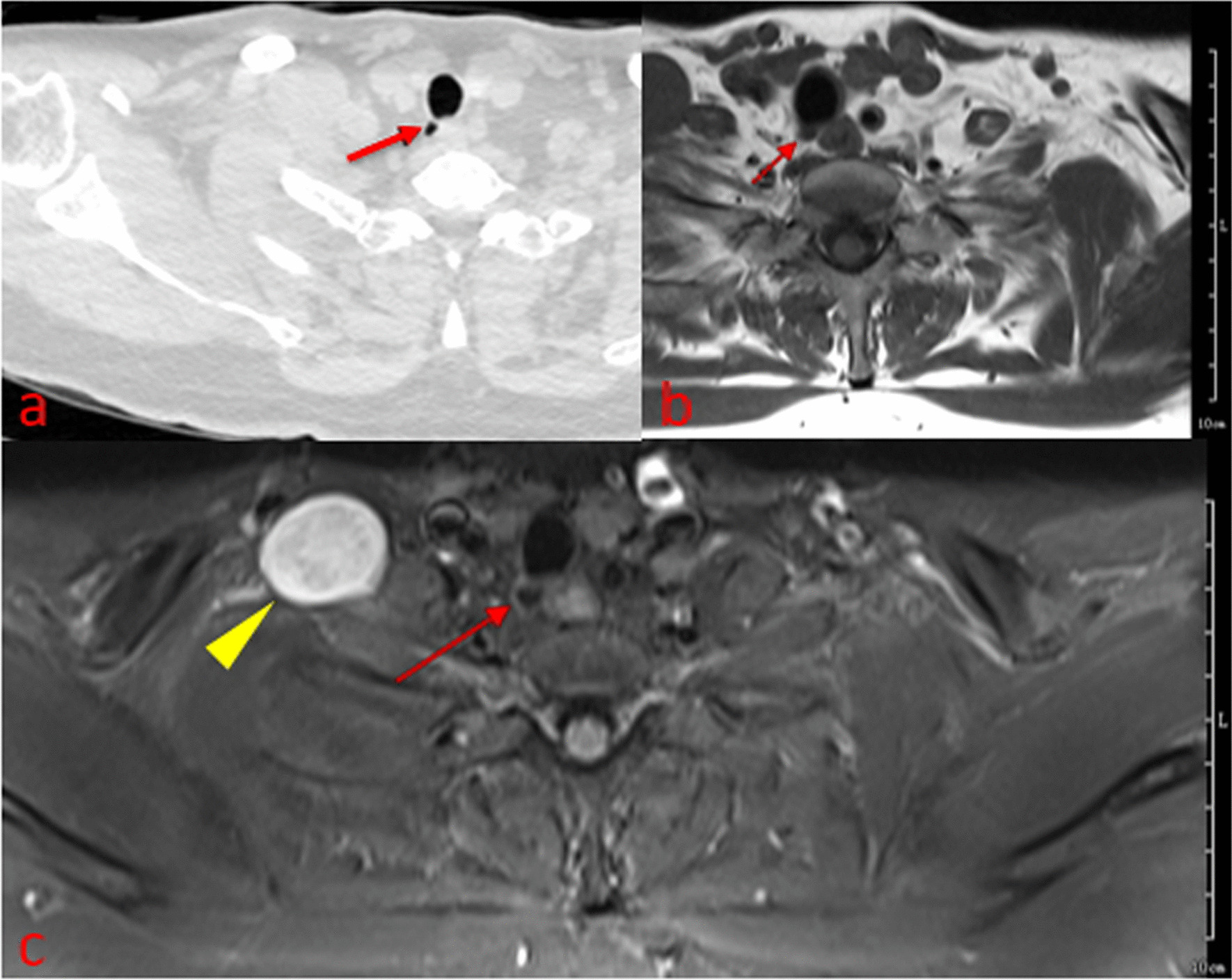
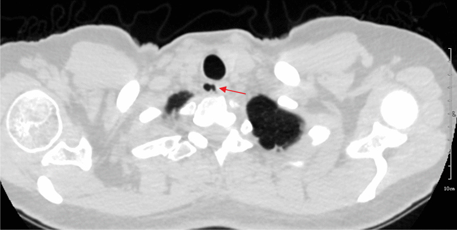
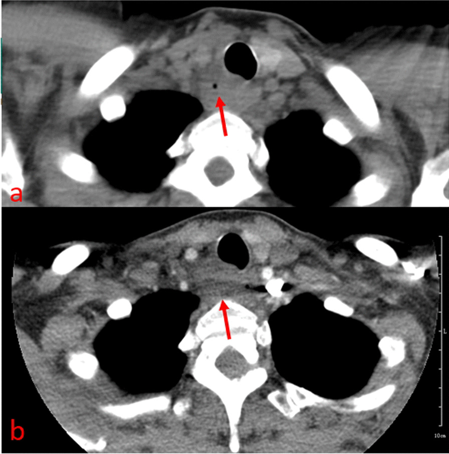
The uninfected group comprised 25 cases with 25 lesions confirmed by CT, including 23 lesions (92%) detected by MRI, with an average diameter of 8.5 mm (range from 3 to 15 mm). Meanwhile, the average diameter was 7.8 mm as measured by CT (range from 2.8 mm to 14.7 mm). The lesion diameters of the two cases not detected by MRI were 2.3 mm and 2 mm. MRI detected walls of all the 23 lesions (23/23), while CT detected no wall (0/23). CT showed channels in 18 lesions (18/23) versus3 for MRI (3/23). The infected case presented with a paratracheal abscess; MRI clearly showed a relationship between the abscess and the trachea, while CT could not show the lesion source. MRI also sensitively showed the whole process of lesion absorption.
MRI can be used as a supplementary method for TD diagnosis, providing information about the wall that cannot be obtained by CT. MRI is superior to CT in diagnosing infected TD cases presenting with a paratracheal abscess, and in monitoring therapeutic efficacy in these patients.
多排 CT 是目前诊断气管憩室(TD)的最佳影像学方法。与 CT 相比,MRI 无辐射,分辨率更高。然而,该疾病的 MRI 特征尚未有报道。本回顾性研究比较了 TD 的 MR 和 CT 成像特征,旨在探讨 MRI 在 TD 诊断和管理中的作用。
收集了 26 例 TD 患者的影像学资料,分为未感染组和感染组。比较了未感染患者的 MR 和 CT 成像特征(大小/壁/通道)。评估了 MRI 和 CT 在诊断和监测感染性 TD 患者治疗效果方面的表现。
未感染组包括 25 例 CT 证实的 25 个病变,其中 23 个病变(92%)被 MRI 检测到,平均直径为 8.5mm(范围 3-15mm)。同时,CT 测量的平均直径为 7.8mm(范围 2.8-14.7mm)。2 例未被 MRI 检测到的病变直径分别为 2.3mm 和 2mm。MRI 检测到所有 23 个病变的壁(23/23),而 CT 则未检测到壁(0/23)。CT 显示 18 个病变(18/23)有通道,而 MRI 显示 3 个(3/23)。感染病例表现为气管旁脓肿;MRI 清晰显示脓肿与气管的关系,而 CT 无法显示病变源。MRI 还敏感地显示了病变吸收的全过程。
MRI 可作为 TD 诊断的补充方法,提供 CT 无法获得的壁信息。MRI 在诊断表现为气管旁脓肿的感染性 TD 病例以及监测这些患者的治疗效果方面优于 CT。