Baines Kiren, Yoshioka Kazuaki, Takuwa Yoh, Lane Jon D
Cell Biology Laboratories, School of Biochemistry, University of Bristol, University Walk, Bristol, BS81TD, UK.
Department of Physiology, Kanazawa University Graduate School of Medical Sciences, 13-1 Takara-machi, Kanazawa Ishikawa 920-8640, Japan.
Autophagy Rep. 2022 Apr 7;1(1):88-118. doi: 10.1080/27694127.2022.2042054. eCollection 2022.
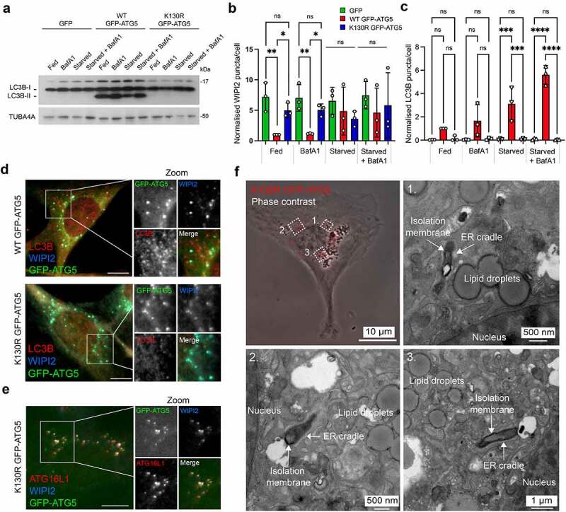
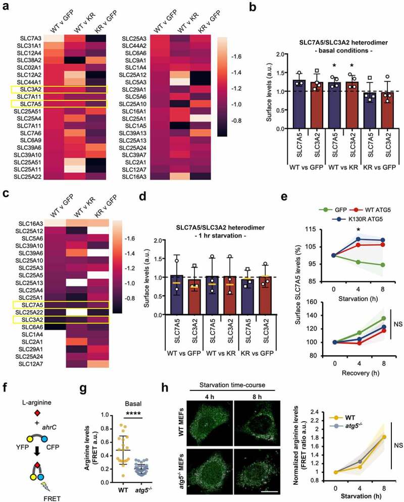
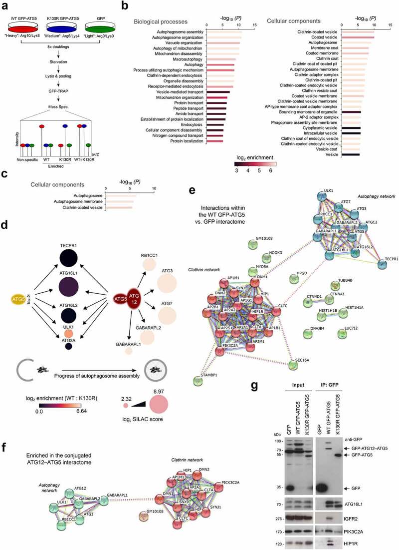
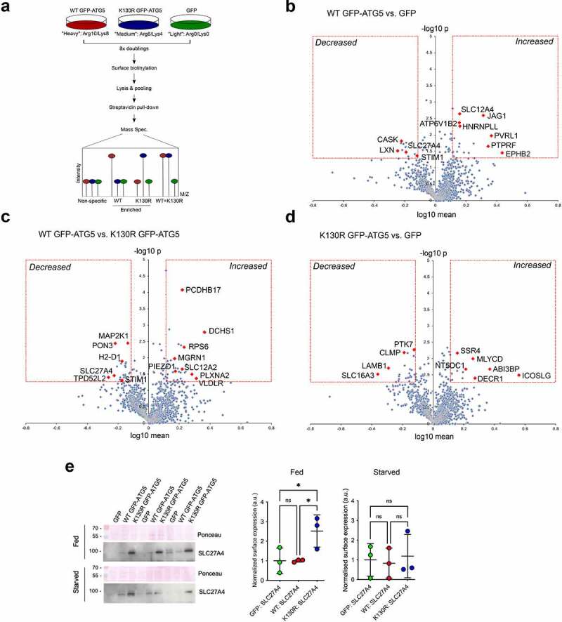
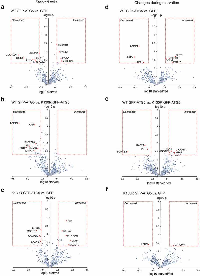
Autophagosome formation involves the sequential actions of conserved ATG proteins to coordinate the lipidation of the ubiquitin-like modifier Atg8-family proteins at the nascent phagophore membrane. Although the molecular steps driving this process are well understood, the source of membranes for the expanding phagophore and their mode of delivery are only now beginning to be revealed. Here, we have used quantitative SILAC-based proteomics to identify proteins that associate with the ATG12-ATG5 conjugate, a crucial player during Atg8-family protein lipidation. Our datasets reveal a strong enrichment of regulators of clathrin-mediated vesicular trafficking, including clathrin heavy and light chains, and several clathrin adaptors. Also identified were PIK3C2A (a phosphoinositide 3-kinase involved in clathrin-mediated endocytosis) and HIP1R (a component of clathrin vesicles), and the absence of either of these proteins alters autophagic flux in cell-based starvation assays. To determine whether the ATG12-ATG5 conjugate reciprocally influences trafficking within the endocytic compartment, we captured the cell surface proteomes of autophagy-competent and autophagy-incompetent mouse embryonic fibroblasts under fed and starved conditions. We report changes in the relative proportions of individual cell surface proteins and show that cell surface levels of the SLC7A5-SLC3A2 amino acid transporter are influenced by autophagy capability. Our data provide evidence for direct regulatory coupling between the ATG12-ATG5 conjugate and the clathrin membrane trafficking system and suggest candidate membrane proteins whose trafficking within the cell may be modulated by the autophagy machinery. ATG, autophagy related; BafA1, bafilomycin A; GFP, green fluorescent protein; HIP1R, huntingtin interacting protein 1 related; MEF, mouse embryo fibroblast; PIK3C2A, phosphatidylinositol-4-phosphate 3-kinase catalytic subunit type 2 alpha; SILAC, stable isotope labelling with amino acids in culture; SQSTM1, sequestosome 1; STRING, search tool for the retrieval of interacting genes/proteins.
自噬体的形成涉及保守的自噬相关(ATG)蛋白的一系列作用,以协调泛素样修饰因子Atg8家族蛋白在新生吞噬泡膜上的脂化过程。尽管驱动这一过程的分子步骤已被充分了解,但不断扩展的吞噬泡的膜来源及其递送方式直到现在才开始被揭示。在这里,我们使用基于定量稳定同位素标记氨基酸培养法(SILAC)的蛋白质组学技术来鉴定与ATG12-ATG5共轭物相关的蛋白质,ATG12-ATG5共轭物是Atg8家族蛋白脂化过程中的关键参与者。我们的数据集显示,网格蛋白介导的囊泡运输的调节因子高度富集,包括网格蛋白重链和轻链,以及几种网格蛋白衔接蛋白。还鉴定出了PIK3C2A(一种参与网格蛋白介导的内吞作用的磷脂酰肌醇-4-磷酸3-激酶)和HIP1R(网格蛋白囊泡的一个组分),在基于细胞的饥饿试验中,缺失这两种蛋白中的任何一种都会改变自噬通量。为了确定ATG12-ATG5共轭物是否会相互影响内吞区室中的运输,我们捕获了在进食和饥饿条件下具有自噬能力和无自噬能力的小鼠胚胎成纤维细胞的细胞表面蛋白质组。我们报告了单个细胞表面蛋白相对比例的变化,并表明SLC7A5-SLC3A2氨基酸转运体的细胞表面水平受自噬能力的影响。我们的数据为ATG12-ATG5共轭物与网格蛋白膜运输系统之间的直接调节偶联提供了证据,并提出了细胞内运输可能受自噬机制调节的候选膜蛋白。ATG,自噬相关;BafA1,巴弗洛霉素A;GFP,绿色荧光蛋白;HIP1R,亨廷顿相互作用蛋白1相关;MEF,小鼠胚胎成纤维细胞;PIK3C2A,磷脂酰肌醇-4-磷酸3-激酶催化亚基2α型;SILAC,稳定同位素标记氨基酸培养法;SQSTM1,聚集体蛋白1;STRING,检索相互作用基因/蛋白的搜索工具