Gallego Miller, López Catalina, Carmona Jorge U
Grupo de Investigación Terapia Regenerativa, Departamento de Salud Animal, Universidad de Caldas, Manizales, Colombia.
Grupo de Investigación Patología Clínica Veterinaria, Departamento de Salud Animal, Universidad de Caldas, Manizales, Colombia.
Vet Med Int. 2022 Apr 11;2022:3377680. doi: 10.1155/2022/3377680. eCollection 2022.
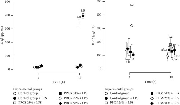
There are scarce studies indicating the basic mechanisms of why platelet-rich plasma (PRP) is useful in the clinical management of dogs with naturally occurring OA. . Cartilage and synovial membrane explants from six dogs were challenged with lipopolysaccharide (LPS) and cultured for 48 h with platelet-poor gel supernatant (PPGS) and platelet-rich gel supernatant (PRGS) at concentrations of 25 and 50%, respectively. The tissue explants challenged with LPS were cocultured over 48 h and culture media were sampled at 1 and 48 h for the determination of IL-1, IL-10, hyaluronan, TGF-1, and PDGF-BB by ELISA. . IL-1 concentrations were significantly higher in tissue explant groups cultured for 48 h with PRGS at 50% and with PPGS at 25% when compared to the remaining experimental groups at any time. IL-10 and HA presented similar concentrations in all evaluated groups at any time. TGF-1 and PDGF-BB presented higher concentrations in the culture media of tissue explants cultured with PPGS and PRGS at 50%, which diminished with time. . Both PPGS and PRGS at both concentrations showed a limited biological effect on cartilage and synovial membrane explants in coculture with LPS. Even PPGS at 25% and PRGS at 50% exhibited proinflammatory effects on these tissues at 48 h.
很少有研究表明富含血小板血浆(PRP)在自然发生骨关节炎的犬临床治疗中发挥作用的基本机制。从六只犬获取软骨和滑膜外植体,用脂多糖(LPS)进行刺激,并分别在浓度为25%和50%的贫血小板凝胶上清液(PPGS)和富血小板凝胶上清液(PRGS)中培养48小时。用LPS刺激的组织外植体共培养48小时,并在1小时和48小时采集培养基样本,通过酶联免疫吸附测定法(ELISA)测定白细胞介素-1(IL-1)、白细胞介素-10(IL-10)、透明质酸、转化生长因子-1(TGF-1)和血小板衍生生长因子BB(PDGF-BB)。与其他实验组相比,在任何时间,用50%的PRGS和25%的PPGS培养48小时的组织外植体组中IL-1浓度显著更高。在任何时间,所有评估组中的IL-10和透明质酸浓度相似。在50%的PPGS和PRGS培养的组织外植体培养基中,TGF-1和PDGF-BB浓度更高,且随时间降低。两种浓度的PPGS和PRGS对与LPS共培养的软骨和滑膜外植体均显示出有限的生物学效应。即使是25%的PPGS和50%的PRGS在48小时时对这些组织也表现出促炎作用。