Pavel Dan G, Henderson Theodore A, DeBruin Simon
Pathfinder Brain SPECT Imaging, Deerfield, IL, United States.
The International Society of Applied Neuroimaging (ISAN), Denver, CO, United States.
Front Neurol. 2022 Mar 28;12:749579. doi: 10.3389/fneur.2021.749579. eCollection 2021.
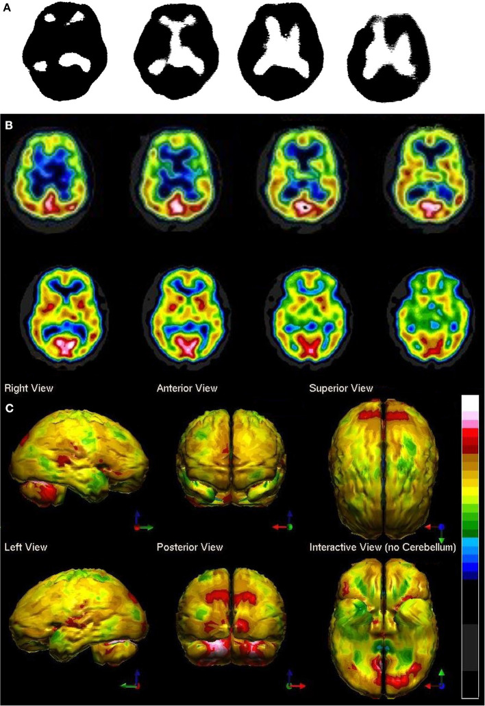
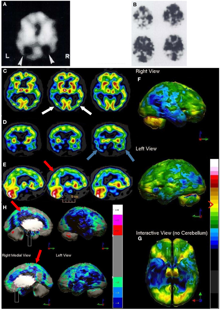
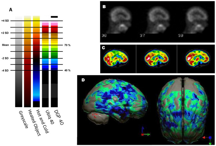
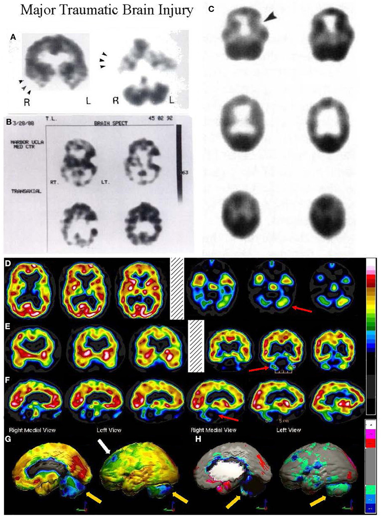
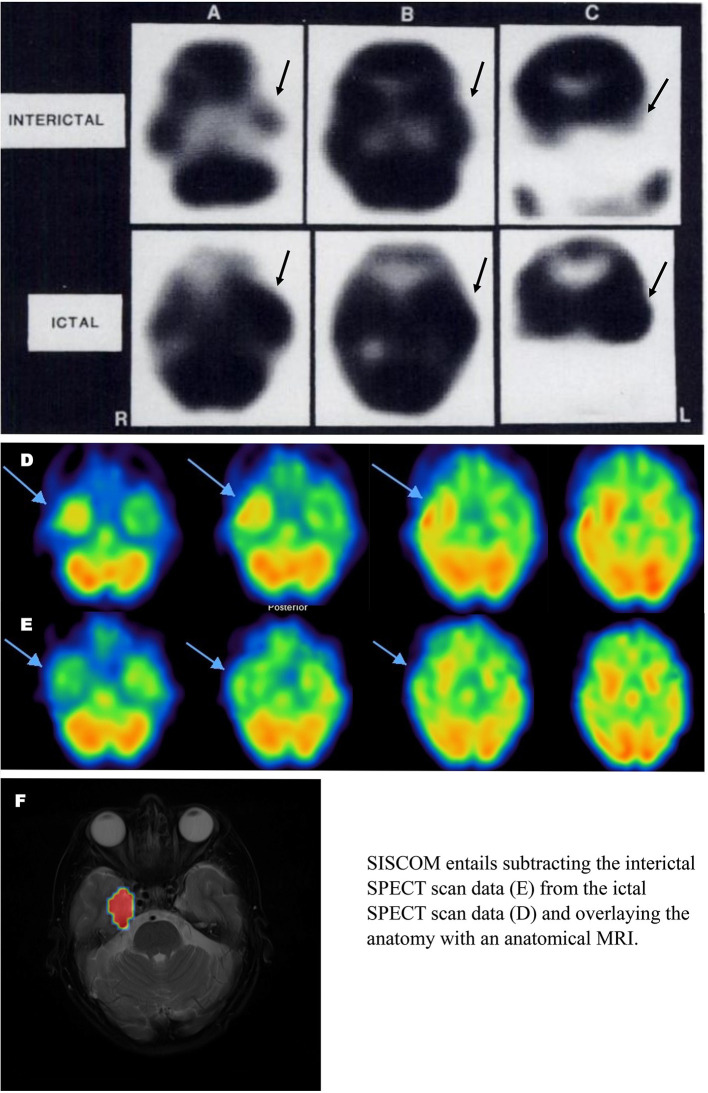
Brain perfusion single photon emission computed tomography (SPECT) scans were initially developed in 1970's. A key radiopharmaceutical, hexamethylpropyleneamine oxime (HMPAO), was originally approved in 1988, but was unstable. As a result, the quality of SPECT images varied greatly based on technique until 1993, when a method of stabilizing HMPAO was developed. In addition, most SPECT perfusion studies pre-1996 were performed on single-head gamma cameras. In 1996, the Therapeutics and Technology Assessment Subcommittee of the American Academy of Neurology (TTASAAN) issued a report regarding the use of SPECT in the evaluation of neurological disorders. Although the TTASAAN report was published in January 1996, it was approved for publication in October 1994. Consequently, the reported brain SPECT studies relied upon to derive the conclusions of the TTASAAN report largely pre-date the introduction of stabilized HMPAO. While only 12% of the studies on traumatic brain injury (TBI) in the TTASAAN report utilized stable tracers and multi-head cameras, 69 subsequent studies with more than 23,000 subjects describe the utility of perfusion SPECT scans in the evaluation of TBI. Similarly, dementia SPECT imaging has improved. Modern SPECT utilizing multi-headed gamma cameras and quantitative analysis has a sensitivity of 86% and a specificity of 89% for the diagnosis of mild to moderate Alzheimer's disease-comparable to fluorodeoxyglucose positron emission tomography. Advances also have occurred in seizure neuroimaging. Lastly, developments in SPECT imaging of neurotoxicity and neuropsychiatric disorders have been striking. At the 25-year anniversary of the publication of the TTASAAN report, it is time to re-examine the utility of perfusion SPECT brain imaging. Herein, we review studies cited by the TTASAAN report vs. current brain SPECT imaging research literature for the major indications addressed in the report, as well as for emerging indications. In Part II, we elaborate technical aspects of SPECT neuroimaging and discuss scan interpretation for the clinician.
脑灌注单光子发射计算机断层扫描(SPECT)最初是在20世纪70年代开发的。一种关键的放射性药物,六甲基丙烯胺肟(HMPAO),最初于1988年获批,但不稳定。因此,直到1993年开发出一种稳定HMPAO的方法之前,SPECT图像的质量因技术不同而有很大差异。此外,1996年以前的大多数SPECT灌注研究是在单头伽马相机上进行的。1996年,美国神经病学学会治疗与技术评估小组委员会(TTASAAN)发布了一份关于SPECT在神经系统疾病评估中应用的报告。尽管TTASAAN报告于1996年1月发表,但它于1994年10月被批准发表。因此,TTASAAN报告中用于得出结论的脑SPECT研究大多早于稳定HMPAO的引入时间。虽然TTASAAN报告中关于创伤性脑损伤(TBI)的研究只有12%使用了稳定示踪剂和多头相机,但随后有69项研究涉及超过23000名受试者,描述了灌注SPECT扫描在TBI评估中的效用。同样,痴呆症的SPECT成像也有所改进。利用多头伽马相机和定量分析的现代SPECT对轻度至中度阿尔茨海默病诊断的敏感性为86%,特异性为89%,与氟脱氧葡萄糖正电子发射断层扫描相当。癫痫神经成像也取得了进展。最后,神经毒性和神经精神疾病的SPECT成像发展显著。在TTASAAN报告发表25周年之际,是时候重新审视灌注SPECT脑成像的效用了。在此,我们回顾了TTASAAN报告引用的研究与当前脑SPECT成像研究文献,涉及该报告中讨论的主要适应症以及新出现的适应症。在第二部分中,我们详细阐述了SPECT神经成像的技术方面,并讨论了临床医生对扫描结果的解读。