Voisin Audrey, Gaillard Afsaneh, Balbous Anaïs, Leveziel Nicolas
Laboratoire de Neurosciences Expérimentales et Cliniques, Equipe Thérapie Cellulaire dans les Pathologies Cérébrales, INSERM, Université de Poitiers, F-86073 Poitiers, France.
CHU Poitiers, F-86021 Poitiers, France.
Antioxidants (Basel). 2022 Apr 5;11(4):713. doi: 10.3390/antiox11040713.
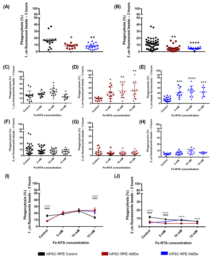
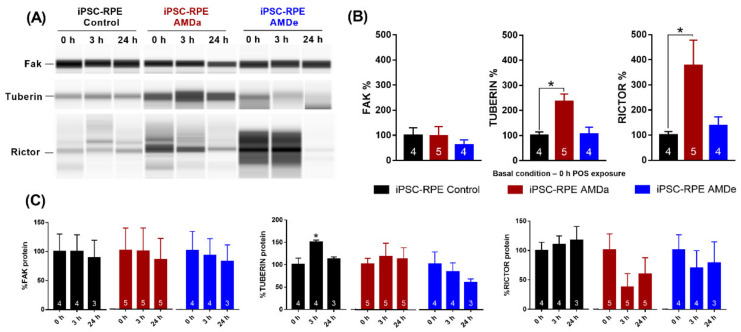
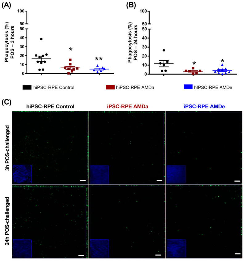
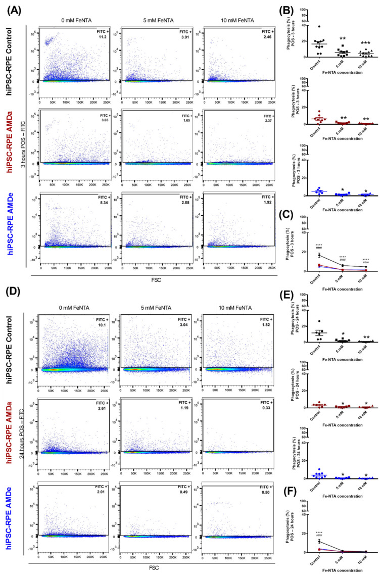
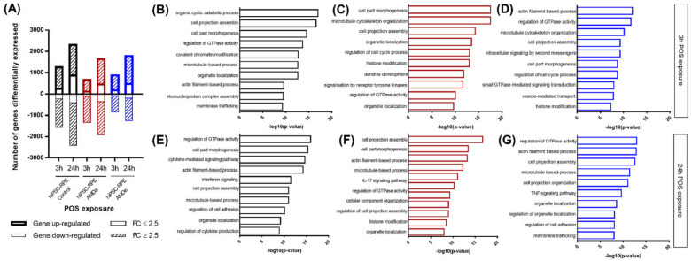
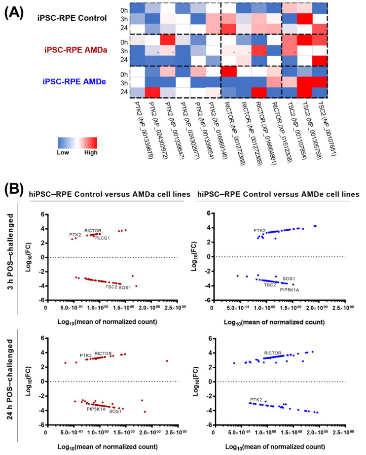
Age-related macular degeneration (AMD) is partially characterized by retinal pigment epithelial (RPE) cell dysfunction. This study focused on phagocytosis activity and its involvement in AMD. Phagocytic activity was analyzed by flow cytometry using porcine photoreceptor outer segment (POS) and fluorescent beads in basal and under oxidative stress condition induced by Fe-NTA in fifteen hiPSC-RPE cell lines (six controls, six atrophic AMD and three exudative AMD). Oxidative stress exposure inhibited phagocytosis in the same manner for control, atrophic AMD (AMDa) and exudative AMD (AMDe) cell lines. However, altered phagocytosis in basal condition in hiPSC-RPE AMDa/e was observed compared to control cell lines. Gene expression after 3 or 24 h of POS incubation was analyzed by RNA-Seq based transcriptomic profiling. Differential gene expression was observed by RNA seq after 3 and 24 h POS exposure. We have focused on the genes involved in mTOR/PI3K-AKT/MEK-ERK pathway. We investigated differences in gene expression by analyzing the expression levels and activity of the corresponding proteins by Western blot. We showed the involvement of three proteins essential for phagocytosis activity: fak, tuberin and rictor. These findings demonstrate that hiPSC-RPE AMDa/e cells have a typical disease phenotype characterized by alteration of the main function of RPE cells, phagocytosis activity.
年龄相关性黄斑变性(AMD)的部分特征是视网膜色素上皮(RPE)细胞功能障碍。本研究聚焦于吞噬活性及其在AMD中的作用。在15个人诱导多能干细胞来源的RPE细胞系(6个对照、6个萎缩性AMD和3个渗出性AMD)中,利用猪光感受器外段(POS)和荧光珠,通过流式细胞术分析基础状态下以及在由氮川三乙酸铁(Fe-NTA)诱导的氧化应激条件下的吞噬活性。氧化应激暴露以相同方式抑制对照、萎缩性AMD(AMDa)和渗出性AMD(AMDe)细胞系的吞噬作用。然而,与对照细胞系相比,观察到hiPSC-RPE AMDa/e在基础状态下吞噬作用发生改变。通过基于RNA测序的转录组分析,分析POS孵育3小时或24小时后的基因表达。在POS暴露3小时和24小时后,通过RNA测序观察到差异基因表达。我们重点关注参与mTOR/PI3K-AKT/MEK-ERK途径的基因。通过蛋白质印迹分析相应蛋白质的表达水平和活性,研究基因表达的差异。我们发现了吞噬活性所必需的三种蛋白质的作用:黏着斑激酶(fak)、结节性硬化蛋白(tuberin)和rictor。这些发现表明,hiPSC-RPE AMDa/e细胞具有典型的疾病表型,其特征是RPE细胞的主要功能即吞噬活性发生改变。